Kam Taghizadeh Week 5
Links to Weekly Assignments
Links to Individual Journal Assignments
- Kam Taghizadeh
- Kam Taghizadeh Week 2
- Kam Taghizadeh Week 3
- Kam Taghizadeh Week 4
- Kam Taghizadeh Week 5
- Kam Taghizadeh Week 6
- Kam Taghizadeh Week 7
- BacFITBase Review
- Kam Taghizadeh Week 9
- Kam Taghizadeh Week 10
- Kam Taghizadeh Week 11
- Kam Taghizadeh Week 12
- Kam Taghizadeh Week 14
Links to Shared Journal Assignments
- Class Journal Week 1
- Class Journal Week 2
- Class Journal Week 3
- Class Journal Week 4
- Class Journal Week 5
- Class Journal Week 6
- Class Journal Week 7
- Class Journal Week 8
- Class Journal Week 9
- Class Journal Week 10
- Class Journal Week 11
- Class Journal Week 12
- Class Journal Week 14
Purpose
The purpose of this assignment is to evaluate the structure of the spike protein, ACE2 protein, and the residues in between that promote their binding to one another. Furthermore, we will analyze the different levels of protein structure as well.
Methods/Results
Exploring the Spike protein Structure
Exploring UniProt
- First, I went to UniProt Knowledgebase (UniProt KB) and inserted "SARS-CoV-2" into the main UniProt search field.
- 1,611 results showed up, and the majority of them were viral proteins.
- Next, I clicked on the entry with accession number"P0DTC2" which corresponds to the reference entry for the SARS-CoV-2 spike protein.
- The information included on the SARS-COV-2 spike protein:
- Function
- Names and Taxonomy
- Subcellular location
- Pathology and biotech
- Posttranslational modification and processing
- Interactions
- Structure
- Family and domains
- Sequence
- Similar proteins
- Cross-references
- I then analyzed one of the structures of the SARS-CoV Spike protein from Wan et al. (2020) in the NCBI Structure Database using the web-based iCn3D viewer.
- The structure I chose to view was Figure 1C SARS-CoV RBD (optimized for human ACE2 recognition) and human ACE2: [1]
Recreating Figure 2A from Wan et al. (2020)
- I rotated the protein so that it recreated Figure 1A from the Wan et al. (2020) paper:
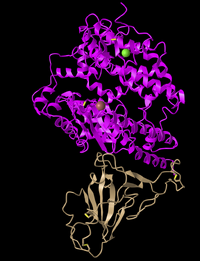
- In this image, the ACE2 protein is in pink while the spike protein is in tan.
- This structure is tertiary as it displays the protein structure in 3D, and is not just the secondary and primary structures.
- There are two domaines in this image, as each protein is its own domain (S protein and ACE 2 protein).
Cylinder and Plate Style
- I clicked on the Style > Proteins menu and selected Cylinder and Plate.

- This style displays alpha-helices as cylinders and beta-sheets as plates.
- The reason this style is unique is that one can easily see the secondary structures.
C Alpha Trace
- I clicked on the Style> Proteins menu and selected C Alpha Trace.
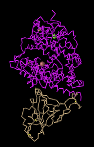
- The reason this style is unique is that it displays the protein backbone, while each bend in the structure represents an alpha carbon.
Lines
- I clicked on the Style > Proteins menu and selected Lines
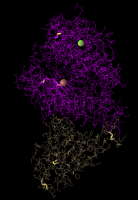
- This style is unique because it shows the amino acid structures throughout the protein.
Ball and Stick
- I clicked on the Style > Proteins menu and selected Ball and Stick.

- This style is unique because it displays the protein backbone and its amino acids in a molecular ball and stick model.
- This style allows one to see the 3D model of the atoms and their bonds that make up the protein.
Spheres
- I clicked on the Style > Proteins menu and selected Spheres.
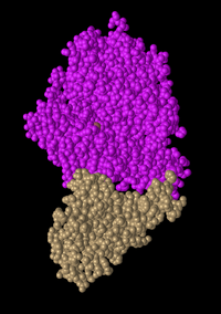
- This style is unique because it displays each amino acid as a sphere.
Spheres Spectrum Color
- In the “Spheres” view, I clicked on the Color menu and selected the Spectrum color scheme.
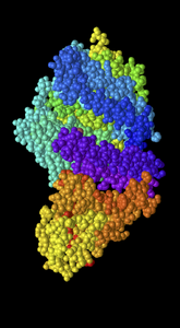
- This color scheme is unique because it displays the ACE2 protein in a color spectrum that includes purple/blue/green/yellow and it shows the Spike protein in a spectrum that includes red and yellow
Spheres Secondary Color
- In the “Spheres” view, I clicked on the Color menu and selected the Secondary color scheme.
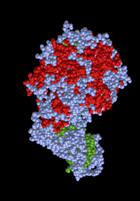
- This color scheme is unique because it displays the residues in alpha helices as red and the residues in beta sheets as green.
Spheres Charge Color
- In the “Spheres” view, I clicked on the Color menu and selected the Charge color scheme.

- This color scheme is unique because it displays the negatively charged residues in red, positively charged residues in blue, and neutrally charged residues in grey.
Spheres Atom Color
- In the “Spheres” view, I clicked on the Color menu and selected the Atom color scheme.
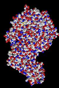
- This style is unique because it displays oxygens as red, carbons as grey, sulfurs as yellow, and nitrogens as blue. and carbons as grey spheres, oxygens as red spheres, nitrogens as blue spheres, and sulfurs as yellow spheres.
Assignment Questions and Answers
N-Terminus and C-Terminus

- The N terminus and C terminus can be seen in the gray boxes
Secondary Structures
- The secondary structures found in the ACE2 protein were only alpha helices
- This could be observed by evaluating the cylinder and plate style, which displayed alpha helices as cylinders in ACE2.
- The secondary structures found in the spike protein were only beta sheets
- This could be observed by evaluating the cylinder and plate style, which displayed the beta sheets as plates in the spike protein.
Exploring the Civet ACE2-Spike Protein Structure
- First, I went to this website
- Next, I clicked on the Windows menu to “View Sequences & Annotations”
- In the window that appeared on the right, I clicked on the “Details” tab to show the actual amino acid sequences
- There were 2 sets of ACE2-spike proteins because of the way the proteins crystallized.
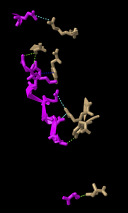
- I focused on the pink and tan chains and oriented them as they were shown in Figure 4B.
- In the sequence window I went to sequence “Protein 3SCK_A” (in pink) and selected the following amino acids
- T31
- E35
- E38
- T82
- K353
- The ribbon that represented these amino acids are highlighted in yellow in the structure
- I went to the Styles menu and selected Proteins > Ball and Stick
- I went to the Color menu and selected Atom
- In the sequence window I went to sequence “Protein 3SCK_A” (in pink) and selected the following amino acids
- I redid this process for the tan spike protein sequence 3SCK_E for the following amino acids:
- T487
- R479
- G480
- Y442
- P472
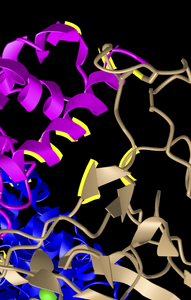
- E35 on ACE2 makes an ionic bond with R479 on spike protein
- It can be concluded that E35 is acidic due to the red oxygens
- It can be concluded that R479 is basic due to the blue nitrogens
- T31 on ACE2 makes a hydrogen bond with Y442 on spike protein
- T is considered polar due to its oxygens
- Y is considered hydrophobic due to its nitrogens
- Next, I went to the View menu and selected H bonds & Interactions
- In part 1 of the window that appeared, I unchecked “Contacts/Interactions” leaving Hydrogen Bonds and Ionic Interaction checked
- In part 2 of the window, I selected the first set “3SCK_A” (pink)
- In part 3 of the window, I selected the second set “3SCK_E” (tan)
- In part 4 of the window, I clicked the button “3D Display”
- This inserted dashed lines representing the ionic bonds and H-bonds between the two polypeptide chains and amino acids that were described above
Beginning your research project
- What question will you answer about sequence-->structure-->function relationships in the spike and/or ACE2 protein?
- The data presented in Wan et al (2020) suggests that a single mutation in N501T which corresponds to the S487T mutation in SARS-CoV
has the potential to greatly enhance the binding affinity between 2019-nCoV RBD and human ACE2. Due to this, patients should be monitored closely incase there is a mutation that occurs at the 501 position. We will answer is why the binding affinity increases between 2019- nCoV RBD and human ACE2, when there is a mutation at position 501.
- What sequences will you use?
- We will use the human ACE2 sequence and 2019-nCoV RBD sequence, along with a mutated sequence, N501T.
Conclusion
Evaluating the structure of the spike protein and Human ACE2 protein via various styles and color combinations on iCn3D has helped distinguish the different components of residues, being polar, uncharged, hydrophobic, or hydrophilic. It has greatly expanded my knowledge on the structure of proteins that are made up of different levels. Furthermore, it was concluded that ACE2 has a high number of alpha helices, while spike protein has beta sheets.
Acknowledgments
- I contacted/discussed with my homework partner, Fatima Alghanem, via text and face time to discuss the our research project and various protein figures
- I copied and paraphrased the protocol on the Week 5 Page
- I utilized the Wan et al. (2020) paper to resemble my protein structures
- I used sequences from GenBank
- I used NCBI Structure Database for protein structures
- Except for what is noted above, this individual journal entry was completed by me and not copied from another source
Kam Taghizadeh (talk) 23:12, 7 October 2020 (PDT)
References
- NCBI Structure (2020). SARS-CoV RBD (year 2002) complexed with human ACE2 complex 2AJF, Retrieved from https://www.ncbi.nlm.nih.gov/Structure/icn3d/full.html?&mmdbid=35213&bu=1&showanno=1.
- NCBI Structure (2020). Civet ACE2-Spike protein structure, Retrieved from https://www.ncbi.nlm.nih.gov/Structure/icn3d/full.html?pdbid=%203SCK.
- OpenWetWare. (2020). BIOL368/F20:Week 1. Retrieved September 22, 2020, from https://openwetware.org/wiki/BIOL368/F20:Week_1
- OpenWetWare. (2020). BIOL368/F20:Week 5. Retrieved October 07, 2020, from https://openwetware.org/wiki/BIOL368/F20:Week_5
- Wan, Y., et al. (2020). Receptor Recognition by the Novel Coronavirus from Wuhan: an Analysis Based on Decade-Long Structural Studies of SARS Coronavirus. Journal of Virology, 54 (7), retrieved from https://doi.org/10.1128/JVI.00127-20.
- Fold it Solving Puzzles for Science (2020). The Science Behind Foldit, retrieved from https://fold.it/portal/info/about#folditpub.