Tissue Engineered Spleen by Ronen Zeidel, John Vetrano
Overview
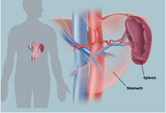
The spleen is located behind the 9th-11th ribs on the left hand side of an individual. The spleen has a variety of niche functions but mainly serves to filter blood. Two predominant types of tissues are found within the spleen: the red pulp and the white pulp. The red pulp is primarily involved in the filtration of red blood cells (RBC) and the white pulp is actively involved in immune response through both humoral and cell-mediated pathways [16].
The spleen is a non-vital organ, meaning it is not imperative for survival; however, individuals without a spleen are subject to elevated risk of contracting common infections that, while not fatal to a typical individual, can prove fatal to these individuals. The most common reason an individual lacks a spleen is due to removal via splenectomy following a splenic rupture from blunt force trauma. Vehicular accidents and contact sports are the most common causes of a ruptured spleen [8].
The most popular method of treating spleen-related problems such as a ruptured spleen or splenic cancer is by partial or complete removal of the spleen (Splenectomy). The spleen’s ability to regenerate allows those who receive partial splenectomy to regrow the spleen, however, those individuals who receive a full splenectomy cannot [8].
Unfortunately, because the spleen is a non-vital organ many patients who undergo full splenectomy do not receive a replacement organ. This increases their susceptibility to various complications such as the fungal infections and septic shock. The susceptibility of the patients to blood-related illness poses a continuous threat which lead to frequent visits to the hospital if they do get infected. Furthermore, since blood-related illnesses are commonly caused by bacterial agents, the patients must be put on antibiotics which can lead to the development of multi-drug resistant pathogens. The fact that the spleen is not a vital organ appears to have impact on research efforts as well. Not as many investments are made toward tissue-engineering spleens as are made to the more necessary, vital organs. However, recent advancements in research have shown promise for both the development of a tissue engineered spleen (TES) and an artificial spleen device. The commercialization of the artificial spleen device may be able to reduce the mortality of Sepsis, which is responsible for 300,000 deaths annually in the US alone [15].
Background
Anatomy and Physiology
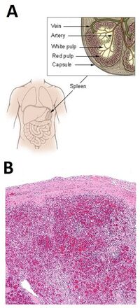
The spleen is an organ found in virtually all vertebrates. In humans, the spleen lies behind and between the 9th and 11th ribs on the left hand side of an individual and weights approximately 7 oz. [17]. The spleen has two ends known as the posterior and anterior ends. The anterior end of the spleen is expanded forwards and downwards towards the center of the body. The posterior end extends in the opposite direction, upwards and towards the back of the body. The spleen also consists of two surfaces: the diaphragmatic and the visceral. The diaphragmatic surface is smooth and convex in contrast to the rougher and concave visceral surface. Both the splenic artery and the splenic vein interface with the organ on the visceral side [6].
The interior of the spleen is composed mainly of two types of tissues: red pulp, and white pulp. Along with these components are supporting tissues and the vasculature. The supporting tissue is fibroelastic and forms the capsule in which the pulp tissues lie. The red pulp is composed of a collection of cells in the cavities within the supporting tissues including lymphocytes, blood cells, and macrophages [8].
The primary role of the spleen is to act as a blood filter. It performs this duty through both the filtration of red blood cells and the filtration of antibody-coated bacteria [16]. The spleen also acts as a storage location of red blood cells which can be released when needed (240 mL RBC storage in humans) [8]. The red pulp is made up of venous sinuses and splenic cords, which are responsible for its hematologic functions, while white pulp is mainly lymphatic tissue, which is responsible for its immune related functions [18].
Additionally, a niche role of the spleen is its responsibility for the production of opsonins, a class of antibodies that serve as biological markers, encapsulating antigens that pass through the spleen and marking them as a target for enhanced immune response. This function is less prominent, especially in healthy adults whose immune systems are already capable of recognizing certain pathogens and have the correct antibodies to respond.

Regeneration
The spleen has the ability to regenerate itself if it is damaged as well as the ability to autotransplant elsewhere in the body, a process known as splenosis. During splenosis a damaged spleen can send cells to regenerate in another area of the body. Splenosis does have its short comings as many of the spleens created via these process either lack a certain pulp or lose functionality [2]. Researchers, however, were able to take advantage of this naturally occurring process in order to regenerate the spleen in full splenectomy cases. The natural regenerative growth of the spleen is often taken advantage of in order to regrow damaged spleens via a partial splenectomy or to grow a new spleen in a patient via the spleen splice method. Both methodologies take advantage of the natural regrowth of splenic tissues, however imperfections in the distribution of red and white pulp tissues have been noted [9].
Motivation
The spleen plays a vital role in blood filtration. One of the largest motivating factors for the artificial spleen device is its potential benefits in the treatment of sepsis which is annually responsible for 300,000 deaths in the US and 8 million deaths worldwide [15]. Sepsis is a life-threatening condition that occurs when the body's response to an infection injures its own tissues and organs. The lack of a spleen, either by birth defect or splenectomy, greatly increases an individual's risk for sepsis. However, even individuals with a healthy spleen are at risk, as sepsis occurs in 1-2% of all hospitalizations. The current treatment of sepsis is to prescribe antibiotics until one is found that selectively targets the pathogen responsible. The downside of this method is that each hour spent giving a patient an ineffective antibiotic greatly increases their risk of death. This risk is already significant as patients with sepsis have a mortality rate above 30% [15]. The artificial spleen device looks to mimic a spleen's ability to remove pathogens without explicitly targeting a single suspect. This can be used in conjunction with current antibiotic therapies to deliver a combination of therapies that both seek out to destroy and remove the pathogen [15].
The motivation for the tissue engineered spleen (TES) was to improve upon current spleen splice methodology of transplant [2]. If protocols of cell culture techniques for spleen tissues can be perfected, this methodology would allow a single donor spleen to serve far more than a lone individual recipient, increasing the availability of donor supply. However, the motivation for artificial splenic tissue regeneration in general is comparatively less than in many other vital organs. The spleen is considered a non-vital organ, as survival without a spleen is possible. The spleen also has the capability to regrow from a partial splenectomy. Although the negative impacts of living without a spleen may pale in comparison to the impacts of living without another vital organ such as the lungs, the impacts on a person's daily life can be profound. An individual lacking a spleen is at a greater risk of infection and as a result must be much more careful about their health and cleanliness. It is recommended that individuals without a spleen always carry antibiotics when they travel and that they receive a flu shot annually [8]. As a result, the greatest weight upon the shoulders of individuals without a spleen is not the direct impact of pain or loss of function but rather the constant threat of common infections that, while harmless to others, can prove to be fatal to these individuals.
History Timeline
1838-1839: Matthias Jakob Schleiden and Theodor Schwann propose cell theory [13]
1897: Jaques Loeb proposes the idea of culturing cells outside of the body [13]
1907: Ross G. Harrison first to grow frog ectodermal cells in vitro [13]
1912: Alexis Carrel grows and maintains chick embryos in vitro for years [13]
1952: John Franklin Enders demonstrates the ability of human embryonic cells to become tissue-specific cells [13]
1998: The use of embryonic and adult stem cells becomes a topic of interest in medicine [13]
2007: First report of tissue-engineered spleen by Tracy C. Grikscheit [2]
2008: first whole organ transplant of tissue-engineered trachea was done by prof. Paolo Macchiarini [7]
2014: First artificial spleen device created [5]
2015: Formation of first commercial entity involved in creation of artificial spleen device [15]
Health Concerns
One of the major concerns for people who lack a spleen are pathogens that cause and can lead to blood-related disease [19]. The pathogen of most concern is Streptococcus pnuemoniae the bacterial agent responsible for pneumonia. In immunocompromised individuals, such as people lacking a spleen, the bacterial agent can penetrate the blood stream in order to spread throughout the body. S. pnuemoniae is of such high interest because of its common rate of infection and the bacteria’s fatal ability of inducing sepsis conditions, such as septic shock. Without the spleen, immune response can be delayed or not occur, which may result in death. Additionally, without the spleen the only course of action is the usage of large amounts of antibiotics, which may result in a multi-drug resistant strain [20].
Past work
Partial Splenectomy
A partial splenectomy involved the removal of some but not all of the splenic tissue. The functionality of a spleen regrown from a partial splenectomy as opposed to a spleen regrown from the spleen slice method has been challenged, but research suggests that it is the most successful at restoring the spleen. While this should be kept in mind, it is important to note that other techniques are geared towards individuals who must receive a full splenectomy.
Spleen Slice
Spleen slicing occurs strictly in vivo and relies on the spleen's natural ability to regenerate through the same mechanism as splenosis. The spleen slices may be introduced to the subject’s body where the spleen is located or in an omental pouch, which is a space that is produced surgically in the omentum (abdomen) close the where the spleen is usually found. Since it can only be done in vivo, the process of spleen slicing proves to be a difficult process to monitor. For example, the development of the new spleen, as it might not occur, and functionality assessments are more difficult to complete. There is no way to guarantee that the right portion of either pulp, or any at all will be created in the subject. The developmental and functionality results can only be accomplished by harvesting the organ.
Current Research
Tissue Engineered Spleen (TES)
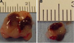
Recent work has highlighted the successful use of tissue engineering techniques for improving the traditional spleen slice method. In a conventional spleen slice, the regeneration of the spleen tissue must first overcome an initial necrosis of the implanted tissue. This reduces the tissue down to a connective tissue structure which subsequently regenerates with regrowth of vascularizartion, red pulp, and white pulp by weeks 5-7 [2]. This regeneration occurs on the connective tissue scaffold resulting from major necrosis; however, Grikscheit et al. have demonstrated an improved tissue-engineered spleen (TES) technique which overcomes this delayed initial growth. The TES methodology involved supplying a framework for regrowth in the form of a biodegradable polyglycolic acid (PGA) scaffold (fiber diameter: 15 μm; mesh thickness: 2 mm; bulk density: 60 mg/cm3; porosity: > 95%) [2]. These polymer scaffolds were formed into 1 cm tubes and sealed with poly-l-lactic acid (PLA) prior to being loaded with splenic units (SU) internally via micropipette. SU are described as multicellular components of juvenile spleen produced by dissecting the spleens of 6-d-old Lewis rat pups. The polymer scaffold, loaded with SU, was then implanted as in a normal spleen slice and allowed to grow for 16 weeks. As can be seen in Figure X, the spleens regrown using the TES technique exemplified much greater regrowth over this time period and exhibit both the yellow and white pulps found in a native spleen [2].
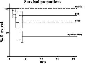
Following this time period weeks, groups of rats who had received no operation, a TES, a spleen slice, or a splenectomy were subsequently inoculated with Streptococcus pneumoniae type 25 (ATCC) via intraperitoneal injection. The survival proportions of these rats over a 21-day period was then recorded. As can be seen in Figure Y, the TES mice exhibited a greater survivability (85.7%) compared to Slice mice (71.43%), demonstrating the improved performance of the TES [2].
Artificial Spleen
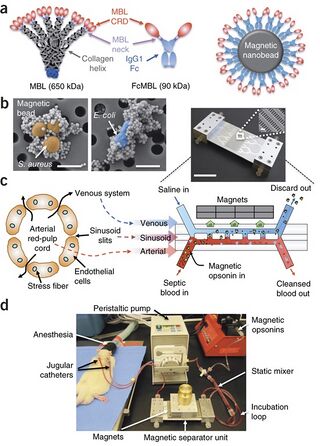
The latest development in the construction of an artificial spleen is work done by Donald Ingber, a bioengineer at the Wyss Institute for Biologically Inspired Engineering in Boston, Massachusetts. The device was originally developed in 2014 and cleansed pathogens and toxins from blood flowing through a dialysis-like microfluidic circuit[5]. It relied on magnetic nanobeads to function as opsonic molecules and filter pathogens and old/damaged erythrocytes from the blood. The magnets were coated with a truncated mannose-binding-lectin (MBL), a human opsonin that captures a multitude of pathogens. The magnetic nanobeads were introduced into the blood flow of mice models and attached to the antigen of interest. They were subsequently filtered out via a magnetic force which pulled the magnetic nanobeads into a parallel flow of saline solution within the microfluidic circuit.
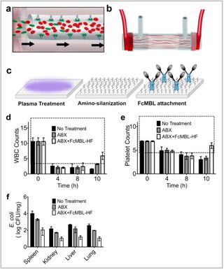
Several concerns with the practicality of this device arose including the cost of the nano-magnets and the risk of not recapturing 100% of the nano-magnets before reentry into the body. Additionally, the device was only capable of processing blood at a rate of 10 mL/hr. The average human contains 5L of blood and would as such require 500 hours or ~21 days of continuous treatment. While the devices could be implemented in parallel, to scale for larger animals, the design challenges and the complexity of the arrangement proposed significant road blocks. As such, the design was simplified and improved to remove the use of nano-magnets by binding a modified version of MBL, FcMBL, to the inner walls of hollow fiber filters found in dialysis cartridges already approved by the FDA [Figure K]. Similar devices have shown practicality in their ability to scale to accommodate larger volumes of blood flow [15]. In small-animal studies, treatment with this improved device removed 99% of E. coli, Staphylococcus aureus, and endotoxins circulating in the bloodstream [15]. The Wyss team is planning to move to large-animal trials in order to demonstrate the device’s proof of concept prior to advancing to human clinical trials. The goal of this improvement is to progress towards commercialization of the treatment, which is intended to be used in conjunction with current anti-biotic therapies.
Future Work
Opsinix Inc, formed by D. Ingber and M. Super [15], launched on October 8, 2015 as the first commercial entity involved in the construction of an artificial spleen-like device. Opsonix is founded by the research team behind the magnetic nanobead technology and seeks to commercialize the product for human treatment in the future. The improved device schematic of using surface bound proteins on conventional dialysis membranes has proved successful in small-animal trials and looks to move forward to large-animal trials in the near future. If the practicality and effectiveness of the device is successfully demonstrated, the device could begin human clinical trials within the next 5-10 years.
Future investigations into tissue engineering the regeneration of the spleen will likely focus on alternative scaffolding methods or the inclusion of growth factors to promote splenic regeneration. Additionally, researchers hope that more attention will be directed towards understanding the mechanisms and steps that take place when a new spleen is introduced into a patient’s body. The information of the dynamics between the host and the transplanted organ will allow for scientists to create a functional spleen with the correct ratio of the white and red pulp regions. Researchers hope that more attention will be directed toward the in-vivo oxygen diffusion limitation of engineered solid organs so that a procedure would be developed in order to counter the harmful effects.
References
[1] Alvarez, F. E., and R. S. Greco. "Regeneration of the Spleen After Ectopic Implantation and Partial Splenectomy." Archives of Surgery 115.6 (1980): 772-75. Web.
[2]Grikscheit, Tracy C., Frédéric G. Sala, Jennifer Ogilvie, Kate A. Bower, Erin R. Ochoa, Eben Alsberg, David Mooney, and Joseph P. Vacanti. "Tissue-Engineered Spleen Protects Against Overwhelming Pneumococcal Sepsis in a Rodent Model." Journal of Surgical Research 149.2 (2008): 214-18. Web.
[3]"Histology Laboratory Manual." Histology Laboratory Manual. Columbia University, n.d. Web. 06 Apr. 2015.
[4]Hoad-Robson, Rachel. "The Spleen | Health | Patient.co.uk." Patient.co.uk. Patient, 16 Oct. 2012. Web. 05 Apr. 2015.
[5]Ingber, Donald E., et al. "An Extracorporeal Blood-cleansing Device for Sepsis Therapy." Nature Medicine (2014): n. pag. Web.
[6]Leal-Calderon, Fernando, and Maud Cansell. "The Design of Emulsions and Their Fate in the Body following Enteral and Parenteral Routes." Soft Matter 8.40 (2012): 10213. Web.
[7]Marchiarrani, Paulo. "First Tissue-engineered Whole-organ Transplant." First Tissue-engineered Whole-organ Transplant. Animal Research, 2008. Web. 05 Apr. 2015.
[8]"Spleen: Information, Surgery and Functions." Spleen: Information, Surgery and Functions. Children's Hospital of Pittsburgh, n.d. Web. 05 Apr. 2015.
[9]"Opsonin." TheFreeDictionary.com. The Free Medical Dicitionary, n.d. Web. 06 Apr. 2015.
[10]Tavassoli, Mehdi, Judith R. Ratzan, and William H. Crosby. "Studies on Regeneration of Heterotopic Splenic Autotransplants." Blood Journal 41.5 (1973): n. pag. Web.
[11]"The Spleen (Human Anatomy): Picture, Location, Function, and Related Conditions." WebMD. WebMD, 2014. Web. 06 Apr. 2015.
[12]Traub, Audrey, Scott Giebink, Clark Smith, Christopher C. Kuni, Mary Lee Brekke, Debbie Edlund, and John F. Perry. "Splenic Reticuloendothelial Function after Splenectomy, Spleen Repair, and Spleen Autotransplantation." New England Journal of Medicine 317.25 (1987): 1559-564. Web.
[13]Van Winterswijk, Peter J., and Erik Nout. "Medscape Log In." Medscape Log In. MedScape, 2007. Web. 05 Apr. 2015.
[14]Wiener, Eugene S. "Preservation of Splenic Function by Autotransplantation of Traumatized Spleen in Man." Journal of Pediatric Surgery 17.4 (1982): 444. Web.
[15] Ingber, Donald E., et al. "Improved treatment of systemic blood infections using antibiotics with extracorporeal opsonin hemoadsorption." Biomaterials 67 (2015): 382-392.
[16] Vellguth, Swantje; Brita von Gaudecker; Hans-Konrad Müller-Hermelink (1985). "The development of the human spleen". Cell and Tissue Research (Springer Berlin / Heidelberg) 242 (3): 579–592. doi:10.1007/BF00225424.
[17] Spielmann, Audrey L.; David M. DeLong; Mark A. Kliewer (1 January 2005). "Sonographic Evaluation of Spleen Size in Tall Healthy Athletes". American Journal of Roentgenology (American Roentgen Ray Society) 2005 (184): 45–49. doi:10.2214/ajr.184.1.01840045. PMID 15615949.
[18] Blackbourne, Lorne H (2008-04-01). Surgical recall. Lippincott Williams & Wilkins. p. 259. ISBN 978-0-7817-7076-7.
[19] Brender, Erin (2005-11-23). Richard M. Glass, ed. Illustrated by Allison Burke. "Spleen Patient Page" (PDF). Journal of the American Medical Association (American Medical Association) 294 (20): 2660. doi:10.1001/jama.294.20.2660. PMID 16304080.
[20] Rubin, LG; Schaffner, W (July 2014). "Clinical practice. Care of the asplenic patient". The New England Journal of Medicine 371 (4): 349–56. doi:10.1056/NEJMcp1314291. PMID 25054718
