User:Anthony Salvagno/Notebook/Research/2009/02/13/Plasmid Prep and Testing
Today we planned out the next set of processes that will take us up to the point of creating tethers.
Plasmid Library Plan

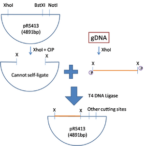
We are choosing to use pRS413 as our plasmid. It has one XhoI site, one BstXI site, and one NotI site. We will implant randomly digested XhoI genomic fragments into the XhoI site of the plasmid. We want to use one of the other sites to get similar template DNA in the tethers. BSTX1 has a very strange cutting pattern, as it recognizes a specific sequence with some number of any nucleotides in the middle. This provides us the opportunity to possibly have a non-palindromic sticky end. In this plasmid the cutting site is palindromic, but a location in the genomic piece may not be. NotI also has its use, but is palindromic. I think we would use this because there are few (not as many as XhoI or other enzymatic sites) in the genome.
Digesting the plasmid in XhoI gives two palindromic sticky ends for ligation. We will use CIP to remove the phosphate group from the exposed sticky ends to prevent self-ligation. We will digest genomic DNA with XhoI to create random fragments of random size. These fragments have the necessary phosphate group to undergo ligation. We will mix the digested components together in the presence of T4 DNA Ligase to insert the fragment into the plasmid. This plasmid will be ready for cloning.
According to Kelly's notes the next steps go as follows:
- Amplify DNA in E. coli
- Isolate DNA
- Test for insert and NotI/BstXI sites by RE digesting
- The digestions will be used for tethering.
Testing for RE sites and digestion
Cory gave us some plasmid DNA for experimentation. Before we perform any cutting, we want to make sure we can cut. To do this we will test for the presence of REs by undergoing digestions. Since there should be only one site in the plasmid, all our DNA should be of the same length. We will digest plasmid DNA with XhoI, BstXI, and NotI. We will also digest gDNA with XhoI.
Below is how we prepared the samples. We needed 1.5ug for each sample of pRs413 and 3ug for each gDNA (we believe that the DNA isn't as pure as it says so we want some extra sample). All final calculations are in the table:
| Tube Number | Vol pRS413 | Vol gDNA | Vol and Type 10x Buffer | Vol BSA 10x | Vol XhoI | Vol NotI | Vol BstXI | Vol H2O |
|---|---|---|---|---|---|---|---|---|
| 1 | 2.8uL | ---- | NE2/2.5uL | 2.5uL | ---- | ---- | ---- | 17.2uL |
| 2 | 2.8uL | ---- | NE2/2.5uL | 2.5uL | 1uL | ---- | ---- | 16.2uL |
| 3 | 2.8uL | ---- | NE3/2.5uL | 2.5uL | ---- | 1uL | ---- | 16.2uL |
| 4 | 2.8uL | ---- | NE3/2.5uL | 2.5uL | ---- | ---- | 1uL | 16.2uL |
| 5 | ---- | 14uL | NE2/2.5uL | 2.5uL | ---- | ---- | ---- | 6uL |
| 6 | ---- | 14uL | NE2/2.5uL | 2.5uL | 2uL | ---- | ---- | 4uL |
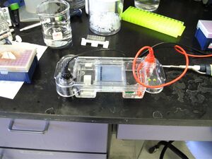
We allowed the digestion to occur. Digestion of BstXI was at 55C while all other enzymes operate at 37C. We allowed to digest for 90 minutes (more cause I went out for lunch). After digestion I added 5uL of loading buffer (with dyes and stuff) to each sample. I loaded 25uL into each well of 1% TAE agarose gel. Gel ran for x minutes.
Results

I have an image of the gel results, but for some reason it didn't save right. Working on it.
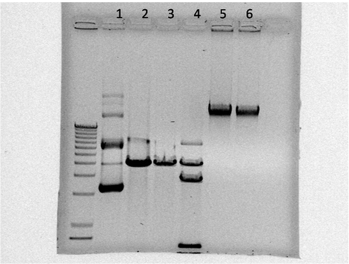
According to the image of the gel, we got some pretty good data from cutting the plasmid with XhoI and NotI. It looks like there is an extra BstXI site in the plasmid which is why there are two bands (lane 4). We checked this on the map and sure enough there was an extra cutting site. Lane 1 provides the expected outcome (supercoiled DNA, nicked DNA, and regular DNA all in the same samples).
When the gDNA was cut with XhoI we didn't get as much a smear as we expected. You can see that the intensity of the fluorescence is not as bright in lane 6 as it is in lane 5 so we assume there was a little bit of cutting. The one solid band in lane 5 leads us to believe that we had some pretty clean DNA to begin with (no particulates, or sheared DNA).

SJK 14:22, 16 February 2009 (EST)

Nice gel! Lanes 1-4 make lots of sense to me and look great. I don't understand how genomic DNA runs, so I'll take your words for it :) . I made a gel labeling template, for sideways gels, if you're interested, I can find it (in Brandon's notebook somewhere).