Biomod/2012/Harvard/BioDesign/methods
<html>
<head>
<link href='http://fonts.googleapis.com/css?family=Open+Sans' rel='stylesheet' type='text/css'>
</head>
<style>
</style>
</html>
Methods
Design Tools
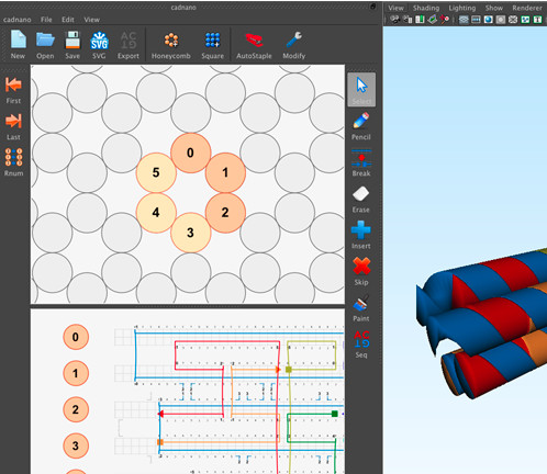
Cadnano2
Cadnano2 is software that we used for designing and visualizing DNA origami nanostructures.
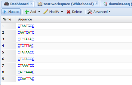
DyNAMiC Workbench
DyNAMic Workbench is an online tool that we used for designing and manipulating our DNA sequences to anneal at specific temperatures.
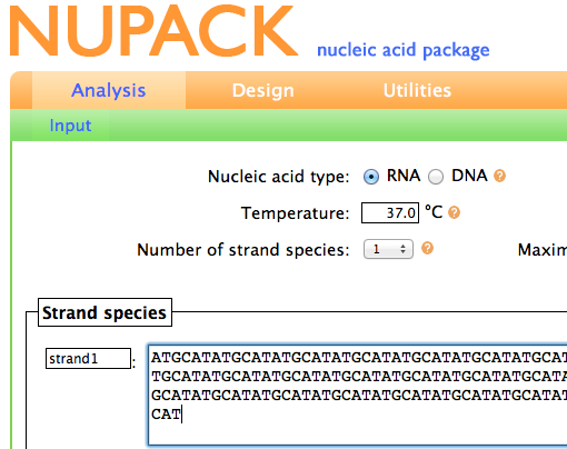
NUPACK
NUPACK is an online tool that we used for computing the temperature at which our SST structures would melt.
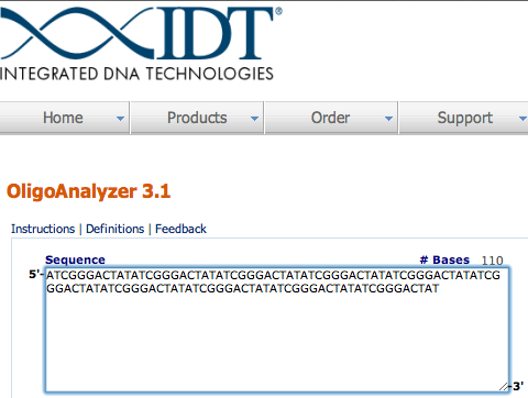
Oligo Analyzer
Oligo Analyzer is an online tool that we used for determining if any of our SST structures would bind complementary to themselves.
Sequence Massager
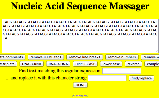
Sequence Massager is an online tool that we used for reversing or finding the complement strands for our SST sequences.
MFold
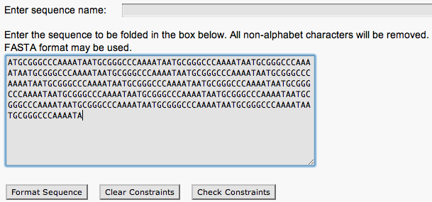
MFold is an online tool that we used for determining the temperatures at which our SST structures would form.
Structure Folding
Thermal Cycler
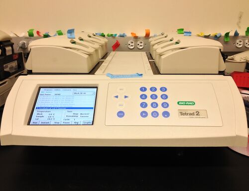
The thermal cycler is a machine which we used to anneal our SST strands to form structures. It works by cycling tubes through predetermined temperatures and times.
Analysis Equipment
Agarose Gel Electrophoresis
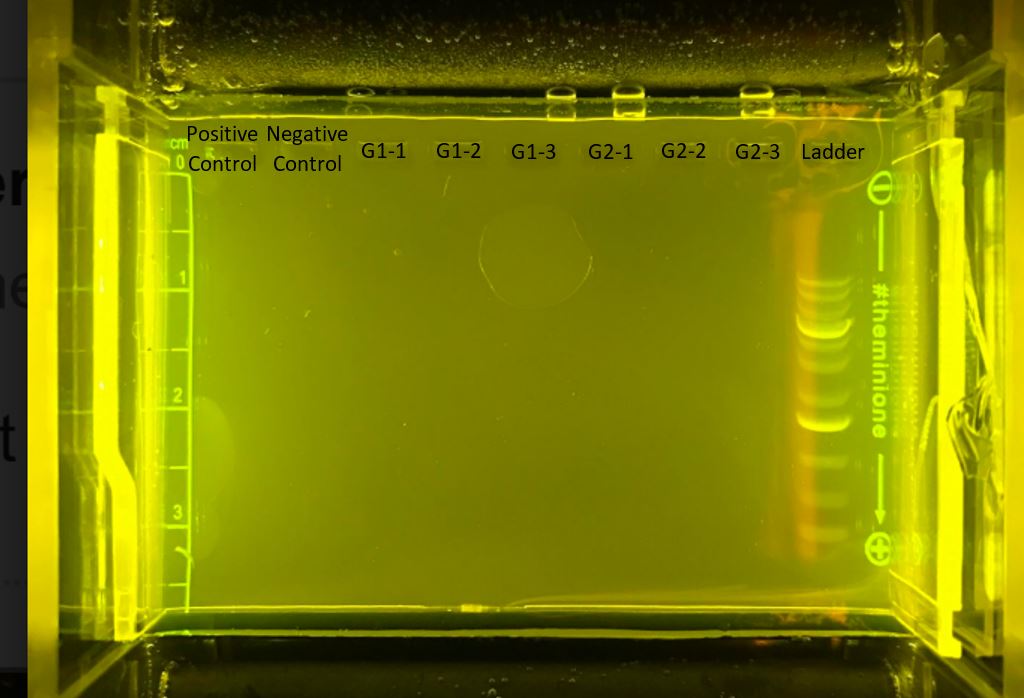
Agarose Gel Electrophoresis is a method which we used in order to view our DNA SST structures separated by size. The agarose gel contains wells along the top in which one can place DNA samples. An electrical current pulls the negatively-charged DNA through the agarose gel. Smaller structures travel through the gel faster than larger structures.
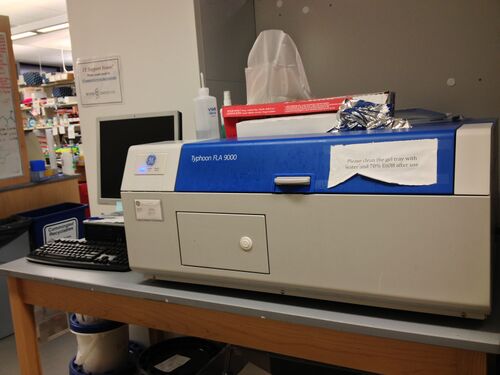
Typhoon Gel Imager
The Typhoon Gel Imager is a machine used to view the DNA run through a gel. It works by scanning the gel with UV light and recording the resulting emissions. Areas containing DNA show up as dark bands on the resulting image.
Gel Purification
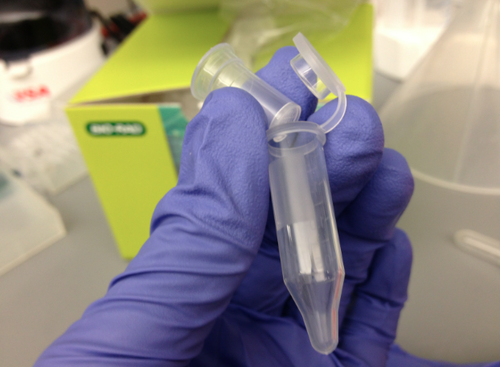
In order to obtain DNA SST scaffolds run on a gel, we performed gel purification with Freeze 'N Squeeze Spin Columns from Bio-Rad. Each Freeze 'N Squeeze Spin Column contains a filter that allows only DNA structures but not the agarose gel through.
Centrifuge
In order to force the DNA through the Freeze 'N Squeeze Spin Column filter, we used a centrifuge. It works by spinning samples at high speeds, exposing them to high centrifugal forces. For our purposes, this helped to force DNA through the filter leaving agarose fragments behind.
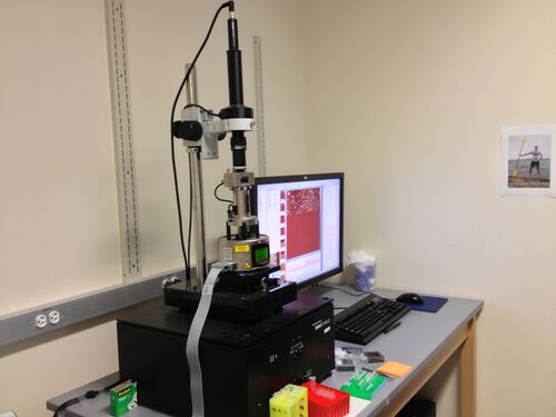
Atomic Force Microscopy
To view the resulting DNA SST structures, we used Atomic Force Microscopy (AFM). This machine works by oscillating a very small tip over a surface. As a result, the tip is able to "feel" any perturbations on the surface on the scale of nanometers. In this case, DNA SST structures were detected as perturbations visible on a computer screen.
