BME100 s2017:Group2 W8AM L6
| Home People Lab Write-Up 1 | Lab Write-Up 2 | Lab Write-Up 3 Lab Write-Up 4 | Lab Write-Up 5 | Lab Write-Up 6 Course Logistics For Instructors Photos Wiki Editing Help | |||||||
OUR COMPANY
Our Brand Name LAB 6 WRITE-UPBayesian StatisticsOverview of the Original Diagnosis System In order to test the 34 patients for the disease-associated SNP gene, the class was divided into 17 groups of 6 students, and each group tested two patients each. Each patient's DNA was replicated three times through PCR to ensure accuracy of the results. Both a positive and negative control were used for comparison and to ensure that there was no cross-contamination. When using the ImageJ software to determine the results of the PCR, calibration controls with varying amounts of DNA were used and each drop had three pictures taken and used for the ImageJ comparisons. The repetition used for each sample was used to help eliminate errors and increase the validity of our results. The final results from the entire class were collected and displayed on a spreadsheet and the final conclusions were compared to the doctor's diagnosis for each patient.
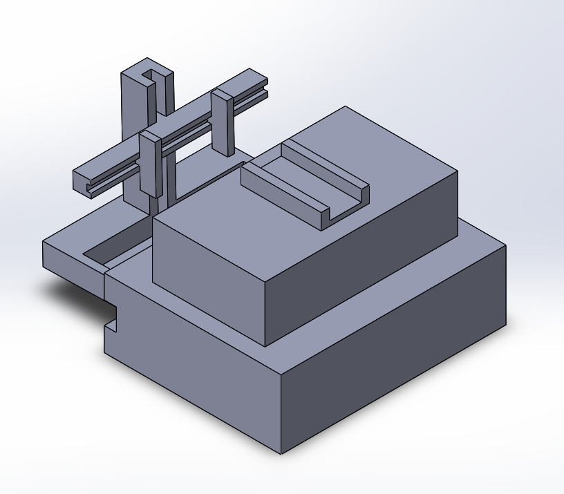
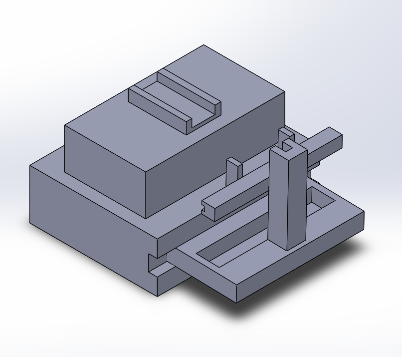
Intro to Computer-Aided Design3D Modeling We decided to use SolidWorks to design our devices. SolidWorks can be difficult to work with because the layout is confusing. There is also a lack of experience with SolidWorks. Overall though, our design turned out well once we had more practice. Our Design
Feature 1: ConsumablesThe consumables involved in this product are standard for fluorimetry. They include:
Included in the packaged kit are the PCR mix, primer solution, SYBR Green dye, buffer solution and tubes, glass slides, micro tubes and micropipette tips, along with an informational manual with step-by-step instructions. In order to cut cost, the micropipette is sold separately, and the DNA sample solution should be acquired according to the experiment.
Feature 2: Hardware - PCR Machine & FluorimeterOur improvement for the PCR machine is to include a pre-made solution of Taq polymerase and Deoxyribonucleotides. The tubes will be standard PCR size and have labels that clearly indicate the amount of solution in the tube. This decreases the chance of cross contamination by decreasing the steps involved in making the solution for the PCR machine. Only the primer and DNA sample need to be added. Our device is a stand for the fluorimeter with adjustable knobs to make it easier to adjust a phone when taking pictures. The fluorimeter will be placed on top of the stand and will have an attachment for a phone to be placed into. The stand for the phone will be able to move vertically and horizontally to allow any type of phone to be used. The stand will also hold the phone and fluorimeter securely so as not to change the image once it is focused.
| |||||||


