|
OUR TEAM
Mohammed Mousa, Maryam Ibrahim, Quintin Woods, Kayla Cummins, Kyndal Sorenson
LAB 5 WRITE-UP
SYBR Green 1 Dye
SYBR Green 1 Dye is used as a nucleic acid stain and has numerous uses in the biology world. It is a small molecular dye that illuminates effectively while in the presence of double-stranded DNA, but does not illuminate as well when mixed with water or single-stranded DNA. The green dye can be placed in a sample with possible DNA and will glow a bright green if DNA is present. This is the most common use of the dye and helps qualitatively analyze if DNA is present in a substance. The dye can also be placed inside a PCR mixture and this allows the dye to bind to the single-stranded DNA. It can still be used with both, and additionally RNA.
Single-Drop Fluorimeter
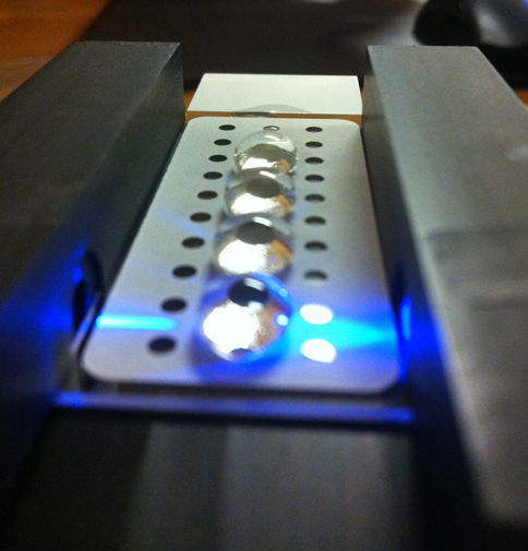
The Single-Drop Fluorimeter is a small and black plastic box with two notches where the multi-welled slide can fit in. It has a switch located on the right hand side that allows for the blue LED light to be turned on or off. The slide of the fluorometer is aligned so that the blue LED shines through a drop placed over two middle wells. The slide has holes along it to allow for a sample to be placed in them. Placing the drops must be carefully done to ensure no overflow.

(This image was borrowed from another group because we did not take a picture of our set-up.)
How the Fluorescence Technique Works
The fluorometer has a slide with small holes where drops of sample DNA were placed. The slide layer is made of Teflon, giving the slide texture to allow the sample drop to maintain its "drop" shape. The reason this is necessary is because the light from the fluorometer can go directly through the drop to increase the intensity of the DNA glow. It also allows for the SYBR Green to move to the surface, which allows for the glow to be seen on the top of the drop. All of this makes it easier to observe and analyze whether the sample has a green glow, and whether there was DNA present in the sample.
Procedure
Smart Phone Camera Settings
- Type of Smartphone: iPhone 4s
- Flash: Off
- ISO setting: NA
- White Balance: NA
- Exposure: NA
- Saturation: NA
- Contrast: NA
Calibration
For the calibration, the phone was placed into the cradle and the self-timer was readied. The flourimeter was 6 cm away from where the drop was going to be placed and the slide was placed into the holder. A sample of DNA was placed onto the slide using the micropipette. The black box was placed over the cradle and flourimeter and the picture was taken. This process was repeated for every DNA sample.
- Distance between the smart phone cradle and drop = 6 cm
Solutions Used for Calibration
| 5 |
80 |
80 |
2.5
|
| 2 |
80 |
80 |
1
|
| 1 |
80 |
80 |
0.5
|
| 0.5 |
80 |
80 |
0.25
|
| 0.25 |
80 |
80 |
0.125
|
| 0 |
80 |
80 |
0
|
Placing Samples onto the Fluorimeter
- Insert the multi-welled slide into the notches on the fluorimeter so that the light passes through the middle of two rows.
- Using the pipette, drop 80 μL of SYBR GREEN onto the slide.
- Drop 80 μL of the DNA sample onto the same droplet, creating a mixture.
- Cover the apparatus using the black box to prevent any light from creeping in and take a timed picture using a smartphone.
Data Analysis
Representative Images of Samples

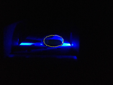
The picture above is a sample with no DNA.

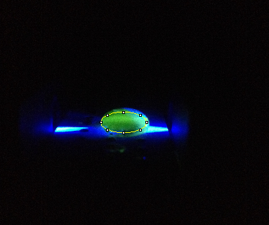
The picture above is a sample with DNA.
Image J Values for All Samples
| Final DNA concentration in SYBR Green I solution (µg/mL)
|
AREA
|
Mean Pixel Value
|
RAWINTDEN
|
RAWINTDEN
|
|
|
|
OF THE DROP |
OF THE BACKGROUND
|
| 1. 0 |
96384 |
7.169 |
690977 |
282517
|
| 2. 0 |
99668 |
6.198 |
617696 |
276082
|
| 3. 0 |
99532 |
7.54 |
750507 |
286709
|
| 4. 0.25 |
90952 |
7.788 |
708329 |
304315
|
| 5.0.25 |
77596 |
15.616 |
1211707 |
236426
|
| 6. 0.25 |
106672 |
4.01 |
427741 |
315565
|
| 7. 0.5 |
74072 |
10.234 |
758023 |
280597
|
| 8. 0.5 |
105444 |
20.949 |
2208918 |
318261
|
| 9. 0.5 |
115664 |
17.723 |
2049928 |
327516
|
| 10. 1 |
123672 |
38.948 |
4816732 |
460646
|
| 11. 1 |
121536 |
39.985 |
4859576 |
447692
|
| 12. 1 |
120572 |
42.031 |
5067789 |
457714
|
| 13. 2 |
123192 |
68.808 |
8476560 |
340345
|
| 14. 2 |
79768 |
149.736 |
11944117 |
269685
|
| 15. 2 |
121772 |
59.096 |
7196188 |
451155
|
| 16. 5 |
155476 |
52.53 |
8167202 |
593385
|
| 17. 5 |
153236 |
53.892 |
8258213 |
562452
|
| 18. 5 |
150292 |
144.603 |
21732671 |
462565
|
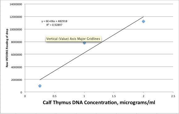
Fitting a Straight Line

PCR Results Summary
Your positive control PCR result was 0.13688 μg/mL
Your negative control PCR result was -0.00149 μg/mL
Patient 34902 :Results were -0.0000335 μg/mL, -0.00174 μg/mL and 0.000359 μg/mL. The sample showed minimal traces of green dye.
Patient 39905 : Results were 0.00525 μg/mL, 0.00425 μg/mL and 0.0194 μg/mL. The sample showed traces of green dye, however, not enough to come up with a conclusion.
The final conclusion was that both patients tested negative. This was determined by comparing the concentrations to the controls.
|


