BME100 s2014:W Group12 L5
| Home People Lab Write-Up 1 | Lab Write-Up 2 | Lab Write-Up 3 Lab Write-Up 4 | Lab Write-Up 5 | Lab Write-Up 6 Course Logistics For Instructors Photos Wiki Editing Help | ||||||||||||||||||||||||||||||||||||||||||||||||||||||||||||||||||||||||||||||||||
|
OUR TEAM
LAB 5 WRITE-UPBackground InformationSYBR Green Dye
How the Fluorescence Technique Works
ProcedureSmart Phone Camera Settings
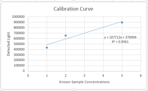
Calibration The phone is placed upright in the cradle with an eraser to help it stand straight. The cradle is moved about 6.5 cm away from the fluorimeter. This set up never moves once the experiment has begun.
Add more rows as needed
 Step 1: Insert the slide, smooth side down
 Step 2: 80 milliliters of the SYBR GREEN on the first slide position
 Step 3: 80 milliliters of the calf thymus (water blank) on top of the "beach ball" of SYBR GREEN
 Step 4: Focus camera on the "beach ball", cover the fluorimeter with the black box, and take a picture in the dark
 Step 5: Repeat this process on different slide placements (at least three) for each sample.
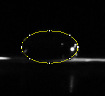
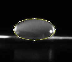
Data AnalysisRepresentative Images of Samples
Image J Values for All Samples
Our positive control PCR result was 30.20 μg/mL. Our negative control PCR result was -36.08 μg/mL Patient 78086 : The images of the three samples of DNA given from Patient 78086 were without color. There was no sign of any SYBR Green 1 in all three images. The amount of corrected PCR product concentration was around -36 μg/mL. The exact values were: -35.89, -36.88 and -36.56 μg/mL. Patient 32445 : The images of the three samples of DNA given from Patient 32445 were without color as well. Again there was no sign of any SYBR Green 1 in all three images. The amount of corrected PCR product concentration was around -35 μg/mL. The exact values were: -35.23, -35.38 and -35.74 μg/mL. Patient 78086 : Negative. We were able to come to this conclusion based on the values of μg/mL for both the positive and negative controls. This patient's corrected PCR product concentration matched that of the negative control. Patient 32445 : Negative. We were able to come to this conclusion based on the values of μg/mL for both the positive and negative controls. This patient's corrected PCR product concentration matched that of the negative control as well. We did not get confused on the results and assume that the results of the patients would be different. They in fact were very similar and we were able to successfully ascertain the desired result.
| ||||||||||||||||||||||||||||||||||||||||||||||||||||||||||||||||||||||||||||||||||


