BME100 s2014:T Group14 L5
| Home People Lab Write-Up 1 | Lab Write-Up 2 | Lab Write-Up 3 Lab Write-Up 4 | Lab Write-Up 5 | Lab Write-Up 6 Course Logistics For Instructors Photos Wiki Editing Help | ||||||||||||||||||||||||||||||||||||||||||
|
OUR TEAM
LAB 5 WRITE-UPBackground InformationSYBR Green Dye SYBR Green I is a dye that is used as a nucleic acid stain in molecular biology. The dye binds to DNA and results in a DNA and dye complex that reflects blue light and emits a green light that binds to double stranded DNA. This can be used in some methods of a quantitative polymerase chain reaction. This dye can also work on RNA, because RNA also is made up of nucleic acids. The dye will bind to the RNA and work in the exact way it would when bound to the DNA. SYBR Green I is declared safe to work with and does not have waste disposal issues. However is has the cabable of binding to DNA with a high affinity. This means that safety gloves must be worn at all times to prent the SYBR Green dye from getting on the skin.
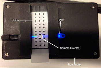
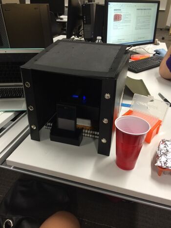
A flourimeter uses optical caustics in applied to flourescence to observe and measure the amount of flourescence. Flourescence can occur when a wavelength excites a molecule causing the emission of light. In this experiment, the SYBR Green when attached to the DNA nucleotides should glow when placed on the Flourimeter. Optical caustics causes light to be concentrated near the surface of the drop in order to increase the intensity of the flourescence. This is what can be eventually captured by our cell phones. The flourimeter is made of generic plastic, and a drop is placed on a special type of slides with a rough surface with a micropipette. Once aligned, a blue light is shown through the drop causing the SYBR Green to flouresce and an image to be captured on a cell phone to later be recorded on the computer program ImageJ.
How the Fluorescence Technique Works First, the flourimeter is set up by turning on the Blue LED and turning on and putting a smart phone on a cradle in front of the flourimeter. From this position, adjust the flourimeter to the height of the cell phone's camera in order to get a picture of the drop sideways and not from any other angle. This can be done by placing books or other materials underneath the flourimeter. After placing the drop of SYBR Green on the slide and adding the DNA to the dye, the dye will bind to the nucletide in the DNA. So, when the SYBR Green is exposed to the blue light, it will start to flouresce, putting a dark container over the reaction will make the flourescence easier to see in the picture taken by your cell phone.
ProcedureSmart Phone Camera Settings
The smart phone to be used in the experiment should be formatted to have the flash off at least. It's also important to have a timer on the cell phone, in order to leave it in the dark and then take the picture. It should be safe to set the timer for 5-10 seconds in order to get good picture. After setting the phone up in the camera program, put the phone in a cradle provided in front of the flourimeter and then measure the centimeters of the phone from the drop.
Placing Samples onto the Fluorimeter
Data AnalysisRepresentative Images of Samples
Image J Values for All Samples PCR Product Table
PCR Product Table Calculations
[Instructions: See worksheet page 8. To save time on typing a new Wiki table from scratch, use THIS TOOL to auto-generate a Wiki table: Excel-to-Wiki Converter. Copy the headers and values from the Excel spreadsheet you made, paste them into the form field, click submit, copy the Wiki code that the tool generated, and replace TABLE GOES HERE (below) with your auto-generated code.]
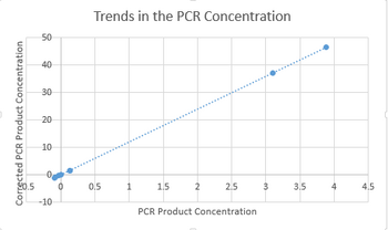
Fitting a Straight Line
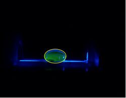
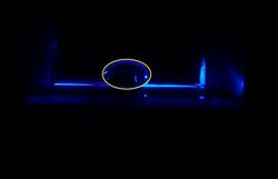
PCR Results Summary Instructor's summary: You completed 8 PCR reactions in a previous lab. You used the SYBR Green I staining and imaging technique to measure the amount of amplified DNA in each PCR reaction. You used a standard curve (based on known concentrations of calf thymus DNA) to convert INTDEN values into DNA concentration. Your positive control and negative control samples should be used as threshold values for determining whether an unknown (patient) sample is truly positive or negative. Your positive control PCR result was 37.16109 μg/mL Your negative control PCR result was -1.06691 μg/mL Write-in each patient ID and give both a qualitative (what the images looked like) and a quantitative description (μg/mL) of what you observed Patient 40197 F : There was no florescence observed from this test drop. The intden values were all around 1.608195. Patient 90029 M : There was no florescence observed from this test drop. The intden values were all around 0.101252. Compare each patient's results to the positive control value and the negative control value. Draw a final conclusion for each patient (positive or negative) and explain why you made that conclusion. Patient 40197 F : When compared to the positive and negative control value, this value seems to be slightly positive although the picture appears negative. Patient 90029 M : When compared to the positive and negative control value, this test seems to be slightly positive as well even though the pictures appear negative.
| ||||||||||||||||||||||||||||||||||||||||||


