BME100 f2017:Group7 W0800 L6
| Home People Lab Write-Up 1 | Lab Write-Up 2 | Lab Write-Up 3 Lab Write-Up 4 | Lab Write-Up 5 | Lab Write-Up 6 Course Logistics For Instructors Photos Wiki Editing Help | |||||||
|
OUR COMPANY
Our Brand Name LAB 6 WRITE-UPBayesian StatisticsOverview of the Original Diagnosis System The organization of this diagnosis system was composed of 15 lab teams each testing 2 patients for the disease-associated SNP. This allowed for 30 total patient samples to be tested for this disease. Each patient sample was tested 3 times to ensure valid results and ultimately reduce the probability of false test results. Additionally, each team had a set of positive and negative controls to ensure the effectiveness of the procedure. The application ImageJ was used to calibrate the measurements for the data collected by the phone camera. The calibration was done in order to compare the fluorescence of the positive and negative controls to the fluorescence of the patient samples. The application ImageJ quantified the fluorescence of the DNA which was used to calculate the relative presence of the target DNA sequence. A low or nonexistent fluorescence indicates a negative result, and a high fluorescence indicated a positive result. Three drop images were taken for each of the patient samples meaning there were 9 total images for each of the unique PCR samples. After applying Bayesian Statistics to the data set, the disease specificity was calculated to be 80.7%. Additionally, the disease sensitivity was calculated to be 95.3%. An issue that may have altered the data retrieved from image J is that the camera was not always a fixed distance away from the drop sample. It was difficult to standardize the camera distance from the sample. A rigid, preset camera holder would be helpful in future experiments to ensure that the data being retrieved is high quality. What Bayes Statistics Imply about This Diagnostic Approach The results for calculations 1 and 2 suggest that using individual PCR replicates is fairly reliable in concluding the presence of the disease SNP. Both of the values were close to .9 or a 90% success rate. Calculation 3 yielded a value close to 1.00 or 100% while calculation 4 gave us a value closer to .80 or 80%. This implies that PCR is very reliable when predicting positive conclusions. The prediction of negative conclusions is slightly less reliable than positive conclusions. This indicates that there will rarely be false positives and a slightly higher chance of false negatives. There are some possible errors that could have affected our Bayes values negatively. The cameras used were iPhone cameras, which did not have the option to change the settings to what was specified in the workbook. Another source of error could stem from the picture analysis. Since we did not have a standardized area, the area of each picture varied. The variation in the area could also be attributed to the fact that the phone camera was not in exactly the same position between trials since there was no standardized permanent rig to hold it in place and we had to rely on a ruler measurement. Some of the pictures were dark and outlines weren't clear. It is possible that we overlapped the background with the water droplet which would lower the rate of detection. Intro to Computer-Aided Design3D Modeling Our Design
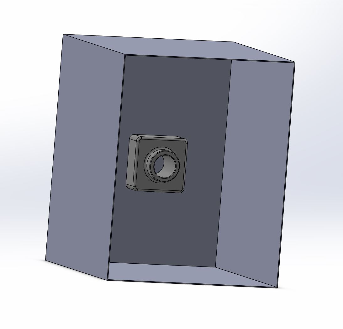
The main factor contributing to the selection of this modification was the group's personal experience when working with various tools and equipment. One of the most difficult sections of the practical experiment was working with the fluorimeter and calibrating the smartphone each time before taking a picture. Using a smartphone camera which was placed outside the fluorimeter box caused various problems in the experiment. First, the device holder was shorter than the inner part of the device which was somewhat resolved by putting notebooks and other equipment under the holder in order to raise it. Secondly, the phone did not stand completely vertical with respect to the box and it needed to be held throughout the experiment. Third, the holder was not connected to the box; thus, with the addition of each sample, it needed to be moved in order to maintain the same distance from all samples. Finally, when placed in dark, the smartphone camera was unable to focus on the sample. All of these issues caused difficulties for the group, increased the time of the experiment and affected the accuracy of the data obtained. In order to solve this issue, a new version of the device was designed. This design included a camera attached to the backside of the fluorimeter. The camera is able to take pictures of the samples placed in the device by the push of the button and share the obtained photos through bluetooth connectivity or LTE. The addition of this camera eliminates the need for camera calibration and focus change and considerably reduces the time required for the experiment while increasing the accuracy of the obtained data.
Feature 1: ConsumablesOur kit will include the most important dry consumables to perform any PCR: a set of 24 plastic tubes, 12 glass slides, 50 pipette tips, with the choice of 50ml of an indicator enzyme such as the SYBR green solution used in lab 5. Customers will have to outsource for their specific PCR mix and primer solution, as well as any specialized buffers required. All of these consumables will come in their own individual bags for packaging to prevent any possible cross contamination. Beyond the standard consumables, our kit will also contain a high quality "black box" where the level of fluorescence measurements can take place. The black box will be made out of Vantablack, essentially made out of aligned carbon nanotubes absorbing up to 99.965% of radiation in the visible spectrum. Within the back face of the black box, there is a high-quality camera where researchers are able to view the fluorescence in real time rather than with a picture of the glow. The camera will also provide the ability to take video, quality pictures, and provide a means of viewing the fluorescence without lifting the door end of the black box, providing lower levels of error and faster analysis.
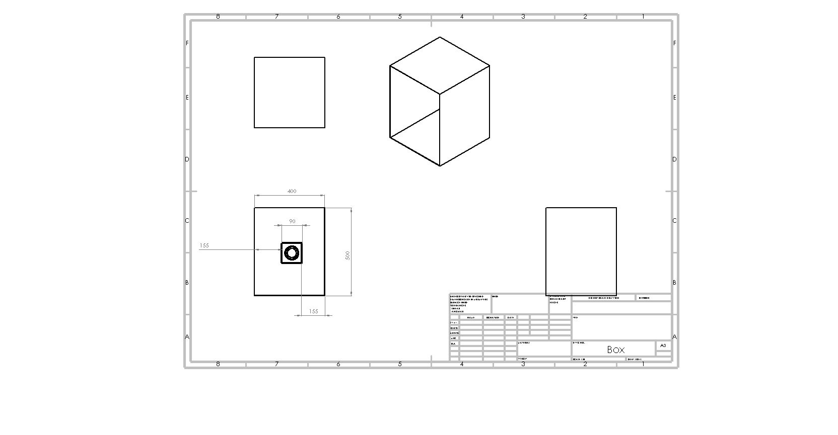
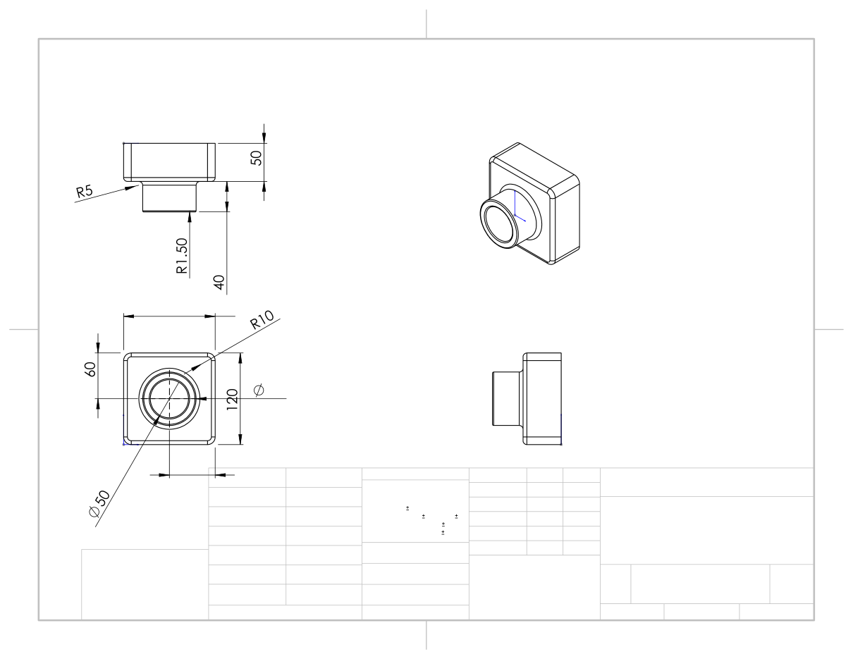
Feature 2: Hardware - PCR Machine & FluorimeterOur system will include both the Open PCR machine and the fluorimeter as separate devices, much like we used in our previous lab in order to find a balance between low cost and accurate and precise results. However our system will include a fluorimeter with an improved version of the black box we used in lab. Our version of the flourimeter setup will encompass a laboratory grade quality camera that will allow the user to view the drop in real time while the picture in taking place, and allow for maximum exclusion of visible light in the black box because it will now be able to completely close. Our black box will also be made of high quality carbon nanotube vantablack material that will absorb 99.965% of radiation in the visible spectrum. These hardware changes will ultimately result in more accurate and quicker fluorimeter analysis. These changes in hardware were chosen upon reflection of the lab group's recent experience in the Open PCR and fluorimeter lab. The section the lab group thought brought the most error into the results was the fluorimeter analysis. This was mainly because the hardware set up was not very standardized; such examples include using a regular smart phone camera to take the pictures to be analyzed and not having a completely enclosed environment for the fluorimeter procedure. Our design will address these issues through the implementation of a high quality camera embedded into a vantablack box that allows for complete enclosure when preforming the fluorimeter procedure and analysis. Schematic of the box and camera can be seen below.
| |||||||


