Biomod/2014/NTU/Result
<html> <head> <title></title> <link href="http://openwetware.org/index.php?title=Biomod/2014/NTU/Templates/css/bootstrap.css&action=raw&ctype=text/css" rel='stylesheet' type='text/css' /> <link href="http://openwetware.org/index.php?title=Biomod/2014/NTU/Templates/css/animate.css&action=raw&ctype=text/css" rel="stylesheet" type="text/css" media="all"> <link href="http://openwetware.org/index.php?title=Biomod/2014/NTU/Templates/css/style.css&action=raw&ctype=text/css" rel='stylesheet' type='text/css' /> <script type="text/javascript" src="http://openwetware.org/index.php?title=Biomod/2014/NTU/Templates/js/jquery.min.js&action=raw&ctype=text/js"></script> <script type="text/javascript" src="http://openwetware.org/index.php?title=Biomod/2014/NTU/Templates/js/slidejs&action=raw&ctype=text/js"></script> <script src="http://openwetware.org/index.php?title=Biomod/2014/NTU/Templates/js/slideexperimnet&action=raw&ctype=text/js"></script> <script> $(function () { $("style[media*='screen']").remove(); $("link[href*='favicon']").remove(); //fix heading var h1 = $(".firstHeading").text().split("/"); $(".firstHeading").text(h1[h1.length-1]); $("tr:odd").addClass("odd"); }); function slideload(){ $('#fadein').fadeIn(6000); }
$(document).ready(function(){ $(".subMenu li ul li").css("background-color","#1E8100"); $("#e1").click(function(){ $("html body").animate({ scrollTop: 1180 },1000) }); $("#e2").click(function(){ $("html body").animate({ scrollTop: 1950 },1000) }); $("#e3").click(function(){ $("html body").animate({ scrollTop: 2780 },1000) }); $("#e4").click(function(){ $("html body").animate({ scrollTop: 3680 },1000) }); $("#e5").click(function(){ $("html body").animate({ scrollTop: 4480 },1000) }); $("#e6").click(function(){ $("html body").animate({ scrollTop: 5610 },1000) }); $("#e9").click(function(){ $("html body").animate({ scrollTop: 6650 },1000) }); $("#e10").click(function(){ $("html body").animate({ scrollTop: 7950 },1000) }); }) </script> <style>
.body {
 width:100%;
 color: #CCCCCC ;
 max-width: 1280px;
 min-width: 0 ;
 padding-top: 55px;
 margin-left: auto;
 margin-right: auto;
}
- column-content #content {
   padding: 0em;
   margin: 0;
}
- content{
 border: 0px;
 float:right;
 padding: 0em;
 width:100%;
}
- contentSub, #search-controls, .firstHeading, #footer-box, #catlinks, #p-logo, #toctitle ,#top-section ,#column-one ,#footer, #siteSub,#jump-to-nav,.printfooter,.visualClear
{
display:none;
}
- globalWrapper {
 background-color:#fff;
 font-size:100%;
 padding-bottom: 0px;
}
.start:hover{ background-color: #1E8100; transition: background-color 0.5s linear; } </style> <link href="http://openwetware.org/index.php?title=Biomod/2014/NTU/Templates/css/slidecss&action=raw&ctype=text/css" rel='stylesheet' type='text/css' /> </head> <body onload="slideload()">
- <a href="http://openwetware.org/wiki/Biomod/2014/NTU"><img src="http://openwetware.org/images/4/4d/Logo1.png" style="left:-50px;"></a>
- <a href="javascript: void(0)">Project</a>
- <a href="http://openwetware.org/wiki/Biomod/2014/NTU/Idea">Idea</a>
- <a href="http://openwetware.org/wiki/Biomod/2014/NTU/Design">Design</a>
- <a href="http://openwetware.org/wiki/Biomod/2014/NTU/Origami">Origami</a>
- <a href="javascript: void(0)">Experiment</a>
- <a href="http://openwetware.org/wiki/Biomod/2014/NTU/Method">Method</a>
- <a href="#">Result</a>
- <a href="javascript: void(0)">Discussion</a>
- <a href="http://openwetware.org/wiki/Biomod/2014/NTU/Review">Review</a>
- <a href="http://openwetware.org/wiki/Biomod/2014/NTU/Future">Future</a>
- <a href="javascript: void(0)">Supplement</a>
- <a href="http://openwetware.org/wiki/Biomod/2014/NTU/Protocol">Protocol</a>
- <a href="http://openwetware.org/wiki/Biomod/2014/NTU/Material">Material</a>
- <a href="http://openwetware.org/wiki/Biomod/2014/NTU/Equipment">Equipment</a>
- <a href="http://openwetware.org/wiki/Biomod/2014/NTU/Member">Member</a>
- <a href="http://openwetware.org/wiki/Biomod/2014/NTU/Acknowledge">Acknowledge</a>
To fulfill our goals of capture, release, and aggregate, we divided our project into 10 components:
√ <a id="e1" href="javascript: void(0)">1. Find the best concentration of magnesium ion in the origami folding process</a>
√ <a id="e2" href="javascript: void(0)">2. Test whether the origami folded into the right structure</a>
√ <a id="e3" href="javascript: void(0)">3. Test whether the guide staple can work</a>
√ <a id="e4" href="javascript: void(0)">4. Test whether the guide staple is removed</a>
√ <a id="e5" href="javascript: void(0)">5. Test whether the lock is tight enough to stay closed without guide staple</a>
√ <a id="e6" href="javascript: void(0)">6. Test whether poly C and poly G will cause monomer aggregation</a>
X <a id="e7" href="javascript: void(0)">7. Test whether protection strand works</a>
X <a id="e8" href="javascript: void(0)">8. Test whether complementary strand of protection strand can be removed successfully</a>
√ <a id="e9" href="javascript: void(0)">9. Test whether virus can open the lock </a>
√ <a id="e10" href="javascript: void(0)">10. HA test: determine whether our product can inactivate virus</a>
We accomplished eight of the above experiments through trials and errors. For the failed part, we provide detail information in our discussion. You can click any components you are interested in and get further explanation.
1. Magnesium ion concentration
Since the concentration level of magnesium ion affects the structure of DNA origami significantly, we test the folding rate of different concentration of magnesium ion.
From left to right
1. marker/8mM/10mM/12mM/14mM/16mM
<img src="http://openwetware.org/images/d/d6/Result1.jpg" style="left:650px; width:400px;">
As you can see, the scaffold and staples folding with 8mM to 12mM of magnesium ion have better structure without the tailing phenomenon. Thus, we chose 10mM of magnesium ion as our subsequent experiment condition.
2. Monomer under TEM
Before we start all our experiment, we have to certify that our origami design can fold into the desired structure.
<img src="http://openwetware.org/images/4/49/Result2.jpg" style="left:120px; top:100px; width:500px;">
This is our monomer under TEM. The radius of the monomer is around 35nm on average, which corresponds to our calculation.
<img src="http://openwetware.org/images/b/b5/Result111111.jpg" style="display:inline-block; left:700px; top:-200px; width:30%;">
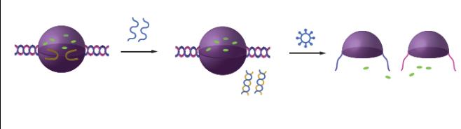
3. GFP packaged with guide staples
We will use the guide staples to package the drug. In our experiment, we use the Green fluorescent protein (GFP) to represent the drugs inside the Dimer. The monomers were mixed with guide staples and GFP at 4°C overnight.
<img src="http://openwetware.org/images/0/06/Result4.jpg" style="left:120px; top:80px; width:40%;">
The black circles are our Dimers. Compared to the previous figure, the center of the circles are black instead of white because GFPs inside absorb the electron beams. From these pictures, we can infer that our guide staple indeed solved the matching problem mentioned in the mechanism and indicated that the suspended drugs will be packaged in the Dimer by random distribution.
<img src="http://openwetware.org/images/1/15/Result22222222.jpg" style=" left:700px; width:30%; top:-100px;" >
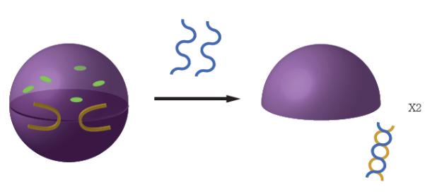
4. Removal of guide staple
In our final Dimer product, the guide staples should be removed so that the H5 protein on the virus can attach to the aptamer lock and open the Dimer.
<img src="http://openwetware.org/images/6/6c/Result7.jpg" style="display:inline-block; left:120px; width:40%; top:-50px;">
This sample was prepared with the following process:
Mix monomer with guide staples and GFP at 4°C overnight
→
Add ten times complementary strand of guide staples.
There is a large number of non-black circles, which indicate that the Dimers in the pictures are mostly opened and have released the packaged drug.
<img src="http://openwetware.org/images/a/a6/Result8.jpg" style="display:inline-block; width:100%; top:20px;">
5. Lock affinity
Previous experiments allowed us to produce our Dimer product. However, we still need to check whether our lock consists of the aptamer and its complementary strand, and whether it could package the drug well.
<img src="http://openwetware.org/images/0/0e/Result9.jpg" style="width:40%;left:120px;">
The sample was prepared with following process:
Mix monomer with guide staples and GFP at 4 overnight
→
Add pre-mixed aptamer-complementary strand complex at 4°C for 6 hours
→
Add ten times complementary strand of guide staples.
You can see clear black spots in the Dimer. This result is exactly the same as that folded with guide staples, implying that our lock functioned quite well.
<img src="http://openwetware.org/images/5/5c/Resulttt.jpg" style=" positione:relative; top:50px; left:120px; width:80%;">
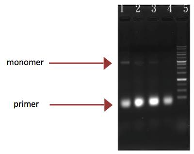
6. Poly C/G aggregation
From left to right:
1.monomer
2.monomer attached with poly C
3.monomer attached with poly G
4.mixed monomer attached with poly C and G
5.Marker
<img src="http://openwetware.org/images/1/18/Result11.jpg" style="display:inline-block; top:-60px;">
In this picture, we can see two major bands. One is the monomer and the other is the primer with staples.
In the first sample, the monomer served as a control group. Monomer attached with poly C or G had the same result as monomer, whereas the band of mixed group seemed much darker than the rest. We assume that it implies that the monomer attached with poly C had some interaction with monomer attached with poly G, which in our experiment will most likely be aggregation.
<img src="http://openwetware.org/images/4/41/Result12.jpg" style="left:350px; top:40px; width:40%;">
To see further discussion, please <a href="http://openwetware.org/wiki/Biomod/2014/NTU/Review#1">click </a>here.
7.Test final product with virus
From left to right:
1.marker
2. dimer + polyC + polyG, react with Allantoic protein for 30 minutes
3. dimer + polyC + polyG, react with H5N2 virus for 30 minutes
4. dimer + polyC, react with H5N2 virus for 30 minutes.
5. dimer + polyG, react with H5N2 virus for 30 minutes.
6. dimer , react with H5N2 virus for 30 minutes
7. dimer , react with Allantoic protein for 30 minutes
8. monomer
<img src="http://openwetware.org/images/c/c7/Result13.jpg" style="display:inline-block; top:-120px; width:40%; left:-130px;">
We believe that the virus could open the dimer and aggregate with the monomers due to the binding between H5 and H5 aptamer. In addition, the sample with poly C and poly G could cause further aggregation. Therefore, we expect that there should be tailing in wells 3 to 6 and the band to have different brightness due to the aggregation of poly C and poly G.
Well 8 is the control. Well 2 and 7 are our negative controls, which contain dimers with allantoic protein. We postulated the band of well 2 and 7 to be like the control. However, the band disappeared and the band of GFP is slower than the rest. We will discuss it further in the discussion part. In addition, we expect tailing in wells 3 to 6 and the band to have different brightness due to further aggregation of poly C and G in well 3. However, we did not see the expected bands but only the vague band that is the virus in the middle.
<img src="http://openwetware.org/images/1/16/Result10.jpg" style=" display:inline-block; width:80%; left:120px; top:0px;">
To see further discussion, please <a href="http://openwetware.org/wiki/Biomod/2014/NTU/Review#3">click</a> here.
8.HA test
The HA test is widely used in the decision of virus activity. The hemagglutinin protein on the virus will cause red blood cell to agglutinate. Hence, if the blood in the well flow down, it means the virus is inactivated. We used two times serial dilution. The first row was the original sample, and the second row was twice thinner as the first row.
from right to left:
1. dimer + polyC + polyG, react with Allantoic protein for 30 minutes
2. dimer + polyC + polyG, react with H5N2 virus for 30 minutes
3. dimer + polyG, react with H5N2 virus for 30 minutes.
4. dimer , react with Allantoic protein for 30 minutes
5. dimer , react with H5N2 virus for 30 minutes
6. H5N2 virus
<img src="http://openwetware.org/images/d/db/Result15.jpg" style="display:inline-block; top:-100px;">
<tbody> </tbody>| sample | 6 | 5 | 4 | 3 | 2 | 1 |
| 1(1X) | O | O | X | O | O | X |
| 2(2-1) | O | O | X | O | O | X |
| 3(2-2) | O | X | X | X | X | X |
| 4(2-3) | X | X | X | X | X | X |
| 5(2-4) | X | X | X | X | X | X |
| 6(2-5) | X | X | X | X | X | X |
| 7(2-6) | X | X | X | X | X | X |
| 8(2-7) | X | X | X | X | X | X |
H5N2 Virus had activity at the concentration of 2-2 compared to original sample, and virus with our dimer had activity at the concentration of 2-1. This suggested that our product can really decrease the activity of the virus.
To see further discussion, please <a href="http://openwetware.org/wiki/Biomod/2014/NTU/Review#2">click </a>here.
