User:Matthewmeisel
Working file scrapbook
Notebook
<calendar> name=User:Matthewmeisel/Notebook date=2006/07/01 view=threemonths format=%name/%year-%month-%day weekstart=14 </calendar>
Literature summaries and brainstorming
Shih's 2004 Nature paper (biblio below)
Shih describes the (rather complex) construction of the DNA octahedron, which consists of two motifs: double-crossover edges ("struts") and paranemic crossover struts. Each edge of the octahedron is combrised of two ds DNAs which are interwoven with these motifs. Edges are connected at vertices by two unpaired thymine residues for flexibility. The structure was formed from a long "heavy chain" (scaffold) and five 40-nt "light chains" (oligos) in two steps: first, by cooling the structure from a denaturation temperatue to form a branched complex, and then further cooling to allow corresponding paranemic crossover areas to associate.
The final formation step is proven through gel electrophoresis. Mg2+ is required for the final folding step, and the product runs faster after Mg2+ has been added, indicating further folding. Other confirmatory steps were complicated: the structures were visualized with cryo-electron microscopy, and a 3d model was formed using microscopy images from 961 particles.
Shih points out that no covalent bonds are created or broken in the formation of the structure, which greatly simplifies the assembly process. He also suggests one possible application/implication of his work, which is that one-dimensional information (the primary DNA sequence of the heavy chain) is effectively encoding 3d positional information (i.e., the final 3d position of the nucleotide).
Yurke's 2000 Nature paper (biblio below)

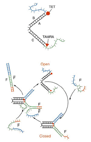
Yurke describes the DNA "tweezers" he engineered, which consist of the following parts:
- a short (~30 nt) ss DNA backbone (A)
- two oligos (B and C) which are each complimentary to half of the backbone and bound to it, and each of which have a short (~20 nt) ss overhang
In this state, the DNA tweezers are in an "open" conformation. A ~50-nt "fuel strand" of ss DNA (F) is added to the tweezers to close them, and this occurs because the fuel strand is complimentary to each of the oligo overhangs, which closes the tweezers. The fuel strand also includes a 8-nt ss overhang, and so when a strand complimentary to the fuel strand ("displacement" strand") (F-bar) is added, the fuel strand is stripped, opening the tweezers.
Yurke reports that the rate-limiting step of this "strand displacement" is the initial binding between the fuel strand and the displacement strand, and the time required for DNA branch migration (on this scale) is negligble. The tweezers open and shut on a time scale of a few minutes after a fuel (or displacement) strand is added.
Thoughts on Shih and Yurke
A convenient way to open a DNA nanobox would be to construct a box held shut by a latch with a DNA tweezer motif in its closed conformation, such that a displacement strand would open the box. Yurke opens his tweezers through the exogenous addition of a displacement strand, but I suggest that we look for a way to bind some sort of ss DNA to the surface of a cell or cellular protein, so that when a nanobox approaches, the strand would unhook the latch.
Given the (hypothesized) large amount of cellular protrusions from E. coli, this could be difficult to engineer, and there may be a kinetic difficulty with the constraining of the displacement strand.
Yurke carried out the reaction in at 20°C SPSC buffer, which contains 50mM sodium biphosphate, 1M NaCl, and pH 6.5. We should investigate if strand displacement will work at physiological pH and lower salt concentrations as well.
Bibliography
- Shih WM, Quispe JD, and Joyce GF. A 1.7-kilobase single-stranded DNA that folds into a nanoscale octahedron. Nature. 2004 Feb 12;427(6975):618-21. DOI:10.1038/nature02307 |
- Shih WM and Spudich JA. The myosin relay helix to converter interface remains intact throughout the actomyosin ATPase cycle. J Biol Chem. 2001 Jun 1;276(22):19491-4. DOI:10.1074/jbc.M010887200 |
- Shih WM, Gryczynski Z, Lakowicz JR, and Spudich JA. A FRET-based sensor reveals large ATP hydrolysis-induced conformational changes and three distinct states of the molecular motor myosin. Cell. 2000 Sep 1;102(5):683-94. DOI:10.1016/s0092-8674(00)00090-8 |
- Yurke B, Turberfield AJ, Mills AP Jr, Simmel FC, and Neumann JL. A DNA-fuelled molecular machine made of DNA. Nature. 2000 Aug 10;406(6796):605-8. DOI:10.1038/35020524 |
Other projects
My work in Biophysics 101
Lachrymosity
- Why Apples make me cry (feel free to contribute)
Contact info
Email me: (my last name) at fas dot harvard dot edu