Biomod/2014/ASU/Results: Difference between revisions
No edit summary |
No edit summary |
||
| Line 1,588: | Line 1,588: | ||
<p style = "color: #000000; font-size: 16px;">The resulting gel image below depicts gel shifts for the positive control tiles PCAB, PC2A, and PC1A, along with control tiles (tiles with filler strands in place of aptamers). The shifts of the bands to higher equivalent base pair values for all three tiles shown when protein was added verify that the protein bound to the tiles and added weight to them as compared to the bands for lanes in which the tiles were not incubated with protein. These shifts are representative of all of the positive control tiles, as they all bound protein.</p> | <p style = "color: #000000; font-size: 16px;">The resulting gel image below depicts gel shifts for the positive control tiles PCAB, PC2A, and PC1A, along with control tiles (tiles with filler strands in place of aptamers). The shifts of the bands to higher equivalent base pair values for all three tiles shown when protein was added verify that the protein bound to the tiles and added weight to them as compared to the bands for lanes in which the tiles were not incubated with protein. These shifts are representative of all of the positive control tiles, as they all bound protein.</p> | ||
<p style = "color: #000000; font-size: 16px;">These particular tiles also show an interesting phenomenon: tile PCAB does not run exactly like PC2A nor PC1A. Both PC2A and PC1A show brighter bands corresponding to the bands in the control lanes; meaning that a much lower percentage of PC2A and PC1A are bound to alpha-thrombin compared to PCAB. The major band of PCAB runs as one band as compared to the two shifted bands of PC2A. We hypothesize that this is because PC2A has two of the same aptamers incorporated into itself, which can each bind a separate protein molecule. In contrast, the form of PCAB bound only to one protein coordinated between the aptamers on the tile is likely much more stable than a form in which each aptamer is bound to a separate protein due to entropic effects. This also explains the dearth of unbound tiles at the control band site: since this complex is more stable, it has a lower KD and fewer tiles exist in an unbound form.</p> | <p style = "color: #000000; font-size: 16px;">These particular tiles also show an interesting phenomenon: tile PCAB does not run exactly like PC2A nor PC1A. Both PC2A and PC1A show brighter bands corresponding to the bands in the control lanes; meaning that a much lower percentage of PC2A and PC1A are bound to alpha-thrombin compared to PCAB. The major band of PCAB runs as one band as compared to the two shifted bands of PC2A. We hypothesize that this is because PC2A has two of the same aptamers incorporated into itself, which can each bind a separate protein molecule. In contrast, the form of PCAB bound only to one protein coordinated between the aptamers on the tile is likely much more stable than a form in which each aptamer is bound to a separate protein due to entropic effects. This also explains the dearth of unbound tiles at the control band site: since this complex is more stable, it has a lower KD and fewer tiles exist in an unbound form.</p> | ||
<img src = "http://openwetware.org/ | <img src = "http://openwetware.org/images/a/a1/Screen_Shot_2014-10-23_at_4.11.42_AM.png"> | ||
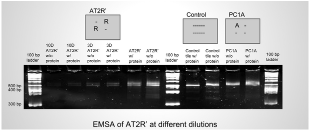
<p style = "color: #000000; font-size: 16px;">This gel is representative of gels run with randomized oligonucleotide inserts. The six lanes on the left between the ladders are different dilutions of the 6th and 7th lanes: the 2nd and 3rd lanes are 10x diluted, while the 4th and 5th lanes are 3x diluted. On the right side of the gel are positive and negative controls for binding alpha-thrombin.</p> | <p style = "color: #000000; font-size: 16px;">This gel is representative of gels run with randomized oligonucleotide inserts. The six lanes on the left between the ladders are different dilutions of the 6th and 7th lanes: the 2nd and 3rd lanes are 10x diluted, while the 4th and 5th lanes are 3x diluted. On the right side of the gel are positive and negative controls for binding alpha-thrombin.</p> | ||
<p style = "color: #000000; font-size: 16px;">It is difficult to tell whether or not a small amount of the AT2R’ tiles in the protein-positive lanes have shifted. Optimisation is currently being done to try to recover any tiles that may be bound to protein but are too few in number to see with gel stain. This process involves cutting and eluting an area of the protein-positive lane above any visible signal, doing PCR for the randomized oligonucleotide insert strands, and comparing this to a control run with gel from the same area in the control lane. It is important to note that, though this procedure is susceptible to human error due to the difficulty of cutting the gel congruently in each lane, the overall outcome of the process can be tested in other ways. If the pool of randomized oligonucleotides is enriched, for example, assaying a new generation of tiles made with the enriched pool should show stronger shifts. </p> | <p style = "color: #000000; font-size: 16px;">It is difficult to tell whether or not a small amount of the AT2R’ tiles in the protein-positive lanes have shifted. Optimisation is currently being done to try to recover any tiles that may be bound to protein but are too few in number to see with gel stain. This process involves cutting and eluting an area of the protein-positive lane above any visible signal, doing PCR for the randomized oligonucleotide insert strands, and comparing this to a control run with gel from the same area in the control lane. It is important to note that, though this procedure is susceptible to human error due to the difficulty of cutting the gel congruently in each lane, the overall outcome of the process can be tested in other ways. If the pool of randomized oligonucleotides is enriched, for example, assaying a new generation of tiles made with the enriched pool should show stronger shifts. </p> | ||
<p style = "color: #000000; font-size: 16px;">The double nature of the bands in AT2R’ is characteristic of tiles of this nature: another previously-made tile, AT2R -- which incorporated two randomized oligonucleotides into the opposite sites as AT2R’ -- also showed such a signal in the control lanes. We suspect that this phenomenon is occurring due to some tiles failing to incorporate the aptamer strands. This should not be an issue, however, as the remaining tiles which have the inserts incorporated appear to be present in approximately equal amount, which should provide in itself a large enough selectable pool. Nonetheless, we are continuing to investigate the cause of the phenomenon and how it might possibly be prevented.</p> | <p style = "color: #000000; font-size: 16px;">The double nature of the bands in AT2R’ is characteristic of tiles of this nature: another previously-made tile, AT2R -- which incorporated two randomized oligonucleotides into the opposite sites as AT2R’ -- also showed such a signal in the control lanes. We suspect that this phenomenon is occurring due to some tiles failing to incorporate the aptamer strands. This should not be an issue, however, as the remaining tiles which have the inserts incorporated appear to be present in approximately equal amount, which should provide in itself a large enough selectable pool. Nonetheless, we are continuing to investigate the cause of the phenomenon and how it might possibly be prevented.</p> | ||
<img src = "http://openwetware.org/ | <img src = "http://openwetware.org/images/5/56/Screen_Shot_2014-10-23_at_4.13.19_AM.png"> | ||
</div> | </div> | ||
Revision as of 11:17, 23 October 2014
<html xmlns="http://www.w3.org/1999/xhtml" xmlns:v="urn:schemas-microsoft-com:vml" xml:lang="en" lang="en" dir="ltr">
	<head>
		<title>Nanodevils - OpenWetWare</title>
		<style type="text/css" media="screen, projection">/*<![CDATA[*/
			@import "/skins/common/shared.css?164";
			@import "/skins/monobook/main.css?164";
		/*]]>*/</style>
		<link rel="stylesheet" type="text/css" media="print" href="/skins/common/commonPrint.css?164" />
		<script type= "text/javascript">/*<![CDATA[*/
var skin = "monobook";
var stylepath = "/skins";
var wgArticlePath = "/wiki/$1";
var wgScriptPath = "";
var wgScript = "/index.php";
var wgVariantArticlePath = false;
var wgActionPaths = [];
var wgServer = "http://openwetware.org";
var wgCanonicalNamespace = "";
var wgCanonicalSpecialPageName = false;
var wgNamespaceNumber = 0;
var wgPageName = "Biomod/2014/ASU";
var wgTitle = "Biomod/2014/ASU";
var wgAction = "view";
var wgArticleId = "136275";
var wgIsArticle = true;
var wgUserName = null;
var wgUserGroups = null;
var wgUserLanguage = "en";
var wgContentLanguage = "en";
var wgBreakFrames = false;
var wgCurRevisionId = "750826";
var wgVersion = "1.13.2";
var wgEnableAPI = true;
var wgEnableWriteAPI = false;
var wgMWSuggestTemplate = "http://openwetware.org/api.php?action=opensearch\x26search={searchTerms}\x26namespace={namespaces}";
var wgDBname = "owwdb";
var wgSearchNamespaces = [0, 1, 2, 3, 4, 5, 6, 7, 8, 9, 10, 11, 12, 13, 14, 15, 100, 101, 110, 111];
var wgMWSuggestMessages = ["with suggestions", "no suggestions"];
var wgRestrictionEdit = [];
var wgRestrictionMove = [];
/*]]>*/</script>
		<script type="text/javascript" src="/skins/common/wikibits.js?164"></script>
		<script type="text/javascript" src="/skins/common/ajax.js?164"></script>
		<script type="text/javascript" src="/ext/AjaxShowEditors/AjaxShowEditors.js"></script>
		<script type="text/javascript" src="/skins/common/mwsuggest.js?164"></script>
		<script type="text/javascript" src="/index.php?title=-&action=raw&gen=js&useskin=monobook"></script>
		<style type="text/css">/*<![CDATA[*/
@import "/index.php?title=MediaWiki:Common.css&usemsgcache=yes&action=raw&ctype=text/css&smaxage=18000";
@import "/index.php?title=MediaWiki:Monobook.css&usemsgcache=yes&action=raw&ctype=text/css&smaxage=18000";
@import "/index.php?title=-&action=raw&gen=css&maxage=18000&useskin=monobook";
/*]]>*/</style>
	</head>
<body class="mediawiki ns-0 ltr page-Biomod_2014_ASU">
<a name="top" id="top"></a>
Biomod/2014/ASU
From OpenWetWare
< <a href="/wiki/Biomod" title="Biomod">Biomod</a> | <a href="/wiki/Biomod/2014" title="Biomod/2014">2014</a>
<link rel="stylesheet" href="http://fonts.googleapis.com/css?family=Lato:300,100&subset=latin">
<script src="//ajax.googleapis.com/ajax/libs/jquery/1.10.2/jquery.min.js" ></script>
<script type"text/javascript">
$(function () {
$("style[media*='screen']").remove();
$("link[href*='favicon']").remove();
//fix heading
var h1 = $(".firstHeading").text().split("/");
$(".firstHeading").text(h1[h1.length-1]);
$("tr:odd").addClass("odd");
});
</script>
<style type="text/css">
/**** Base styles ****/
- column-one, #footer, div#sidebar-main, #contentSub, .firstHeading, #siteSub, #jump-to-nav, .printfooter{
display: none;
}
- content {
margin: 0;
padding: 0;
background: #fff;
border: none;
}
html, body, div, span, object, iframe,
h1, h2, h3, h4, h5, h6, p, blockquote, pre,
abbr, address, cite, code, del, dfn, em, img, ins, kbd, q, samp,
small, strong, sub, sup, var, b, i, dl, dt, dd, ol, ul, li,
fieldset, form, label, legend,
table, caption, tbody, tfoot, thead, tr, th, td,
article, aside, canvas, details, figcaption, figure,
footer, header, hgroup, menu, nav, section, summary,
time, mark, audio, video {
margin: 0;
padding: 0;
border: 0;
font: inherit;
}
article, aside, details, figcaption, figure,
footer, header, hgroup, menu, nav, section {
display: block;
}
body {
/*padding: 15px;*/
font-family: 'Lato', 'Lucida Sans Regular', 'Lucida Grande', 'Lucida Sans Unicode', Arial, sans-serif;
font-size: 2rem;
font-weight: 200;
line-height: 1.4;
background: #fff;
color: #000000;
max-width: 1280px;
padding-top: 55px;
margin-left: auto;
margin-right: auto;
}
}
h1, h2, h3, p, ul, ol, pre, dl {
font-weight: 100;
}
h1, h2, #super-list, .box, .tagline, #index-list {
font-family: 'Lato', 'Helvetica Neue', Arial, sans-serif;
}
h1, h2, h3 { font-weight: 300; }
h1 {
font-size: 16px;
line-height: 1.1em;
}
h2 {
font-size: 22px;
}
a,
a code {
color: #FB4;
text-decoration: none;
}
a:hover,
a:hover code {
color: #FFCC00;
}
a:active,
a:active code {
color: #000000;
/*background: black;*/
}
a img { border: none; }
a.anchor{display: block; position: relative; visibility: hidden;}
p{
text-align:left;
}
em { color: #00EF00; }
strong { font-weight: bold; }
blockquote {
padding-left: 1.0em;
margin-left: 1.0em;
border-left: 1px solid #333;
font-style: italic;
}
nav {
background: rgba(25, 25, 25, 0.85);
padding: 0px;
position: fixed;
top: 0px;
left: 0px;
bottom: 0px;
right: 0px;
z-index: 100;
}
nav ul {
width: 100%;
margin: 0px auto;
padding: 0px;
list-style-type: none;
}
nav ul li {
float: left;
line-height:2.5;
}
nav ul li.selected {
border-bottom: solid 10px #580000;
}
nav ul li.home {
padding: 0px;
}
nav ul a {
float: left;
text-decoration: none;
color: #F2F2F2;
text-transform: uppercase;
font-size: 20px;
font-weight: 300;
padding: 0px 20px 0;
}
- content {
margin-top: 60px;
}
- filters > li{
margin: 0px;
display: inline-block;
}
.box.clickable:hover{
background: none repeat scroll 0 0 #fff;
}
.clickable img {
transition: 0.3s ease;
}
.clickable img:hover {
opacity:0.9;
transition: 0.3s ease;
}
.background {
left: 0;
margin: 0;
max-width: 100%;
padding: 0;
}
/*the boxes*/
.box.b2x2{
height: 300px;
width: 300px;
position: fixed;
}
.box.b2x2 > img{
display: block;
margin-left: auto;
margin-right: auto;
margin-top: 10px;
height: 300px;
width: 300px;
position: fixed;
}
.box.b2x1{
height: 360px;
}
.box.b1x2{
width: 930px;
height: 2500px;
}
.box.b1x3{
width: 700px;
}
/*start page*/
.box.intro { font-size:7.2rem;}
.box > p {
font-size: 16px;
padding: 0 20px;
margin-top: 10px;
text-align: justify;
font-weight: 300;
}
.box > h2 {
font-size: 20px;
font-weight: 100;
text-align:left;
margin-left: 20px;
margin-top: 15px;
}
.tease > h2 {
font-size: 40px;
font-weight: 100;
margin-top: 80px;
}
/*project*/
.project{
background-attachment: fixed;
width: 100%;
padding-bottom: 40px;
}
.project h2 {
color: #FFFFFF;
font-weight: 300;
margin-bottom: 30px;
margin-left: 180px;
padding-top: 20px;
position: relative;
}
.project h3 {
font-size: 26px;
}
.project .box {
margin-bottom: 20px;
margin-top: 0;
}
.interlude{
background: none repeat scroll 0 0 #2A2A2A;
box-shadow: 0 0 25px rgba(0, 0, 0, 0.8);
border: 1px solid rgba(0, 0, 0, 0.3);
height: 150px;
position: relative;
z-index: 3;
}
.interlude *{
float:left;
}
.interlude h2{
color: #FFFFFF;
display: block;
font-weight: 300;
line-height: 150px;
margin-left: 24%;
margin-right: -14%;
width: 50%;
}
.interlude img{
float: left;
line-height: 150px;
margin-left: 10%;
margin-top: 25px;
vertical-align: middle;
}
.clear{
clear: both;
}
.project_box{
background: none repeat scroll 0 0 #FFFFFF;
color: #000000;
display: block;
float: left;
font-size: 18px;
font-weight: 300;
line-height: 1.6;
margin-left: auto;
margin-right: auto;
overflow: hidden;
padding: 10px;
width: 50%;
}
.figure_box{
display: block;
float: left;
margin-left: 20px;
overflow: hidden;
width: 230px;
}
/*se*/
.project_box h2{
color: #1A1A1A;
display: block;
font-weight: 300;
margin-left: 0%;
margin-right: -10%;
width: 90%;
}
.project_box p{
text-align: justify;
margin-bottom: 18px;
}
.project_box li {
margin-left:50px
}
- pb_mot.project_box{
height: 350px;
}
- pb_dna_scaff.project_box{
height: 400px;
}
- pb_dna_req.project_box{
height: 250px;
}
- pb_poly_intro.project_box{
/*right: -20%;*/
height: 200px;
}
- pb_poly_pmoxa.project_box{
/*right: -20%;*/
height: 600px;
width: 1000px;
overflow:scroll;
}
- pb_ir.project_box{
/*right: -20%;*/
height: 1300px;
}
- pb_poly_cp.project_box{
/*right: -20%;*/
height: 600px;
}
/*team page*/
.bio_box {
background: none repeat scroll 0 0 #E74C3C;
float: left;
font-size: 15px;
height: 440px;
padding: 15px;
text-align: justify;
width: 210px;
}
.bio_box > .name{
font-size: 24px;
font-weight: 300;
margin-bottom: 25px;
text-align: center;
width: 100%;
}
.bio_box > p{
text-align: justify;
font-weight: 300;
font-size: 16px;
}
.box.big img{
opacity:1;
}
.flag > *{
float:left;
}
.flag > p{
font-size: 18px;
position: relative;
text-align: center;
top: -6px;
width: 75%;
margin-bottom: 10px;
}
- team .big{
opacity:1;
}
.head{
width:220px;
float:left;
}
/*sponsor page*/
- sponsors .box {
background: none repeat scroll 0 0 white;
}
- sponsors figcaption {
height: 65px;
width: 100%;
font-size: 15px;
font-weight: 300;
top: auto;
bottom: 0;
opacity: 0;
transform: translateY(100%);
transition: transform 0.4s, opacity 0.1s 0.3s;
-webkit-transform: translateY(100%);
-webkit-transition: -webkit-transform 0.4s, opacity 0.1s 0.3s;
}
- sponsors .descr{
background: none repeat scroll 0 0 rgba(0, 0, 0, 0.4);
font-size: 12px;
font-weight: 300;
height: 60px;
margin: 0;
padding-left: 10px;
padding-right: 10px;
padding-top: 10px;
text-align: justify;
top: -155px;
line-height: 1.3;
}
- sponsors .descr p{
width:90%;
margin-left:auto;
margin-right:auto;
}
- sponsors figure.clickable:hover figcaption{
opacity: 1;
transform: translateY(0px);
transition: transform 0.4s, opacity 0.1s;
-webkit-transform: translateY(0px);
-webkit-transition: -webkit-transform 0.4s, opacity 0.1s;
}
- sponsors figure:hover .descr{
opacity: 1;
transform: translateY(155px);
transition: transform 0.4s, opacity 0.1s;
-webkit-transform: translateY(155px);
-webkit-transition: -webkit-transform 0.4s, opacity 0.1s;
}
/*gallery*/
- gallery .box img{
min-height: 220px;
min-width: 220px;
}
/*ptocols*/
.protocol_box{
background: none repeat scroll 0 0 #FFFFFF;
color: #000000;
display: block;
float: left;
font-size: 18px;
font-weight: 300;
line-height: 1.6;
margin-left: 40px;
margin-right: auto;
overflow: hidden;
padding: 10px;
width: 66%;
}
.protocol_box li {
margin-left:50px
}
.protocol_box p{
text-align: justify;
margin-top: 18px;
}
.protocol_box h1 {
font-size: 30px;
}
.protocol_box h2 {
font-size: 24px;
}
.protocol_box h3 {
font-size: 22px;
}
/*Outreach*/
.outreach_box{
background: none repeat scroll 0 0 #FFFFFF;
color: #000000;
display: block;
float: left;
font-size: 18px;
font-weight: 300;
line-height: 1.6;
margin-left: 10px;
margin-right: auto;
overflow: hidden;
padding: 10px;
width: 70%;
}
.outreach_box li {
margin-left:50px
}
/*Acknowlegement*/
.ack_box{
background: none repeat scroll 0 0 #FFFFFF;
color: #000000;
text-align: center;
display: block;
float: left;
font-size: 18px;
font-weight: 300;
line-height: 1.6;
margin-left: 10px;
margin-right: auto;
overflow: hidden;
padding: 10px;
width: 80%;
}
.ack_box p {
text-align: center;
}
.next, .prev{
z-index: 99;
background-image: url("http://openwetware.org/images/5/55/Fancybox_sprite.png");
width: 36px;
height: 36px;
top: 200px;
}
figure.box > .next {
left: 425px;
background-position: 0 -72px;
}
figure.box > .prev {
background-position: 0 -36px;
}
/**** Isotope styles ****/
/* required for containers to inherit vertical size from window */
html,
body {
height: 100%;
}
- container {
padding: 0px;
bottom-margin: 10px;
}
.box {
width: 220px;
height: 220px;
margin: 10px;
float: left;
overflow: hidden;
position: fixed;
background: #fff;
color: #000000;
display: table-cell;
text-align: center;
vertical-align: middle;
overflow:hidden;
}
div#b2x2{ position:fixed;}
figure.box > *{
left: 0;
position: absolute;
right: 0;
}
.box figure{
overflow: hidden;
}
.box figcaption {
background: none repeat scroll 0 0 rgba(0, 0, 0, 0.4);
bottom: 0;
font-size: 20px;
font-weight: 300;
padding-left: 5px;
text-align: center;
width: 100%;
z-index: 4;
}
.clickable .box:hover {
cursor: pointer;
}
/* The Magnificent Clearfix: nicolasgallagher.com/micro-clearfix-hack/ */
.clearfix:before, .clearfix:after { content: ""; display: table; }
.clearfix:after { clear: both; }
.clearfix { zoom: 1; }
/* Start: Recommended Isotope styles */
/**** Isotope Filtering ****/
.isotope-item {
z-index: 2;
}
.isotope-hidden.isotope-item {
pointer-events: none;
z-index: 1;
}
/**** Isotope CSS3 transitions ****/
.isotope,
.isotope .isotope-item {
-webkit-transition-duration: 0.8s;
-moz-transition-duration: 0.8s;
-ms-transition-duration: 0.8s;
-o-transition-duration: 0.8s;
transition-duration: 0.8s;
}
.isotope {
-webkit-transition-property: height, width;
-moz-transition-property: height, width;
-ms-transition-property: height, width;
-o-transition-property: height, width;
transition-property: height, width;
}
.isotope .isotope-item {
-webkit-transition-property: -webkit-transform, opacity;
-moz-transition-property: -moz-transform, opacity;
-ms-transition-property: -ms-transform, opacity;
-o-transition-property: -o-transform, opacity;
transition-property: transform, opacity;
}
.rs-wrap:after,
.rs-slider:after,
.rs-thumb-wrap:after,
.rs-arrows:after,
.rs-caption:after {
content: ".";
display: block;
height: 0;
clear: both;
line-height: 0;
visibility: hidden;
}
/* ===[ Slider ]=== */
.rs-wrap {
position: relative;
max-width: 100%;
}
.rs-slide-bg { *zoom: 1 }
.rs-slider > li > a { display: block }
.rs-slider > li {
list-style: none;
filter: alpha(opacity=0);
opacity: 0;
width: 100%;
height: 100%;
margin: 0 -100% 0 0;
padding: 0;
float: left;
position: relative;
}
.rs-slider > li > a {
padding: 0;
background: none;
-webkit-border-radius: 0;
-moz-border-radius: 0;
border-radius: 0;
}
.rs-slider > li img {
display: block;
max-width: 100%;
max-height: 100%;
-ms-interpolation-mode: bicubic;
}
/* ===[ Thumbnails ]=== */
.rs-thumb-wrap { *zoom: 1 }
.rs-thumb-wrap > a {
display: block;
float: left;
position: relative;
-moz-box-sizing: border-box;
-webkit-box-sizing: border-box;
box-sizing: border-box;
-webkit-backface-visibility: hidden; /* Hardware accelerate to prevent jumps on transition */
}
.rs-thumb-wrap > a > img {
max-width: 100%;
max-height: 100%;
display: block;
-ms-interpolation-mode: bicubic;
}
.rs-thumb-wrap > a:first-child { margin-left: 0!important }
/* ===[ Arrows ]=== */
.rs-arrows .rs-next,
.rs-arrows .rs-prev { z-index: 1; background-image: url("fancybox_sprite.png");}
.rs-arrows .rs-next,
.rs-arrows .rs-prev { z-index: 1; background-image: url("fancybox_sprite.png");}
.rs-arrows:hover .rs-next,
.rs-arrows:hover .rs-prev { z-index: 2; }
/* ===[ Captions ]=== */
.rs-caption {
position: absolute;
max-height: 100%;
overflow: auto;
-moz-box-sizing: border-box;
-webkit-box-sizing: border-box;
box-sizing: border-box;
bottom: 0;
left: 0;
}
.rs-caption.rs-top-left {
top: 0;
bottom: auto;
}
.rs-caption.rs-top-right {
top: 0;
right: 0;
left: auto;
bottom: auto;
}
.rs-caption.rs-bottom-left {
bottom: 0;
left: 0;
}
.rs-caption.rs-bottom-right {
right: 0;
left: auto;
border-bottom: none;
border-right: none;
}
.rs-caption.rs-top {
top: 0;
bottom: auto;
width: 100%!important;
}
.rs-caption.rs-bottom { width: 100%!important }
.rs-caption.rs-left {
top: 0;
height: 100%;
}
.rs-caption.rs-right {
top: 0;
left: auto;
right: 0;
height: 100%;
}
/* ===[ Grid ]=== */
.rs-grid {
position: absolute;
overflow: hidden;
width: 100%;
height: 100%;
display: none;
}
.rs-gridlet {
position: absolute;
opacity: 1;
}
/* Optional - remove captions at smaller screen widths
@media screen and (max-width: 480px) {
.rs-caption { opacity: 0!important; }
}
- /
.project_box > img {
margin-left: 90px;
}
- protocols, #polymers_protocols, #origami_protocols, #reaction_protocols, #nanocontainer_protocols, #imaging_protocols {
font-size: 20px;
font-weight: 300;
margin-bottom: 30px;
margin-left: 50px;
}
- protocols > h2, #polymers_protocols > h2, #origami_protocols > h2, #reaction_protocols > h2, #nanocontainer_protocols > h2, #imaging_protocols > h2 {
margin-bottom: 20px;
margin-top: 20px;
}
- protocols .interlude, #polymers_protocols .interlude, #origami_protocols .interlude, #reaction_protocols .interlude, #nanocontainer_protocols .interlude, #imaging_protocols .interlude {
margin-left: -50px !important;
}
- protocols > ul {
margin-bottom: 30px;
margin-left: 30px;
margin-top: 20px;
}
li > ul {
margin-left: 10px;
}
/*achievement*/
.achievement_box{
background: none repeat scroll 0 0 #FFFFFF;
color: #000000;
display: block;
float: left;
font-size: 18px;
font-weight: 300;
line-height: 1.6;
margin-left: 180px;
margin-right: auto;
overflow: hidden;
padding: 10px;
width: 50%;
}
- subnav-sticky-wrapper {
height: 5px !important;
}
table {
border-collapse: collapse;
margin: auto auto 40px;
width: 635px;;
}
th {
background-color: #5F5F5F;
border: 1px solid #999999;
color: #FFFFFF;
}
tr td {
border: 1px solid #999999;
text-align: center;
}
tr.odd td {
background-color: #EEEEEE;
color: #000000;
}
.ref li {
font-size: 14px;
font-weight: 300;
}
</style>
<link href="http://openwetware.org/images/2/29/Nano_icon.png" rel="shortcut icon">
<script src="https://biomod2013.googlecode.com/svn/trunk/js/jquery.isotope.min.js"></script>
<script src="https://biomod2013.googlecode.com/svn/trunk/js/jquery.refineslide.min.js"></script>
<script type="text/javascript" src="http://biomod2013.googlecode.com/svn/trunk/js/fb/jquery.fancybox.pack.js"></script>
<script type="text/javascript" src="http://biomod2013.googlecode.com/svn/trunk/js/fb/helpers/jquery.fancybox-buttons.js"></script>
<script type="text/javascript" src="http://biomod2013.googlecode.com/svn/trunk/js/fb/helpers/jquery.fancybox-media.js"></script>
<script type="text/javascript" src="http://biomod2013.googlecode.com/svn/trunk/js/fb/helpers/jquery.fancybox-thumbs.js"></script>
<script type="text/javascript" src="http://biomod2013.googlecode.com/svn/trunk/js/jquery.easing.min.js"></script>
<script type="text/javascript" src="http://biomod2013.googlecode.com/svn/trunk/js/jquery.scrollUp.min.js"></script>
<script type="text/javascript" src="http://biomod2013.googlecode.com/svn/trunk/js/jquery.stellar.min.js"></script>
<script type="text/javascript" src="http://biomod2013.googlecode.com/svn/trunk/js/jquery.sticky.js"></script>
<script type="text/javascript" src="http://biomod2013.googlecode.com/svn/trunk/js/jquery.scrollTo.min.js"></script>
<script type="text/javascript" src="http://biomod2013.googlecode.com/svn/trunk/js/jquery.localscroll.min.js"></script>
<script>
(function(i,s,o,g,r,a,m){i['GoogleAnalyticsObject']=r;i[r]=i[r]||function(){
(i[r].q=i[r].q||[]).push(arguments)},i[r].l=1*new Date();a=s.createElement(o),
m=s.getElementsByTagName(o)[0];a.async=1;a.src=g;m.parentNode.insertBefore(a,m)
})(window,document,'script','//www.google-analytics.com/analytics.js','ga');
ga('create', 'UA-45176973-1', 'openwetware.org');
ga('send', 'pageview');
</script>
<style type="text/css">
/*! fancyBox v2.1.5 fancyapps.com | fancyapps.com/fancybox/#license */
.fancybox-wrap,
.fancybox-skin,
.fancybox-outer,
.fancybox-inner,
.fancybox-image,
.fancybox-wrap iframe,
.fancybox-wrap object,
.fancybox-nav,
.fancybox-nav span,
.fancybox-tmp
{
padding: 0;
margin: 0;
border: 0;
outline: none;
vertical-align: top;
}
.fancybox-wrap {
position: absolute;
top: 0;
left: 0;
z-index: 8020;
}
.fancybox-skin {
position: relative;
background: #f9f9f9;
color: #444;
text-shadow: none;
-webkit-border-radius: 4px;
-moz-border-radius: 4px;
border-radius: 4px;
}
.fancybox-opened {
z-index: 8030;
}
.fancybox-opened .fancybox-skin {
-webkit-box-shadow: 0 10px 25px rgba(0, 0, 0, 0.5);
-moz-box-shadow: 0 10px 25px rgba(0, 0, 0, 0.5);
box-shadow: 0 10px 25px rgba(0, 0, 0, 0.5);
}
.fancybox-outer, .fancybox-inner {
position: relative;
}
.fancybox-inner {
overflow: hidden;
}
.fancybox-type-iframe .fancybox-inner {
-webkit-overflow-scrolling: touch;
}
.fancybox-error {
color: #444;
font: 14px/20px "Helvetica Neue",Helvetica,Arial,sans-serif;
margin: 0;
padding: 15px;
white-space: nowrap;
}
.fancybox-image, .fancybox-iframe {
display: block;
width: 100%;
height: 100%;
}
.fancybox-image {
max-width: 100%;
max-height: 100%;
}
- fancybox-loading, .fancybox-close, .fancybox-prev span, .fancybox-next span {
background-image: url('http://openwetware.org/images/5/55/Fancybox_sprite.png');
}
- fancybox-loading {
position: fixed;
top: 50%;
left: 50%;
margin-top: -22px;
margin-left: -22px;
background-position: 0 -108px;
opacity: 0.8;
cursor: pointer;
z-index: 8060;
}
- fancybox-loading div {
width: 44px;
height: 44px;
background: url('http://openwetware.org/images/d/d0/Fancybox_loading.gif') center center no-repeat;
}
.fancybox-close {
position: absolute;
top: -18px;
right: -18px;
width: 36px;
height: 36px;
cursor: pointer;
z-index: 8040;
}
.fancybox-nav {
position: absolute;
top: 0;
width: 40%;
height: 100%;
cursor: pointer;
text-decoration: none;
background: transparent url('http://openwetware.org/images/c/c0/Blank.gif'); /* helps IE */
-webkit-tap-highlight-color: rgba(0,0,0,0);
z-index: 8040;
}
.fancybox-prev {
left: 0;
}
.fancybox-next {
right: 0;
}
.fancybox-nav span {
position: absolute;
top: 50%;
width: 36px;
height: 34px;
margin-top: -18px;
cursor: pointer;
z-index: 8040;
visibility: hidden;
}
.fancybox-prev span {
left: 10px;
background-position: 0 -36px;
}
.fancybox-next span {
right: 10px;
background-position: 0 -72px;
}
.fancybox-nav:hover span {
visibility: visible;
}
.fancybox-tmp {
position: absolute;
top: -99999px;
left: -99999px;
visibility: hidden;
max-width: 99999px;
max-height: 99999px;
overflow: visible !important;
}
/* Overlay helper */
.fancybox-lock {
overflow: hidden !important;
width: auto;
}
.fancybox-lock body {
overflow: hidden !important;
}
.fancybox-lock-test {
overflow-y: hidden !important;
}
.fancybox-overlay {
position: absolute;
top: 0;
left: 0;
overflow: hidden;
display: none;
z-index: 8010;
background: url('http://openwetware.org/images/e/e0/Fancybox_overlay.png');
}
.fancybox-overlay-fixed {
position: fixed;
bottom: 0;
right: 0;
}
.fancybox-lock .fancybox-overlay {
overflow: auto;
overflow-y: scroll;
}
/* Title helper */
.fancybox-title {
visibility: hidden;
font: normal 13px/20px "Helvetica Neue",Helvetica,Arial,sans-serif;
position: relative;
text-shadow: none;
z-index: 8050;
}
.fancybox-opened .fancybox-title {
visibility: visible;
}
.fancybox-title-float-wrap {
position: absolute;
bottom: 0;
right: 50%;
margin-bottom: -35px;
z-index: 8050;
text-align: center;
}
.fancybox-title-float-wrap .child {
display: inline-block;
margin-right: -100%;
padding: 2px 20px;
background: transparent; /* Fallback for web browsers that doesn't support RGBa */
background: rgba(0, 0, 0, 0.8);
-webkit-border-radius: 15px;
-moz-border-radius: 15px;
border-radius: 15px;
text-shadow: 0 1px 2px #222;
color: #FFF;
font-weight: bold;
line-height: 24px;
white-space: nowrap;
}
.fancybox-title-outside-wrap {
position: relative;
margin-top: 10px;
color: #fff;
}
.fancybox-title-inside-wrap {
padding-top: 10px;
}
.fancybox-title-over-wrap {
position: absolute;
bottom: 0;
left: 0;
color: #fff;
padding: 10px;
background: #000;
background: rgba(0, 0, 0, .8);
}
/*Retina graphics!*/
@media only screen and (-webkit-min-device-pixel-ratio: 1.5),
only screen and (min--moz-device-pixel-ratio: 1.5),
only screen and (min-device-pixel-ratio: 1.5){
#fancybox-loading, .fancybox-close, .fancybox-prev span, .fancybox-next span {
background-image: url('http://openwetware.org/images/b/b8/Fancybox_sprite%402x.png');
background-size: 44px 152px; /*The size of the normal image, half the size of the hi-res image*/
}
#fancybox-loading div {
background-image: url('http://openwetware.org/images/0/01/Fancybox_loading%402x.gif');
background-size: 24px 24px; /*The size of the normal image, half the size of the hi-res image*/
}
}
- fancybox-buttons {
position: fixed;
left: 0;
width: 100%;
z-index: 8050;
}
- fancybox-buttons.top {
top: 10px;
}
- fancybox-buttons.bottom {
bottom: 10px;
}
- fancybox-buttons ul {
display: block;
width: 166px;
height: 30px;
margin: 0 auto;
padding: 0;
list-style: none;
border: 1px solid #111;
border-radius: 3px;
-webkit-box-shadow: inset 0 0 0 1px rgba(255,255,255,.05);
-moz-box-shadow: inset 0 0 0 1px rgba(255,255,255,.05);
box-shadow: inset 0 0 0 1px rgba(255,255,255,.05);
background: rgb(50,50,50);
background: -moz-linear-gradient(top, rgb(68,68,68) 0%, rgb(52,52,52) 50%, rgb(41,41,41) 50%, rgb(51,51,51) 100%);
background: -webkit-gradient(linear, left top, left bottom, color-stop(0%,rgb(68,68,68)), color-stop(50%,rgb(52,52,52)), color-stop(50%,rgb(41,41,41)), color-stop(100%,rgb(51,51,51)));
background: -webkit-linear-gradient(top, rgb(68,68,68) 0%,rgb(52,52,52) 50%,rgb(41,41,41) 50%,rgb(51,51,51) 100%);
background: -o-linear-gradient(top, rgb(68,68,68) 0%,rgb(52,52,52) 50%,rgb(41,41,41) 50%,rgb(51,51,51) 100%);
background: -ms-linear-gradient(top, rgb(68,68,68) 0%,rgb(52,52,52) 50%,rgb(41,41,41) 50%,rgb(51,51,51) 100%);
background: linear-gradient(top, rgb(68,68,68) 0%,rgb(52,52,52) 50%,rgb(41,41,41) 50%,rgb(51,51,51) 100%);
filter: progid:DXImageTransform.Microsoft.gradient( startColorstr='#444444', endColorstr='#222222',GradientType=0 );
}
- fancybox-buttons ul li {
float: left;
margin: 0;
padding: 0;
}
- fancybox-buttons a {
display: block;
width: 30px;
height: 30px;
text-indent: -9999px;
background-color: transparent;
background-image: url('fancybox_buttons.png');
background-repeat: no-repeat;
outline: none;
opacity: 0.8;
}
- fancybox-buttons a:hover {
opacity: 1;
}
- fancybox-buttons a.btnPrev {
background-position: 5px 0;
}
- fancybox-buttons a.btnNext {
background-position: -33px 0;
border-right: 1px solid #3e3e3e;
}
- fancybox-buttons a.btnPlay {
background-position: 0 -30px;
}
- fancybox-buttons a.btnPlayOn {
background-position: -30px -30px;
}
- fancybox-buttons a.btnToggle {
background-position: 3px -60px;
border-left: 1px solid #111;
border-right: 1px solid #3e3e3e;
width: 35px
}
- fancybox-buttons a.btnToggleOn {
background-position: -27px -60px;
}
- fancybox-buttons a.btnClose {
border-left: 1px solid #111;
width: 35px;
background-position: -56px 0px;
}
- fancybox-buttons a.btnDisabled {
opacity : 0.4;
cursor: default;
}
- fancybox-thumbs {
position: fixed;
left: 0;
width: 100%;
overflow: hidden;
z-index: 8050;
}
- fancybox-thumbs.bottom {
bottom: 2px;
}
- fancybox-thumbs.top {
top: 2px;
}
- fancybox-thumbs ul {
position: relative;
list-style: none;
margin: 0;
padding: 0;
}
- fancybox-thumbs ul li {
float: left;
padding: 1px;
opacity: 0.5;
}
- fancybox-thumbs ul li.active {
opacity: 0.75;
padding: 0;
border: 1px solid #fff;
}
- fancybox-thumbs ul li:hover {
opacity: 1;
}
- fancybox-thumbs ul li a {
display: block;
position: relative;
overflow: hidden;
border: 1px solid #222;
background: #111;
outline: none;
}
- fancybox-thumbs ul li img {
display: block;
position: relative;
border: 0;
padding: 0;
max-width: none;
}
p.serif {
font-family: "Times New Roman", Times, serif;
}
.box.b1x3{
width: 853px;
height: 465px;
}
- b2x2
{
position:fixed;
}
- main-nav {
width: 100%;
height: 67px;
background: #f2f2f2;
}
- main-nav .subnav {
display: none;
position: absolute;
top: 67px;
left: 0px;
right:0px;
width: 100%;
list-style-type: none;
background: #f2f2f2;
margin: 0;
border:solid 1px #eeeeee;
z-index:5;
padding:0;
}
- main-nav .subnav li {
display: block;
border-bottom: solid 1px #580000;
margin:0;
}
- main-nav .subnav li a {
color: #333;
height:18px;
font-size:20px;
}
- main-nav .subnav li a:hover {
background:#f9f9f9;
}
- nav-primary {
list-style-type: none;
margin: 0;
float: left;
padding:0;
}
- nav-primary li {
float: left;
position: relative;
}
- nav-primary li a {
float: left;
color: #000;
text-align: center;
font-size: 20px;
height: 40px;
padding-top: 35px;
line-height: 13px;
width:120px;
text-decoration:none;
}
- nav-primary li a:hover {
text-decoration:none;
color:#FFCC00;
}
- nav-primary li:hover .subnav {
display: block;
}
</style>
<nav id = "main-nav">
</nav>
<img src = "http://openwetware.org/images/1/1b/Rsz_ascube.png">
Testing Tile
As a form of positive control, we created tiles of our basic design that could incorporate four random aptamers to specifically incorporate two aptamers known to bind alpha thrombin, called “aptamer A” and “aptamer B”, in specific configurations (shown below). The tiles were created using the tile assembly protocol under the Protocols tab of the Experiment section.
<img src = "http://openwetware.org/images/8/8c/Screen_Shot_2014-10-23_at_4.05.45_AM.png">
<img src = "http://openwetware.org/images/c/c3/Screen_Shot_2014-10-23_at_4.06.24_AM.png">
After the above tiles were created, they were incubated at room temperature with alpha thrombin protein at a ratio of 20nM tile: 1uM protein for one hour with gentle rotation. The samples were then run on an 8% Native PAGE gel with 10x bromophenol blue at 200V for 8 hours at 20 degrees Celcius with stirring. The gel was stained with Sybr Green and imaged by a Biorad Gel Imager. This process is an Electrophoretic Mobility Shift Assay, which is further detailed in the Protocols tab of the Experiment section.
The resulting gel image below depicts gel shifts for the positive control tiles PCAB, PC2A, and PC1A, along with control tiles (tiles with filler strands in place of aptamers). The shifts of the bands to higher equivalent base pair values for all three tiles shown when protein was added verify that the protein bound to the tiles and added weight to them as compared to the bands for lanes in which the tiles were not incubated with protein. These shifts are representative of all of the positive control tiles, as they all bound protein.
These particular tiles also show an interesting phenomenon: tile PCAB does not run exactly like PC2A nor PC1A. Both PC2A and PC1A show brighter bands corresponding to the bands in the control lanes; meaning that a much lower percentage of PC2A and PC1A are bound to alpha-thrombin compared to PCAB. The major band of PCAB runs as one band as compared to the two shifted bands of PC2A. We hypothesize that this is because PC2A has two of the same aptamers incorporated into itself, which can each bind a separate protein molecule. In contrast, the form of PCAB bound only to one protein coordinated between the aptamers on the tile is likely much more stable than a form in which each aptamer is bound to a separate protein due to entropic effects. This also explains the dearth of unbound tiles at the control band site: since this complex is more stable, it has a lower KD and fewer tiles exist in an unbound form.
<img src = "http://openwetware.org/images/a/a1/Screen_Shot_2014-10-23_at_4.11.42_AM.png">
This gel is representative of gels run with randomized oligonucleotide inserts. The six lanes on the left between the ladders are different dilutions of the 6th and 7th lanes: the 2nd and 3rd lanes are 10x diluted, while the 4th and 5th lanes are 3x diluted. On the right side of the gel are positive and negative controls for binding alpha-thrombin.
It is difficult to tell whether or not a small amount of the AT2R’ tiles in the protein-positive lanes have shifted. Optimisation is currently being done to try to recover any tiles that may be bound to protein but are too few in number to see with gel stain. This process involves cutting and eluting an area of the protein-positive lane above any visible signal, doing PCR for the randomized oligonucleotide insert strands, and comparing this to a control run with gel from the same area in the control lane. It is important to note that, though this procedure is susceptible to human error due to the difficulty of cutting the gel congruently in each lane, the overall outcome of the process can be tested in other ways. If the pool of randomized oligonucleotides is enriched, for example, assaying a new generation of tiles made with the enriched pool should show stronger shifts.
The double nature of the bands in AT2R’ is characteristic of tiles of this nature: another previously-made tile, AT2R -- which incorporated two randomized oligonucleotides into the opposite sites as AT2R’ -- also showed such a signal in the control lanes. We suspect that this phenomenon is occurring due to some tiles failing to incorporate the aptamer strands. This should not be an issue, however, as the remaining tiles which have the inserts incorporated appear to be present in approximately equal amount, which should provide in itself a large enough selectable pool. Nonetheless, we are continuing to investigate the cause of the phenomenon and how it might possibly be prevented.
<img src = "http://openwetware.org/images/5/56/Screen_Shot_2014-10-23_at_4.13.19_AM.png">
<script>
$(function(){
var $container = $('#container');
$container.isotope({
itemSelector : '.box',
columnWidth: 220,
sortBy : 'random',
gutterWidth: 8,
cornerStampSelector: '.logo',
category : function( $elem ) {
return $elem.attr('data-category');
},
sortBy: 'category'
});
});
</script>
<script>
$(function () {
$('.rs-slider').refineSlide({
transition : 'fade',
useThumbs : false,
autoplay: false,
maxWidth: 460,
onInit : function () {
var slider = this.slider;
$('.next').on('click', function (e) {
e.preventDefault();
slider.next()
});
$('.prev').on('click', function (e) {
e.preventDefault();
slider.prev()
});
}
});
});
</script>
<script>
$(document).ready(function() {
$(".yt").fancybox({
maxWidth : 800,
maxHeight : 600,
fitToView : false,
width : '70%',
height : '70%',
autoSize : false,
closeClick : false,
openEffect : 'none',
closeEffect : 'none'
});
});
</script>
Views
- <a href="/wiki/Biomod/2014/ASU" title="View the content page [c]" accesskey="c">Page</a>
- <a href="/index.php?title=Talk:Biomod/2014/ASU&action=edit" title="Discussion about the content page [t]" accesskey="t">Talk</a>
- <a href="/index.php?title=Biomod/2014/ASU&action=edit" title="This page is protected.
You can view its source. [e]" accesskey="e">View source</a>
- <a href="/index.php?title=Biomod/2014/ASU&action=history" title="Past versions of this page. [h]" accesskey="h">History</a>
Personal tools
- <a href="/wiki/User:209.147.144.5" title="The user page for the ip you're editing as [.]" accesskey="." class="new">209.147.144.5</a>
- <a href="/wiki/User_talk:209.147.144.5" title="Discussion about edits from this ip address [n]" accesskey="n" class="new">Talk for this IP</a>
- <a href="/index.php?title=Special:UserLogin&returnto=Biomod/2014/ASU" title="You are encouraged to log in, it is not mandatory however. [o]" accesskey="o">Log in</a>
<script type="text/javascript">if (window.runOnloadHook) runOnloadHook();</script>
<script src="/js/Urchin/urchin.js" type="text/javascript">
</script>
<script type="text/javascript">
_uacct = "UA-2860391-2";
urchinTracker();
</script>
</body></html>
