Microfluidics for DNA Sequencing - Khiem Le
Overview of DNA Sequencing
DNA is a carrier of genetic information and includes the four bases, or nucleotides: adenine, guanine, cytosine, and thymine. DNA sequencing is the process of determining the ordering of nucleotides within a DNA molecule. This technology has enabled the extensive study of biological sciences, enabling breakthroughs in genetics, medicines, and various aspects of evolutionary biology. Through DNA sequencing, scientists can identify genetic disorders, track disease outbreaks, and trace genetic ancestry. Traditional sequencing methods utilize chain-terminating inhibitors to synthesize and label DNA fragments. Modern techniques often involve amplifying DNA fragments to create enough material for analysis and employing various methods to detect the sequence of nucleotides quickly and accurately. These fragments are then visualized through gel or capillary electrophoresis in order to determine its specific sequence. [1]
History
The history of DNA sequencing begins with the development of two primary methods in the 1970s: the Sanger method or chain termination method by Frederick Sanger and colleagues, and the Maxam-Gilbert method by Allan Maxam and Walter Gilbert. Both techniques were published in 1977, marking a significant milestone in molecular biology. The Sanger method, in particular, became more widely adopted due to its simplicity and reliability.[1] Advancements in DNA sequencing technologies over the next decades were primarily focused on increasing throughput, reducing costs, and improving accuracy. The introduction of automated sequencers in the 1980s significantly reduced the required labor-intensive steps within the Sanger method. In the 1990s, the development of capillary electrophoresis, an analytical method that separates molecules based on ionic charge, further enhanced the efficiency and simplicity of DNA sequencing.
In the early 2000s, new sequencing methods, called next-generation sequencing (NGS) technologies, massively revolutionized genetic research by allowing millions of fragments to be sequenced simultaneously. This shift was driven by biotech companies like Illumina and 454 Life Sciences, which developed techniques such as sequencing by synthesis and pyrosequencing, respectively. These advancements greatly reduced the required time and cost compared to traditional methods, enabling DNA sequencing of entire genomes within days rather than years.[2] Rapid evolution of these sequencing technologies and methods has had profound impacts within the field of genomics. Gradually, scientists were able to transition from gene-centric studies to whole-genome analysis. Notably, the Human Genome Project, completed in 2003, was an international effort to sequence and map the entire human genome through the use of NGS technologies. Since then, DNA sequencing has become an indispensable tool for daily research in fields such as medical diagnostics, forensic biology, and evolutionary biology.[3]
Sanger Method
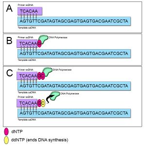
The labor-intensive Sanger method utilized special chain-terminating inhibitors to selectively terminate DNA synthesis during replication, followed by imaging via gel electrophoresis. A denatured double stranded DNA (dsDNA) template is first prepared using heat treatment. This template would then be amplified in a chain-terminated PCR process. Chain-terminated PCR is similar to standard polymerase chain reaction (PCR), but with the addition of modified fluorescent-labeled nucleotides called di-deoxynucleotide (ddNTPs). The ddNTPs serve as a chain-terminating inhibitor, selectively stopping the DNA replication at one of the four nucleotide sites (A, T, C, G). As shown (Figure 1), when incorporated into the PCR process, depending on the specific ddNTPs used, the produced DNA fragments would be produced at varying length depending on the nucleotide sites of termination. By analyzing the lengths of known fluorescently labeled nucleotides fragments on gel electrophoresis, the sequence of the original DNA strand could be systematically deduced. Despite its labor-intensive process, Sanger sequencing's accuracy and reliability made it the gold standard for many years. [1]
NGS Technologies
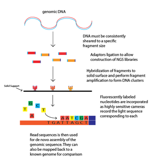
NGS technologies involve several major steps: DNA fragmentation, library preparation, sequencing, and data analysis through bioinformatics. NGS technologies marked a departure from traditional sequencing methods by allowing for the parallel sequencing of millions of DNA fragments. Specifically, these technologies employ different methods such as sequencing by synthesis (Illumina), ion semiconductor sequencing (Ion Torrent), and single-molecule real-time sequencing (Pacific Biosciences). NGS platforms use massively parallel processing to generate vast amounts of data, enabling entire genomes to be sequenced in a fraction of the time and cost required by traditional methods. The main advantages of NGS include its scalability, speed, and the ability to detect single-nucleotide polymorphisms, insertions, deletions, and other genetic variations with high precision.[4]
The core mechanism behind NGS starts with breaking down DNA into smaller fragments that can be efficiently processed. These fragments are then ligated with adaptor sequences, which are short, known DNA sequences that provide a binding platform for DNA to attach to the sequencing instrument. This step also includes a barcode sequence unique for each sample, allowing multiple samples to be sequenced together in one run. [8] Once the library is prepared, it is loaded onto a flow cell where each fragment is clonally amplified. One common method of amplification is bridge amplification, where each DNA molecule bends over and attaches to a nearby complementary oligo, creating a localized clonal amplification of the DNA fragment directly on the flow cell. This amplification is critical as it makes the minute signal from each fragment detectable. [9]
For the sequencing step, one method involves the cyclic addition of differently-colored fluorescently labeled nucleotides. As each nucleotide is incorporated into the growing DNA strand, a camera captures the color emitted by the fluorescent tag, which identifies the base. After each cycle, the fluorescent dye is cleaved to prepare for the next cycle of nucleotide incorporation. Another method employs semiconductor-based technology that detects pH changes instead of fluorescent signals. This method skips the optical detection step, potentially reducing run times and costs. After sequencing, the resulting data undergo complex bioinformatic analysis, where sequences are aligned, indexed, and analyzed to identify genetic variants and structural variations. This step is powered by sophisticated algorithms that handle vast datasets, correcting errors and calling variants with high accuracy, pushing the boundaries of genetic research and medical diagnostics.
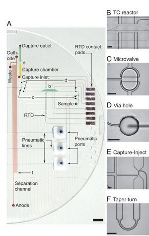
Capillary Electrophoresis-based Sequencing
Capillary electrophoresis (CE) in microfluidics utilizes narrow, fluid-filled channels to separate DNA fragments based on size and charge, under an electric field. This microfluidic application enhances the resolution and speed of traditional CE, making it highly suitable for high-throughput sequencing tasks. The integration of microfluidics with CE has been critical in developing portable, low-cost, and efficient sequencing devices. A significant advancement was documented by Blazej et al., who demonstrated the potential of microfluidic devices to perform rapid DNA sequencing using capillary electrophoresis.[5] In their work, the author fabricated 250-nL reactors, affinity-capture purification chambers, high performance capillary electrophoresis channels, and pneumatic valves and pumps onto a single device (Figure 3).
DNA Microarray Processing
Microfluidic platforms have been adapted to work with DNA microarrays, allowing for simultaneous analysis of thousands of DNA sequences. DNA microarrays function by attaching a large number of different DNA probes to a solid surface, creating an array. Each probe corresponds to a specific DNA sequence that researchers are interested in detecting. When a sample containing DNA is applied to the microarray, complementary sequences in the sample will hybridize—or bind—to their matching probes on the array. This hybridization is typically detected and quantified using fluorescence labeling of the sample DNA, allowing for the analysis of gene expression or genetic variations across many sequences simultaneously. The integration of DNA microarrays in sequencing microfluidics greatly enhances the microarrays capabilities by reducing sample and reagent volumes. Additionally, DNA hybridization, a process that allows the identification and cloning of specific genes, could precisely be controlled in a microfluidic chip. Huang et al. provided insights into microfluidic integration with DNA microarray technology, showing improvements in detection sensitivity and throughput efficiency. [6] Specifically, an automated microfluidic DNA microarray (AMDM) platform was developed for use in mutation detection of genetic variants in arrhythmic diseases. To enable precise control of hybridization, real time flow and temperature control units were fabricated throughout the sequencing process (Figure 4). With the aid of the ADMD platform, the main workflow for microarray DNA hybridization process contained three major steps: DNA hybridization, DNA staining (through SYBR solutions), and graphene oxide (GO) quenching.
DNA Hybridization: This step involves the binding of complementary DNA strands. When the target DNA in the sample comes into contact with probe DNA fixed on the microarray, they pair due to their complementary base sequences. This process is fundamental in microarray technology as it directly impacts the specificity and efficiency of the detection.
DNA Staining: After hybridization, the microarray is stained with a fluorescent dye, such as SYBR Green, which binds to the double-stranded DNA. This dye is highly specific for double-stranded DNA and emits fluorescence when excited by light at a specific wavelength. The intensity of the fluorescence correlates with the amount of target DNA hybridized to the probes, allowing quantitative analysis of gene expression or genetic variations.
Graphene Oxide (GO) Quenching: This innovative step involves the use of graphene oxide to improve the signal-to-noise ratio in microarray experiments. GO can quench excess fluorescent dye that has not bound to target DNA, thereby reducing background fluorescence. This selective quenching enhances the contrast and clarity of the signals from the bound targets, improving the accuracy of the results.
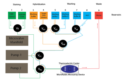
These enhancements in the microarray process, driven by microfluidic integration, optimize the precision and reliability of genetic analysis, making technologies such as the AMDM platform invaluable in the detection and study of genetic variants associated with diseases.
Microdroplet-Based Technology
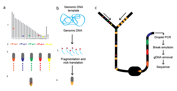
Microdroplet-based technology leverages the precision and sensitivity of PCR to allow for parallel amplification of complex DNA sequences. This method uses microfluidic droplets to encapsulate each primer pair, effectively preventing unwanted primer interactions and enabling the accurate and simultaneous amplification of up to 4,000 targeted DNA sequences. Such encapsulation provides a controlled reaction environment that enhances both specificity and efficiency significantly. This technology shines particularly in large-scale targeted sequencing efforts, demonstrating its ability to manage 1.5 million amplifications simultaneously while maintaining high reproducibility and accuracy across various samples. The approach has been compared to traditional PCR methods, consistently producing high-quality data. Notably, the microdroplet method supports uniform coverage across all targeted sequences and is perfect for a variety of uses as a DNA sequencing tool (Figure 5). [7]
References
1. Sanger, F., Nicklen, S., & Coulson, A. R. (1977). DNA sequencing with chain-terminating inhibitors. Proceedings of the National Academy of Sciences, 74(12), 5463–5467. https://doi.org/10.1073/pnas.74.12.5463
2. Mardis, E. R. (2008). Next-Generation DNA Sequencing Methods. Annual Review of Genomics and Human Genetics, 9(1), 387–402. https://doi.org/10.1146/annurev.genom.9.081307.164359
3. Green, E. D., Watson, J. D., & Collins, F. S. (2015). Human Genome Project: Twenty-five years of big biology. Nature, 526(7571), 29–31. https://doi.org/10.1038/526029a
4. Mardis, E. R. (2011). A decade’s perspective on DNA sequencing technology. Nature, 470(7333), 198–203. https://doi.org/10.1038/nature09796
5. Blazej, R. G., Kumaresan, P., & Mathies, R. A. (2006). Microfabricated bioprocessor for integrated nanoliter-scale Sanger DNA sequencing. Proceedings of the National Academy of Sciences, 103(19), 7240–7245. https://doi.org/10.1073/pnas.0602476103
6. Huang, S.-H., Chang, Y.-S., Jyh-Ming Jimmy Juang, Chang, K.-W., Tsai, M.-H., Lu, T.-P., Lai, L.-C., Chuang, E. Y., & Huang, N.-T. (2018). An automated microfluidic DNA microarray platform for genetic variant detection in inherited arrhythmic diseases. Analyst (London. 1877. Print), 143(6), 1367–1377. https://doi.org/10.1039/c7an01648d
7. Tewhey, R., Warner, J. B., Nakano, M., Libby, B., Medkova, M., David, P. H., Kotsopoulos, S. K., Samuels, M. L., Hutchison, J. B., Larson, J. W., Topol, E. J., Weiner, M. P., Harismendy, O., Olson, J., Link, D. R., & Frazer, K. A. (2009). Microdroplet-based PCR enrichment for large-scale targeted sequencing. Nature Biotechnology, 27(11), 1025–1031. https://doi.org/10.1038/nbt.1583
8. Robin, J. D., Ludlow, A. T., LaRanger, R., Wright, W. E., & Shay, J. W. (2016). Comparison of DNA Quantification Methods for Next Generation Sequencing. Scientific Reports, 6(1). https://doi.org/10.1038/srep24067
9. Mardis, E. R. (2008). Next-Generation DNA Sequencing Methods. Annual Review of Genomics and Human Genetics, 9(1), 387–402. https://doi.org/10.1146/annurev.genom.9.081307.164359
10. Bunnik, E. M., & Le Roch, K. G. (2013). An Introduction to Functional Genomics and Systems Biology. Advances in Wound Care, 2(9), 490–498. https://doi.org/10.1089/wound.2012.0379
