Biomod/2013/Titech/methods&results
<!DOCTYPE> <html> <head> <title>project</title> <style type='text/css'> h1.firstHeading{display:none;}
- jump-to-nav{display:none;}
- siteSub{display:none;}
- contentSub{display:none;}
- column-one { display:none; width:0px;}
- haikei{
width:900px;
margin-right:200px; margin-left:330px;
margin-top:-290px;
background-color:#ffffff;
box-shadow:#000000 2px 2px, gray 5px 5px 5px 5px ;}
- globalWrapper{background-color:green; display:none;}
- column-content{background-color:yellow; display:none;}
- bodyContent{display:none;}
- column-one { display:none; width:0px;}
.OWWNBcpCurrentDateFilled {display: none;}
- footer{margin-left:330px;; width:900px}
body{
 background-color:#ffffff;
 font-size:18px;
 font-family:arial;
text-decoration:none;
 }
h1{ text-align:left; font-style: italic; font-size:50px; text-decoration:none; }
h2{ font-style: italic; font-size:40px; text-align:center; text-decoration:none; }
- haikei p span {
font-weight: bold; }
table{ border:1px black solid; } table tr td{ border:1px black solid; }
.fig{ font-weight:bold; font-size:20px; text-align:center; } .blue{
color:red;
background-color:#ffffff; font-style:italic; }
.itimojime{ font-size:36px; font-family:arial light; }
.menu{
text-decoration:none;
font-family:Arial, Helvetica, sans-serif;
list-style-type:none;
display:block;
position:fixed;
}
a.menu:link{
color:#4e90ff;
}
a.menu:visited{
color:#4e90ff;
}
a.menu:active{ color:#4e90ff; } a.menu:hover{ font-size:24px; text-shadow:#e77070 2px 2px; }
- bar{
list-style-type: none }
- bar li {
text-decoration:none; } .risuto{ font-size:24px; } .risuto2{ font-size:18px; list-style-type: square; } </style> </head> <body>
- <a href = "http://openwetware.org/wiki/Biomod/2013/Titech" class = "menu">Home</a>
- <a href = "http://openwetware.org/wiki/Biomod/2013/Titech/project" class = "menu">Project</a>
- <a href = "http://openwetware.org/wiki/Biomod/2013/Titech/design" class = "menu">Design</a>
- Methods&Results
- <a href="http://openwetware.org/wiki/Biomod/2013/Titech/methods&results" class="menu"> UV-Tuning Nano- Parasol</a>
- <a href="http://openwetware.org/wiki/Biomod/2013/Titech/M%26R_Controlable_Optical_Makeup" class="menu"> Controllable Optical Makeup</a>
- <a href = "http://openwetware.org/wiki/Biomod/2013/Titech/Achievements" class = "menu">Achievements</a>
- <a href = "http://openwetware.org/wiki/Biomod/2013/Titech/" class = "menu">supl.info </a>
- <a href = "http://openwetware.org/wiki/Biomod/2013/Titech/team" class = "menu">member</a>
- <a href = "http://openwetware.org/wiki/Biomod/2013/Titech/acknowledgement" class = "menu">Acknowledgement </a>
<img src="https://upload.wikimedia.org/wikipedia/commons/thumb/5/50/%E3%83%AD%E3%82%B42%E3%81%AE%E3%82%B3%E3%83%94%E3%83%BC_2.jpg/197px-%E3%83%AD%E3%82%B42%E3%81%AE%E3%82%B3%E3%83%94%E3%83%BC_2.jpg" width="250px" height="750px">
<a href="http://openwetware.org/wiki/Biomod">Biomod<a/>|<a href="http://openwetware.org/wiki/Biomod/2013">2013</a>
<img src="https://upload.wikimedia.org/wikipedia/commons/3/34/%E3%81%A8%E3%81%A3%E3%81%B7%E3%81%8C.png" width="900">
<UV-Tuning Nano-Parasol>
<a href ="http://openwetware.org/wiki/Biomod/2013/Titech/M&R Controlable Optical Makeup">>>Controllable Optical Makeup</a>
UV-Tuning Nano-parasol system has four target functions as the component of this system.
0.Making of the Nano-Parasol
The nano-parasol is made of gold nanoparticles and DNA. We confirmed two things, first, gold nanoparticles absorbe UV light, and second, DNA connect with gold nanoparticles.
<img src="https://upload.wikimedia.org/wikipedia/commons/thumb/3/3d/Resolt%EF%BC%86Method_P_%E7%AD%8B%E3%83%8A%E3%83%8E.png/800px-Resolt%EF%BC%86Method_P_%E7%AD%8B%E3%83%8A%E3%83%8E.png" width="400" align="center" >
<a href="http://openwetware.org/wiki/Biomod/2013/Titech/see more 0">>>see more details</a>
1 . UV-DNA converter
By using a DNA containing azobenzene, “Converter complex”, this system can convert the total amount of UV exposure into the amount of DNA signal.
<a href="http://openwetware.org/wiki/Biomod/2013/Titech/see more 1">>>see more deatails</a>
To confirm the proposed function, we implemented the following experiment.
・Release of DNA signal by UV irradiation
We confirmed that DNA siganl is released accordimg to the amount of UV irradiation from“Converter complex”.
We observed that the hybridization of a DNA containing azobenzene is destabilized by UV irradiation by mesuring an absorption spectrum of a DNA containing azobenzene. The detail of the materials we used is mentionded in suple.info.
Fig. 8. (a) shows a graph of an absorbance spectrum of the sample when it is irradiated with UV for 0 minute, 1 minute, 5minutes and 10 minutes. In this spectrum, the peak absorbance at 260nm is DNA’s absorbance, at 350nm is trans-azobenzene’s absorbance and at 440nm is cis-azobenzene’s absorbance.
And Fig. 8. (b) shows a graph of the chronological change of the absorbance at 260nm. This graph shows that the longer UV irradiation time is, the bigger absorbance is. This means that single strand DNA increases according to UV irradiation time. That is to say, it shows that DNA signal is released according to the amount of UV irradiation.
We concluded that DNA signal is released according to the amount of UV irradiation from Converter complex.
|
(a) <img src="https://upload.wikimedia.org/wikipedia/commons/1/11/Resolt%EF%BC%86Method_P_Fig1%28a%29.png" width="300" > |
(b) <img src="https://upload.wikimedia.org/wikipedia/commons/3/35/Resolt%EF%BC%86Method_P_Fig1%28b%29.png" width="300" > |
| Fig. 8. The transformation of a DNA containing azobenzene under UV irradiation (a) The absorbance spectrum of the sample when it is irradiated with UV for |
|
And we also did a supplimentary experiment using a DNA containing azobenzene to make this function certain.
Fig. 9. (a) shows a graph of the chronological change of the absorbance at 350nm. This graph shows that the longer UV irradiation time is, the smaller the absorbance is.
Fig. 9. (b) shoes a graph of the chronological change of the absorbance at 440nm. This graph shows that the longer UV irradiation time is, the bigger the absorbance is. This means that azobenzen changes from trans to cis according to UV irradiation time.
These results also suport this function.
|
(c) <img src="https://upload.wikimedia.org/wikipedia/commons/d/de/Resolt%EF%BC%86Method_P_Fig1%28c%29.png" width="300"> |
(d) <img src="https://upload.wikimedia.org/wikipedia/commons/3/32/Resolt%EF%BC%86Method_P_Fig1%28d%29.png" width="300"> |
|
Fig. 9. Supplimentary information : The transformation of a DNA |
|
<a href="http://openwetware.org/wiki/Biomod/2013/Titech/see more 2">>>see more details</a>
2. Threshold processor
By introducing Threshold complex, this system can process the amount of DNA signal with a threshold.
Here we confirmed the following point to achieve UV-Tuning Nano-Parasol system.
・Thresholding the amount of DNA signal
We succeeded in processing the amount of DNA signal with a threshold using Threshold complex.
<img src="https://upload.wikimedia.org/wikipedia/commons/thumb/a/a1/Design_10-25_%E9%96%BE%E5%80%A4%E3%82%B2%E3%83%BC%E3%83%882.png/800px-Design_10-25_%E9%96%BE%E5%80%A4%E3%82%B2%E3%83%BC%E3%83%882.png" width="550">
Fig. 10. (a) shows the transition of the reaction rate of Linker-output complex and Threshold complex to DNA signal. The reaction rate of each complex was calculated on the basis of fluorescence intensity observed upon strand displacement reaction. To make it easy to understand when these reactions are facilitated, we took the slope of the two curves in vertical axis and the concentration of DNA signal in horizontal axis, which is the next Fig. 10. (b) . From these two graphs, we confirmed that DNA signal primarily reacts with Threshold complex, and then reacts with Linker-output complex.
In this experiment, we set the concentration of Threshold complex at 0.2μM, so ideally the reaction of DNA signal with Threshold Complex ends at this point and that with Linker-output complex should start. It is not an ideal result, but as Fig. 10. (b) shows, a tendency was observed that the reaction of Linker-output complex with DNA signal accelerated around the DNA signal concentration of 0.2μM.
We succeeded in processing the amount of DNA signal with a threshold using Threshold complex.
| (a)
<img src="https://upload.wikimedia.org/wikipedia/commons/c/c8/Resolt%EF%BC%86Method_P_Fig2%28a%29.png" width="320"> |
(b)
<img src="https://upload.wikimedia.org/wikipedia/commons/0/02/Resolt%EF%BC%86Method_P_Fig2%28b%29.png" width="320" > |
|
Fig. 10. The reaction of DNA signal with Linker-output complex and Threshold complex. |
|
<a href="http://openwetware.org/wiki/Biomod/2013/Titech/see more 4">>>see more details</a>
3. Nano-parasol
The strand-displacement reaction between DNA signal and “Linker-output complex” happen, and “Linker strand” is released. This “Linker strand” hybridize with both gold nanoparticles’ “Handle DNA” and micro beads’ “Port DNA” and gold nanoparticles connect with model cells.
To confirm the proposed function, we implemented the following experiment.
3.1. Modelling of skin cells
3.2. Connection between gold nanoparticles and model cells
3.1. Modelling of skin cells
We succeeded in connecting DNA with polystyrene micro beads and modeling skin cells
We used polystyrene micro beads as a model cells, and used DNA as a model of cell membrene protein which connects with “Linker strand”, and confirmed that DNA connects with polystyrene micro beads.
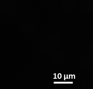
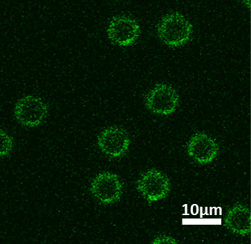
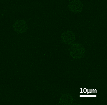
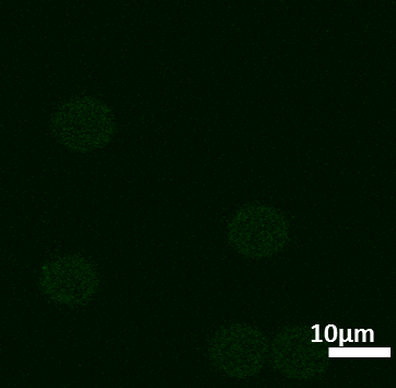
By using fluorophore-modified DNA (FAM-DNA), we confirmed that Port DNA connects with micro-sized bead. We hybridized fluorophore-modified DNA with Port DNA, and observed its fluorescence by fluorescence microscope. Fig. 11.1. shows the two kinds of condition of micro-sized beads (1 : Port DNA+ fluorophore-modified DNA , 2 : micro-sized beads+fluorophore-modified DNA ). First, we confirmed the position of the micro-sized beads by transmitted light, and next, we observed micro-sized beads’ fluorescence by fluorescence light.
We concluded that Port DNA can be attached to micro-sized beads.
| 1 | 2 |
|
(a) <img src="https://upload.wikimedia.org/wikipedia/commons/0/02/Resolt%EF%BC%86Method_P_Fig3%28e%29.png" width="200" height="250"> |
(b) <img src="https://upload.wikimedia.org/wikipedia/commons/4/41/Resolt%EF%BC%86Method_P_Fig3%28f%29.png" width="200" height="250"> |
|
(c) <img src="https://upload.wikimedia.org/wikipedia/commons/a/a2/Resolt%EF%BC%86Method_P_Fig3%28a%29.png" width="200"> |
(d) <img src="https://upload.wikimedia.org/wikipedia/commons/c/c4/Resolt%EF%BC%86Method_P_Fig3%28b%29.png" width="200"> |
|
(e) <img src="https://upload.wikimedia.org/wikipedia/commons/1/1e/Resolt%EF%BC%86Method_P_Fig3%28c%29.png" width="200"> |
(f) <img src="https://upload.wikimedia.org/wikipedia/commons/b/b5/Resolt%EF%BC%86Method_P_Fig3%28d%29.png" width="200"> |
|
Fig. 11.1. The micro-sized beads connected with Port DNA |
|
3.2. Connection between gold nanoparticles and model cells
We succeeded in connecting gold nanoparticles to model cells by “Linker strand”.
First, we confirmed that Linker strand crosslinks Handle DNA and Port DNA. We hybridized these three DNAs and observed the result by electrophoresis.
Experiments in this section are conducted with a substitutional DNA linker whose sequence corresponds to the RNA portion of Linker strand to save cost.
Fig. 11.2.1. shows the result of the electrophoresis. We concluded that three DNA are tightly hybridyzed.
|
<img src="https://upload.wikimedia.org/wikipedia/commons/f/fa/%E3%81%93%E3%82%8C%E3%81%8C%E6%9C%AC%E5%BD%93%E2%91%A0.png" width="600"> |
|
Fig. 11.2.1: The result of electrophoresis which confirmed that Linker strand crosslinks Handle DNA and Port DNA. |
Second, we confirmed that gold nanoparticles connect with micro-sized beads by Linker strand using fluorephor- modified DNA. First, we prepared two solutions, one contains gold nanoparticles connected with Handle DNA, the other contains micro-sized beads connected with Handle DNA. We mixed these two solutions and added Linker Strand. And we also added fluorephor-modified DNA which connects with gold nanoparticles , so if gold nanoparticles connect with micro-sized beads, a fluorecence is observed on the circumference of micro-sized beads.
Fig. 11.2.2. shows three kinds of the scheme, the microscopic image of micro-sized beads by transmitted light and by fluorecence. We compared three kinds of solutions, 1 contains gold nanoparticles with Handle DNA, micro-sized beads with Port DNA, Linker Strand, and fluorephor- modified DNA, 2 contains the same things as 1 except fluorephor- modified DNA , 3 contans only micro-sized beads.
Fig. 11.2.2. (g) shows that there is green fluorecence on the circumference of micro-sized beads. On the other hand, there is no fluorecence in the microscopic image in Fig. 11.2.2. (h) and (i).
We concluded that gold nanoparticles connects with micro-sized beads by Linker strand.
4. Reset mechanism
<img src="https://upload.wikimedia.org/wikipedia/commons/thumb/2/26/%EF%BC%88a%EF%BC%89%E3%81%AA%E3%81%97.png/800px-%EF%BC%88a%EF%BC%89%E3%81%AA%E3%81%97.png" width="500">
UV-Tuning Nano-Parasol system is reset when the crosslinking structutres between gold nanoparticles and micro-sized beads are broken by DNA-RNA double strand degredation reaction caused by RNaseH. Moreover, RNA part of the Linker-output complex is designed to keep a single stranded loop structure so that it is not degraded by RNaseH.
To confirm the proposed function, we implemented the following experiments.
・Break the connection between gold nanoparticles and micro-sized beads.
We confirmed that Linker strand is degrated by RNaseH.
Using polyacrylamide gel electrophoresis, we confirmed that a RNA portion of Linker strand was degraded by RNaseH.
We conducted the reaction by adding 0.5U of RNaseH to each 1μM DNA sample. The reaction times of each sample are the following: 0min, 1min, 2min, 3min, 4min, 5min, 10min, 20min and 6h.
Fig. 12.1. shows the result of the electrophoresis. You can see that the band of degraded RNA is getting darker as reaction time becomes longer. At the same time, the band of crosslinking structure is getting lighter.
We concluded that RNaseH can successfully degrade the crosslinking of handle DNA and port DNA and reset UV-Tuning Nano-Parasol system.
|
<img src="https://upload.wikimedia.org/wikipedia/commons/e/ea/%E3%81%93%E3%82%8C%E3%81%8C%E6%9C%AC%E5%BD%93%E2%91%A4.png" width="660"> |
|
Fig. 12.1. The reaction times between RNaseH and DNA sample of each sample are the following: 0min, 1min, 2min, 3min, 4min, 5min, 10min, 20min and 6h (1-9). |
We also confirmed that the single-stranded RNA portion in Linker-output complex is not degraded by RNaseH.
Fig. 12.2 shows the result of the comparison of Linker-output complex with/without RNaseH. The bands of these two samples almost coincided. In addition, a band that appears when the sigle-stranded RNA portion is degraded by RNaseH didn’t appear. Therefore, we succeeded in confirming that degradation of Linker-output complex by RNaseH doesn’t occur.
We concluded that Linker-output complex is not degraded when it is in inactivated condition, and is degraded when it is forming a crosslinking structure.
|
<img src="https://upload.wikimedia.org/wikipedia/commons/thumb/0/0a/Resolt%EF%BC%86Method_P_Fig_4.2.png/718px-Resolt%EF%BC%86Method_P_Fig_4.2.png" width="500"> |
|
Fig. 12.2. The result of the comparison of Linker-output complex with/without RNaseH |
<a href="http://openwetware.org/wiki/Biomod/2013/Titech/see more 9">>>see more details</a>
</body> </html>
