BME100 s2016:Group6 W1030AM L6
| Home People Lab Write-Up 1 | Lab Write-Up 2 | Lab Write-Up 3 Lab Write-Up 4 | Lab Write-Up 5 | Lab Write-Up 6 Course Logistics For Instructors Photos Wiki Editing Help | |||||||
OUR COMPANY
LAB 6 WRITE-UPBayesian StatisticsOverview of the Original Diagnosis System In BME 100, patients were given to 17 groups of 6 students each, testing for the disease associated SNP. 3 samples of each patient were given to the groups as well as buffer solution, calibration solutions, a positive control, a negative control, and green fluorescence. Firstly, the calibration solutions were imaged through PCR and analyzed in imageJ. The actual patient DNA samples were then imaged through PCR and analyzed in image J. In order to prevent error when combining the DNA with other solutions, the pipette tips were constantly changed, preventing contamination. The green fluorescence was also covered immediately after use to prevent it from absorbing the room's lighting. In total, there were 96 PCR's with 36 total positive PCR's. Out of the total PCR's, there were 56 negative results. In total COMPLETE PCR's, there were 32 tests. Out of those 32 tests, 12 were positive, 18 were negative, and 2 were inconclusive. Some challenges that may have occurred were that the images were not analyzed in imageJ accurately. When drawing the oval around the droplet, there could have been some error when sizing it, affecting the other data measurements as well. Also, the images may have not been at the same angle in all of the pictures taken so this, again, would change results by changing the droplets size and shape. What Bayes Statistics Imply about This Diagnostic Approach
Some possible sources of error that could potentially affect the Bayes values are: incorrect use of the pipet, the limitation of the measurement technology used to only be accurate to a certain degree, and rounding errors. Intro to Computer-Aided DesignTinkerCAD Using the TInkerCAD tool, a new design for the box that covers the fluorimeter was created. In comparison to Solid Works, TInkerCAD is more user friendly and only requires a short video to get a general understanding of the program. TinkerCAD is easier in the sense of moving objects, sizing objects, and assembling overall parts together. Solid Works is very difficult because of the programs errors in measurements and the difficulty in assembling parts in a uniform way. TInkerCAD was able to create our brainstormed device improvement easily. Our Design
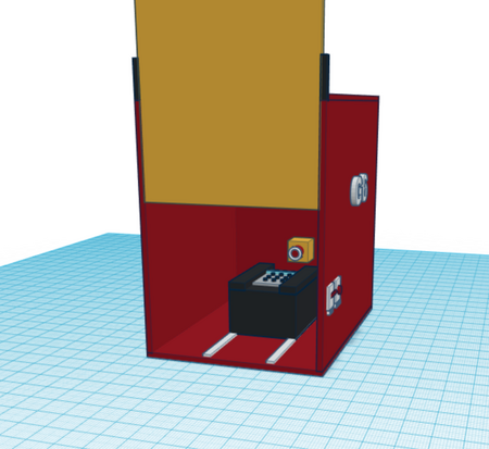
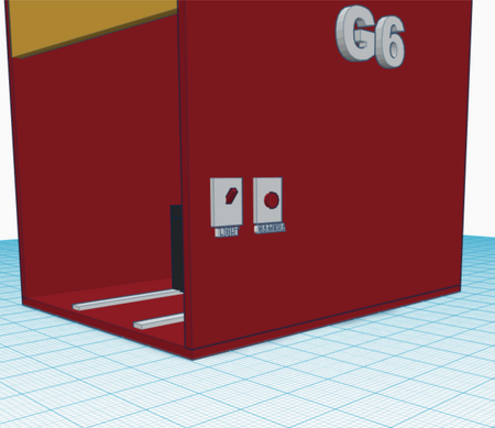
There was no change in the OpenPCR design. The new design is attempting to modify the fluorimeter design and make it more user friendly. This design features an built in camera into the box that covers the fluorimeter. This improves the fluorimeter design by reducing the error in different angles of camera pictures. Therefore, this would assist in making the data more accurate through more accurate pictures from the results. The fluorimeter design will also feature a built-in light switch and camera button. This will reduce chance of knocking over the PCR sample. The design will also include a door which can slide up and down. This will reduce the chance of any outside light affecting the PCR sample. In addition, the box contains sliders underneath the fluorimeter device in order to allow for easy changing of the sample slides.
Feature 1: ConsumablesIn our kit, standard tubes will be provided that will fit into our PCR machine. Primer solution, SYBR Green solution, and buffer will be included as well, making sure that the Green solution is in a closed container to reduce errors. These consumables are added to this kit in order to be convenient to the user/buyer. Another consumable that our product will have a built in camera box in order for easy image use. This camera will be a part of the box that will complement the PCR machine. Weaknesses: This camera will be unique to the box, therefore if the camera were to break, a whole new box with the built in camera will be needed to be purchased.
Feature 2: Hardware - PCR Machine & FluorimeterFluorimeter: Our design changes the fluorimeter's outer box, intending to make imaging more user friendly. Only the outer box covering the fluorimeter will be changed with a built in camera to create level, clear images to be analyzed in ImageJ. The camera is aimed to be level with the fluorimeter in order to create identical results. The camera will be small enough not to bulge out of the back of the box entirely but large enough to hold and store images
The Open PCR machine will not be included because nothing is changed to the PCR machine. The focus of this design is aimed around benefitting the fluorimeter to make it more user friendly.
| |||||||


