BME100 s2016:Group5 W1030AM L6
| Home People Lab Write-Up 1 | Lab Write-Up 2 | Lab Write-Up 3 Lab Write-Up 4 | Lab Write-Up 5 | Lab Write-Up 6 Course Logistics For Instructors Photos Wiki Editing Help | |||||||
OUR COMPANY
LAB 6 WRITE-UPBayesian StatisticsOverview of the Original Diagnosis System In BME 100, 17 groups of six students each, tested for the disease associated SNP. Three samples of each patient were given to the 17 groups, along with a buffer, calibration solutions, a positive and negative control, and green fluorescence. The calibration solutions were imaged through PCR and analyzed in the software imageJ. The patient DNA samples were then imaged through PCR and put in imageJ. To prevent error, the pipette tips were changed every time they came in contact with one of the solutions. The green fluorescence was covered immediately to prevent it from absorbing light. In total, 96 PCRs with 36 total positive PCRs and 56 negative PCRs. In total, 32 tests were run with 12 positive, 18 negative, and 2 inconclusives out of those 32 tests. Some problems that occurred were that the images were tough to analyze in imageJ. Also, not all the pictures were probably taken at the exact same angle. This could be another source of error. What Bayes Statistics Imply about This Diagnostic Approach Calculation 1 was relatively close to 1. This means that if a patient has a positive test diagnosis, then they will have a positive pcr reaction. The patient will develop the disease. Calculation 2 shows that if a patient has a negative test result, they will have a negative pcr reaction. So the patient will not develop the disease. Calculation 3 shows us that if a patient received a positive test diagnosis, there is still a low chance of developing a disease, less than 50%. The result for calculation 4 indicates that if a patient recieved a negative diagnosis, there is no chance of developing the disease. 0% chance of developing the disease. Some possible sources of error could be rounding errors, incorrect use of the pipette, and the limitation of the measurement. Another possible error could be an incorrect interpretation when analyzing the tests.
Intro to Computer-Aided DesignTinkerCAD Our Design
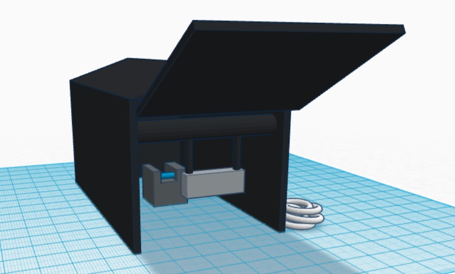
Feature 1: ConsumablesOur Consumables kit : PCR mix, primer, pre-labeled plastic tubes and SYBR Green solution The biggest weakness for the general consumables kit is in the plastic tubes. Our team encountered difficulties to stay organized because of the plastic tubes, therefore, we thought it would be a good idea to improve them by pre-labeling so there will be no confusion or accidents like clearing the marker from the plastic material that they are made of. We thought increasing the size with 15% will make them easier to operate. The reagents are playing the main part of the PCR reaction, therefore these reagents will have their own space in the kit and will have the name written above them, to keep things organized. Feature 2: Hardware - PCR Machine & FluorimeterThe Open PCR machine will not see any changes in our system but there will be changes made to the light box and camera cradle for the fluorimeter. Both fluorimeter and the Open PCR will be included in our system, but the main focus will be the easier to use fluorimeter. The biggest change comes from the light box used with the fluorimeter. The light box now includes brackets that hold a camera mounted in place and it remains focused on the slide stand. This will allow for much easier and more consistent set up, as well as easier calibration. There is a USB cable connected to the box that will allow for faster image transfer into the computer software program.
| |||||||


