BME100 s2016:Group12 W1030AM L5

From OpenWetWare
Jump to navigationJump to search
BME 100 Spring 2016 Home
People
Lab Write-Up 1 | Lab Write-Up 2 | Lab Write-Up 3
Lab Write-Up 4 | Lab Write-Up 5 | Lab Write-Up 6
Course Logistics For Instructors
Photos
Wiki Editing Help

OUR TEAM

Error creating thumbnail: Unable to save thumbnail to destination
Name: Elizabeth Delgado
Error creating thumbnail: Unable to save thumbnail to destination
Name: Allan Santos
Error creating thumbnail: Unable to save thumbnail to destination
Name: Ryan Male
Error creating thumbnail: Unable to save thumbnail to destination
Name: Antonio Lopez
Error creating thumbnail: Unable to save thumbnail to destination
Name: Stephanie Vargas
Error creating thumbnail: Unable to save thumbnail to destination
Name: Aderonke Adewuyi


LAB 5 WRITE-UP

PCR Reaction Report

Last weeks experiments involved pipetting samples to set up the reaction, the pre-lab material was essential for this lab because it explained how to properly pipet the samples and how the instrument should be set up. The difference between the first and second pipettor is important because the first pipettor grabs the sample while pushing the pipet down to the second releases the sample. The main objective of this lab was to test if dsDNA is present in either one of the two patient samples. In order to being this pipetting lab we collecting all materials such as, test tubes, positive/negative samples, patient samples, pippettor, pippetor tips, lab coat, gloves, and patient samples. First we dived up the given tubes in designated groups then labeled them accordingly to keep track of each sample. Then we used pippetting to form each reaction, while doing this we used a new tip for each sample. After all the samples were mixed the eight test tubes were placed in thePCR machine where the actual reaction took place.

Fluorimeter Procedure

Setting up the smart phone camera
STEP 1: Put on gloves, then find which side of the slide is smooth STEP 2: Turn on fluorimeter STEP 3: Place smooth side of slide facing down in the fluorimeter STEP 4: Set camera timer to 3 seconds STEP 5:Open camera application then place smart phone in the cradle STEP 6: Adjust height of the fluorimeter so that the camera view is at the edge of fluorimeter STEP 7: Place 80 uL drops of SYBR green solution onto the center of slide STEP 8: Add calibration solution on top of SYBR green solution again on the center of the slide STEP 9: Adjust lights so it is aligned with the solution STEP 10: Adjust the distance between the smart phone and fluorimeter so the drop is clear and vivid STEP 11: Cover the fluorimeter with the light box keeping one side open STEP 12: Readjust the drop for accuracy STEP 13: Start timer and close the side STEP 14: Take picture STEP 15: Remove drops from slide and discard in waste STEP 16: Shift slide to the center of the next two circles REPEAT STEPS


Placing Samples onto the Fluorimeter

  1. [Instructions: Step one, in your OWN words]
  2. [Instructions: Step two, in your own words]
  3. [Instructions: Step three, in your own words]
  4. [Instructions: Step etc., in your own words]


Data Collection and Analysis

Images of High, Low, and Zero Calf Thymus DNA


Calibrator Mean Values

TABLE GOES HERE


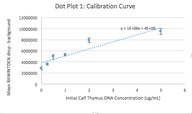
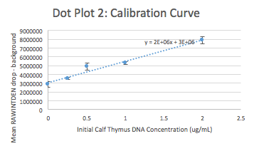
Calibration curves
Error creating thumbnail: Unable to save thumbnail to destination Error creating thumbnail: Unable to save thumbnail to destination


Images of Our PCR Negative and Positive Controls


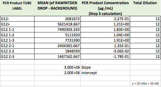
PCR Results: PCR concentrations solved


PCR Results: Summary

  • Our positive control PCR result was ____ μg/mL
  • Our negative control PCR result was ____ μg/mL


Observed results

  • Patient _____ :
  • Patient _____ :


Conclusions

  • Patient _____ :
  • Patient _____ :