BME100 f2013:W1200 Group1 L5
| Home People Lab Write-Up 1 | Lab Write-Up 2 | Lab Write-Up 3 Lab Write-Up 4 | Lab Write-Up 5 | Lab Write-Up 6 Course Logistics For Instructors Photos Wiki Editing Help | ||||||||||||||||||||||||||||||||||
OUR TEAM
LAB 5 WRITE UPBackground InformationSYBR Green Dye
ProcedureSmart Phone Camera Settings
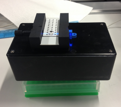
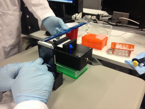
1. Switch the blue light on on the fluorimeter. 2. Adjust camera settings on smart phone to match the above settings. 3. Place smart phone in cradle so that it is perpendicular to the table and the camera lens is level with the glass slide and the drop. 4. Place cradle and phone as close as possible to the drop so that the drop remains in sharp focus. 5. Use the provided ruler to measure the distance between the drop and the camera lens. In our case, this distance was 8.5 cm.
Solutions Used for Calibration
Placing Samples onto the Fluorimeter 1. Insert the slide into the fluorimeter, smooth side down. 2. Use the micropipette to place 80µL of the SYBR green dye in the second row of the middle column of the slide. 3. Micropipette 80µL of the specific calf thymus DNA on top of the SYBR green dye. 4. Ensure that the light is turned on. Then start self timer on phone and place light box over entire set up.
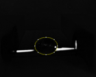
Data AnalysisRepresentative Images of Samples
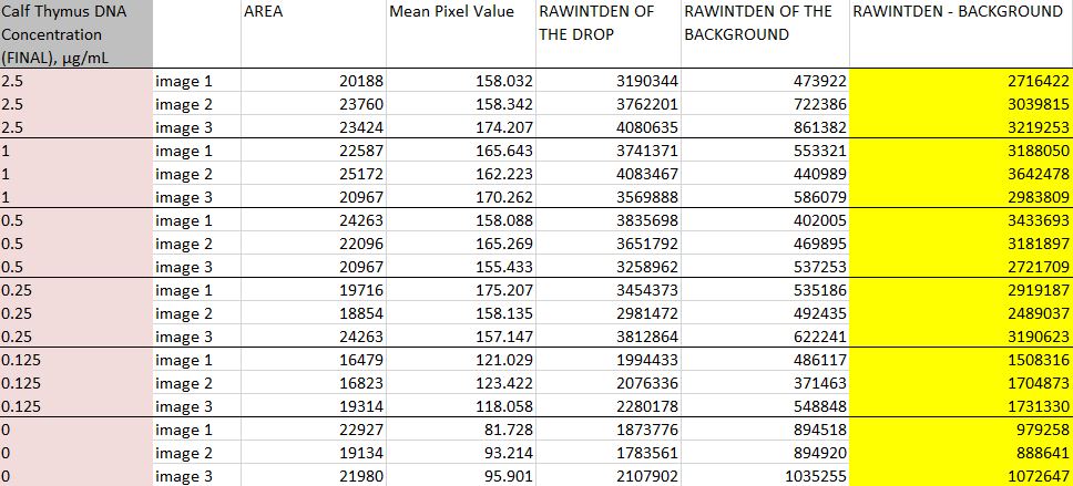
Image J Values for All Samples | ||||||||||||||||||||||||||||||||||
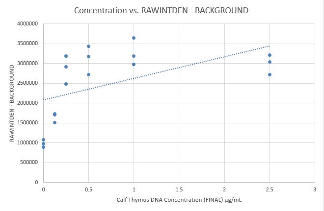
Fitting a Straight Line
