HRV:Phase One

Home Lab Members Phase One Physiological Systems Monitoring Parameters in the ICU ECG HRV Cardiorespiratory Monitors Phase Two Deliverables Journal Abbreviations
Introduction
Phase 1 of the project comprises a “library” investigation into the physiological systems of respiration, cardiovascular and blood pressure control, and thermoregulation. Our team investigated the basis of these physiological systems with an aim to understand their importance. Having established their basic physiological behaviour, the next step was to identify the parameters that were paramount to clinicians when monitoring the critically ill in an ICU/ITU setting.
One of the physiological signals that are monitored on a continuous basis is the ECG. We investigated the clinical significance of ECG in by identifying two important aspects of the ECG, in terms of medicine, the first is the morphology and the second is the variations in heart rate.
Over the last 20 years, the beat by beat variation in heart rate (HRV) has become an increasingly important indicator of underlying physiological state. We studied the reason for this, particularly in relation to respiration, blood pressure, and temperature regulation. Our team performed a detailed analysis of how HRV is currently used in clinical medicine and established a link to cardiorespiratory monitoring.
Finally, we explored the importance of cardiorespiratory monitoring in relation to defining patient state in the ICU/ITU. We looked into the whole area of cardiorespiratory monitors developed by various companies, their features and their function within an ITU.
Physiological systems
Respiratory System
Respiratory diseases affect more than 1 billion people[1], with more than 3 million dying from chronic obstructive pulmonary diseases (COPD) per year.[2]
Background
The respiratory system (Figure 2.1a) is responsible for the exchange of gases to provide oxygen for ATP production and remove carbon dioxide from the body. Oxygen diffuses from the alveoli into the capillaries and then erythrocytes, where it binds reversibly to haemoglobin. Oxygen saturation (SpO2) is the percentage of oxygen that has bound to heamoglobin and can be measured using a pulse oximeter.

In cells, respiration is fundamentally the exchange of glucose and oxygen into water, carbon dioxide and energy. This is summarised by the following equation:
C6H12O6 + 6O2 → 6H2O + 6CO2 (+ ATP) — (1)
ATP is needed for various active processes occurring in internal organs, keeping an organism alive.
Respiration is measured in respiratory rate (RR), representing the ventilation in and out of the lungs. If not kept in check, there is a higher risk of a malfunction in the body. Without gas exchange (essentially oxygen intake and excretion of carbon dioxide), no energy is produced. As a result, organs would fail and no longer support the life of an organism.
Cardiovascular System
Cardiovascular diseases (CVDs) are the leading cause of death worldwide, contributing to 31% of all annual deaths according to the World Health Organisation (WHO)[4], which highlights the importance of the cardiovascular system.
Background
The cardiovascular system (Fig 2.2a) is separated into the pulmonary and systemic circulation. The heart is divided into 4 chambers, with the upper chambers known as the atria and the lower chambers known as the ventricles. There are several types of blood vessels, namely the arteries, veins and capillaries, which serve different functions. The arteries carry blood away from the heart, which branch into arterioles and then capillaries. The capillaries rejoin to form venules and finally veins, which transport blood back to the heart. The right side of the heart pumps deoxygenated blood through the pulmonary artery to the lungs for gaseous exchange. Oxygenated blood is pumped by the left side of the heart to other parts of the body.
![Parts of the Cardiovascular System [1]](/wiki/File:HRV_Cardiovascular_system.png)
Figure 2.2a: Parts of the Cardiovascular System[5]
The crucial functions of the cardiovascular system for survival are as follows[6]:
- Transport of substances
- Control by transport of hormones
- Body temperature regulation
An electrocardiogram (ECG) measures the electrical activity of the heart, which in turn reflects the current condition of the cardiovascular system. The workings and real-life applications of the ECG are extensively explained in the section "ECG".
If the cardiovascular system does not function properly, substances cannot be moved to the appropriate places of the body. Regardless of respiration, cardiovascular failure is fatal. This is evident in cardiac arrest where beforehand, the absence of blood flow causes symptoms such as irregular breathing and a loss of consciousness.
Transport of substances
The cardiovascular system is required to transport essential substances throughout the body. With reference to equation (1):
- Nutrients are transported to cells in the rest of the body for energy (ATP) production. These include:
- Glucose from the digestive system, and
- Oxygen from the respiratory system.
- Waste products are transported to the excretory system to be removed from the body. These include:
- Water from the digestive system, and
- Carbon dioxide from the respiratory system.
Blood Pressure Control
Blood pressure represents the force exerted by the heart to pump blood throughout the body. This is measured by wearing a cuff around the upper arm, and the apparatus is inflated until no blood can flow through the brachial artery[7]
If blood pressure is too high, arteries will thicken to adapt to this change over time. Thus the inner diameter decreases and less blood can flow per unit time. With a smaller diameter, the resultant surface area decreases and there is a higher risk of clogging by plaque (atherosclerosis)[8] As a result the cardiovascular system would have a reduced blood circulation of nutrients and waste. There is a higher risk of plaque buildup with certain lifestyles or traits, including:
On the other hand, if blood pressure is too low, nutrients cannot be transported quickly enough to critical organs such as muscles and the brain. This leads to a high risk of fainting or generally feeling weak, i.e. symptoms of anemia.
Thermoregulation
The body needs to operate at a specific range of temperatures to function properly: in humans, this is typically defined to be 36.5-37.5˚C[12].
Heat transfer between the body and surrounding environment occurs through conduction, convection, radiation and evaporation.
If the internal body temperature is too high, vasodilatation occurs and sweat is produced on the skin. The process of sweat evaporation cools the body. In contrast, a low internal temperature causes vasoconstriction to reduce blood flow, thus reducing heat loss. Additionally, hairs on the skin trap a layer of hair above the latter. These processes aid the maintenance of heat (and thus temperature) in the body.
There are a range of methods to measure thermoregulation. For example:
- Oral or tympanic thermometers[13]
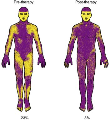
- Thermoregulatory sweat tests (TST)[14]. This involves applying a powdered substance throughout the skin and watching its changes in colour while sweat is produced on different body parts, under controlled conditions (Figure 2.2.4a).

Whether too high or too low, an abnormal body temperature causes fatalities. For instance, heat stroke can happen under prolonged periods of time in hot weather, which is a cause for heart failure[16].
Monitoring Parameters in the ICU
Overview of Respiratory Monitoring in the ICU
Respiratory monitoring is vital in the current management of patients suffering from acute respiratory failure. Clinicians understanding of disease processes and effects of clinical interventions can be improved through the appropriate use of various available monitoring techniques and correct interpretation of data. A table summarizing the current modes of respiratory monitoring and their potential usefulness in the clinical setting is available here.[17] These techniques are essentially categorized into timing (continuous/intermittent), specific situations to be used in, potential usefulness, and their limitations.
Overview of key parameters that can and should be monitored in the critically ill patient with respiratory failure:
| Category | Parameters |
|---|---|
| Gas Exchange | |
| Respiratory Mechanics | |
| Lung Volumes |
Overview of Hemodynamic Monitoring in the ICU
Hemodynamic monitoring is crucial in critically ill patients as they are often hemodynamically unstable due to hypovolemia, cardiac dysfunction and or alterations of vasomotor function which all leads to organ failure as there is a mismatch between oxygen delivery and demand. Monitoring of the cardiovascular system allows clinicians to guide their medical management to prevent or treat organ failure. A summary table of currently available monitoring techniques and its advantages and disadvantages, categorized into the degree of invasiveness of the techniques as well as whether or not they are calibrated can be found here.[18]
The basic vital parameters that can and should be monitored for patients who hemodynamically unstable:
- Heart rate
- Blood pressure
- CVP
- Peripheral and central venous oxygen saturation
- Respiratory variables
- Urine output
When the basic parameters fall out of a safe range, there is an increased for hemodynamic monitoring of:
- CO
- PAOP or wedge pressure
- PAP
- SvO2
- SVV
- Extravascular water
Overview of various cardiac output monitoring techniques and other key parameters that need to be monitored for patients who are suffering from organ failure or are at risk:
| Degree of Invasiveness | Monitoring Technique | Parameters |
|---|---|---|
| Invasive | Pulmonary Artery Catheter (PAC) |
|
| Transpulmonary thermodilution |
| |
| Transpulmonary dye dilution |
| |
| Ultrasound flow dilution |
| |
| Respiratory derived cardiac output monitoring |
| |
| Pulse contour and pulse pressure analysis |
| |
| Transesophageal echocardiography |
| |
| Esophageal Doppler |
| |
| Estimated continuous cardiac output |
| |
| Ultrasonic cardiac output monitoring |
|
Heart Rate
Heart rate (HR) is one of the most basic cardiovascular parameters. The average resting heart rate (HR) is 72 beats per minute (bpm) but varies depending on age, physical activity and diet. HR can be measured by manually counting the number of pulses by placing your fingers on your wrist or neck. In clinical settings, HR is measured using HR monitors or ECG. In the last few years, many wearable devices such as sports watches can measure HR to track fitness during exercise and rest.
An abnormal heart rhythm can lead to more serious complications. Arryhthmia is the condition in which the patient's heart rate is irregular[19] and has many different forms:
Tachycardia
Tachycardia is the condition for a resting HR that is too quick, typically above 100 bpm[19].
Types of tachycardia[20]:
- Supraventricular tachycardia: occurs in the atria
- Ventricular tachycardia: occurs in the ventricles
- Sinus tachycardia: increase in HR during excitement or illness, can return to normal
Bradycardia
When the resting HR is too slow (below 60 bpm), the patient is said to have bradycardia[19]. Bradycardia occurs when there is disruption of the electrical signals as they travel frmo the atria to the ventricles[20]. Most athletes will have slower heart rates due to their exercise or training regimes, though this is not a clinical condition.
Atrial Fibrillation
Atrial fibrillation is one form of arrhythmia where the atria contract randomly and too quickly[19].
Ventricular Fibrillation
Cardiac Output
Cardiac Output (CO) is the amount of blood in litres that the heart pumps per minute and is calculated by equation (2) below[21]:
CO = HR x SV — (2)
where CO is the cardiac output, HR is the heart rate and SV is the stroke volume.
A high cardiac output could indicate sepsis[21] while a low cardiac output could result from heart failure.
Blood Pressure (BP)
Blood pressure is made of the systolic and diastolic pressures, which are the pressure during the contraction and relaxation of the heart respectively[22]. Normal BP is in the range of 90/60 mmHg to 120/80 mmHg[23]. High BP or hypertension can lead to heart attacks and strokes[22].

Mean Arterial Pressure (MAP)
Septic shock is caused by a low BP. It is more common to track MAP in a patient for the potential onset of septic shock, however.
MAP is more efficiently measured through invasive monitoring by providing up-to-date measurements. Below is a procedure outlined by the Association of Anaesthesists[25]:
- The ulnar artery is located by taking Allen's test, which simply involves clenching the fist. An arterial cannula (needle) is then inserted into the appropriate site where the arterial lumen should be located.
- The cannula is connected to a saline column enabling electrical conduction to an adjacent transducer. The transducer contains a diaphragm that alters electrical output depending on BP levels.
- The outputs of the transducer are measured visually on a connected display.
The invasive system also features a "flushing" system consisting of a bag of saline pressurised at 300mmHg. This flushing can be done manually or automatically, and allows the ejection of blood samples and air at certain intervals.
Such a method brings very accurate output measurements and graphical charts conveniently tracking the progress of MAP over time. There is a risk of several complications arising (listed below), but this should be very low if the flushing system is adequate and the initial procedure of cannula insertion is done carefully.
- Thrombosis (blood clot) can develop within the artery
- Ischaemia (restricted blood supply to tissues) upon delivery of external drugs to the blood
If measurements are taken non-invasively, MAP is calculated through the formula[26]:
MAP=DP+1⁄3(SP-DP) or MAP=DP+1⁄3(PP)
Where DP is the diastolic blood pressure, SP is the systolic blood pressure, and PP is the pulse pressure. It is straightforward to calculate this as all the variables can be tracked in real time.

Non-invasive BP monitors on the market are very diverse. Prices can start from £30 (Figures 3.5.1c-d) for monitors that are hassle-free to set up, however they do not calculate MAP automatically. This is problematic if this is exact parameter needs to be tracked over time. On the other hand, monitors priced from £1200 (Figure 3.5.1e) features integration of EMRs from partner companies (TPP, EMIS, etc.) and technical support.

All BP monitors score in terms of portability, regardless of price. The major difference is the more expensive monitors have more reliable and efficient methods for recording measurements.
By comparing invasive and non-invasive monitors, it is evident that there is a trade-off between comfort and efficiency of obtaining measurements. With non-invasive monitors, patients do not need to worry about procedures involving the insertion of needles into their skin. However, time has to be spent manually calculating MAP. This type of monitor can be improved by having the calculations done automatically, but it should be noted that 3 variables (1 of SP and 2 of DP) are involved in this calculation. Without invasive measuring, each variable would introduce a margin of error, and this margin would be even larger upon calculating MAP. The advantages of invasive monitors seem to outweigh those of non-invasive monitors.
Oxygen Saturation
SpO2 is normally between 95% - 100% and, if it is <90%, the patient requires immediate treatment[30]. The Oxygen-haemoglobin dissocation curve (Figure 3.6a) shows the relationship between SpO2 and the partial pressure of Oxygen (PO2). The S-shape of the curve is a safety factor, as a large decrease in PO2 results in a relatively small decrease in SpO2 (for higher levels of PO2).The Bohr effect explains how several factors such as pH, temperature and CO shifts the curve to the left or right.

Temperature
The normal body temperature is between 36 to 37 degrees Celsius. High body temperatures are an indication of an infection or sepsis.
Challenges of Monitoring in the ICU
In the ICU, nurses are responsible for the primary monitoring of patients which includes checking monitors. While doctors oversee patients by assessing their progress, it is nurses who predominantly look at monitors. Nurses identify early changes in a patient’s conditions and alert the relevant physicians. Nurses in ICUs normally only have 1 or 2 patients at a time, in order to closely follow their progress. [32] In ICU, the interface for health care providers to monitors the various physiological parameters is a large monitor which displays either graphically or numerically the different variables. These monitors have various functions which can include alarm systems to warn nurses when a particular parameter has gone out of the ‘healthy’ range. Nurses in ICUs have reported problems they experience when using these monitors [33]:
- False Alarms- there is a high frequency of alarms of which are high proportion are false, a suggestion has been made that alarm thresholds should be adjustable depending on the individual. This has been implemented in a number of monitors; however nurses cite the complexity of menu structure of monitors meant they were not able to change alarm parameters.
- Integration of Information- one such example is that oxygen saturation falls when body temperature lowers, this in itself is not a reason for concern. However, alarms sound for the decreased oxygen saturation and nurses then have to check other parameters to see whether it has fallen due to a decline in health or just do to fluctuations in other parameters.
- Lack of standardisation in display- different monitors have different displays which require training in order to use to the best of its functionality. Nurses lack the time needed in order to properly understand all the different menu pathways which leads in inefficient use of the devices.
ECG
Overview
An ECG is a recording of the electrical signals produced by the heart[34]. There are several different types of ECG monitors, their differences mainly being how many leads they operate with, and how long they monitor an individual for. The ECG (Fig 4a) consists of a P wave, QRS complex and T wave which are caused by atrial depolarisation, ventricular depolarisation and ventricular repolarisation respectively[6]. Variations in ECG readings can be used to detect arrhythmias, coronary heart disease, heart attacks and cardiomyopathy[21], although this proves to be challenging. A study conducted by the Heart Rhythm Society (HRS) found that less than 1/4 of 800 physicians distinguished the length of all QT intervals correctly[35].
![A typical ECG recording (Friederich,Patrick)[5]](/wiki/File:HRV_ECG_recording.png)
![Abnormal ECG recordings and corresponding disease type [6]](/wiki/File:HRV_ECG_disease.png)
Clinical Importance of ECG
ECGs are used in hospitals for the following reasons [37]:
- Looking for the cause of chest pain
- Identifying irregular heartbeats
- Determining overall health of heart before procedures, for example surgery
- Tracking heart health after treatment for conditions such as myocardial infarction or endocarditis
- Used to obtain a baseline of regular heart function to compare with future examinations
Variations in frequency, amplitude and shape are examined to diagnose issues. It is important to note that age, sex and race can also affect the morphology of an ECG [38].
Morphologies
Some examples of changes in the morphology which can indicate a possible medical condition include [39]:
- Changes in the shape of P wave – mitral or pulmonary stenosis, this is where the mitral or pulmonary valves become narrower meaning less blood is pumped through the heart.

- Changes in the interval between P and R waves- first degree block atrioventricular block, this is where conduction through the atrioventricular node is delayed, so there is a delay in the contraction of the ventricles.
- Loss of R wave amplitude- myocardial infarction, which is a heart attack, this occurs when blood through to the heart in greatly reduced causing damage to heart muscle.

.
- Increased frequency of the entire PQRST segment- tachycardia, the heart is beating to fast generally classified with a resting rate is above 100bpm.
HRV
HRV is the beat-by-beat variation in heart rate. The autonomic nervous system (ANS) controls HRV via the sympathetic and parasympathetic nervous system, which increase and decrease the heart rate respectively[42]. A high HRV means that the body responds to inputs from both systems and can adapt easily to stimuli[43].
![Typical HRV[43]](/wiki/File:HRV.png)
![Relationship between HRV and age(Deusen,Mark van)[2]](/wiki/File:HRVandAge.png)
HRV is a critical marker with vast existing applications, and countless potential applications, some of which are very relevant in the current COVID-19 pandemic. Investigating HRV is hence not only important, but crucial. Our dual-phase project will initially focus on exploring the physiological basis behind HRV and which systems influence it.
Relationship to ANS
During chronic stress - both psychological and physical – the parasympathetic nervous system (PNS) is inhibited, and the sympathetic nervous system (SNS) goes into overdrive. This imbalance between the two branches of the autonomic nervous system (ANS) can therefore be a good indicator of stress, and vulnerability to stress [44], and can be measured using heart rate variability (HRV).
ANS function is invaluable in the prognosis of various conditions, including depression, heart failure (HF) and peripheral vascular disease (PVD) [45]. Increased sympathetic outflow has been linked to worse prognosis of conditions such as renal damage and heart failure [46], and these comorbidities increase your risk of mortality with COVID-19 [47]. By inference, HRV could be a useful indicator of prognosis in COVID-19 patients with these comorbidities.
As the ANS is a branch of the peripheral nervous system, it can therefore be argued that disturbances in the ANS reflect the condition of the peripheral nervous system. Neurological symptoms affect about 36% of COVID-19 patients [48], and can manifest in both the central nervous system (CNS) or the peripheral nervous system. In theory, HRV could therefore also indicate the degree to which the peripheral nervous system has been affected in COVID-19 patients.

Figure 5.1a: Relationship of ANS to HRV [49]
Measuring HRV
Domains
Time Domain
HRV is considered a non-invasive marker of the activity of the vagal and sympathetic function [50]. The gold standard for measuring HRV is contact ECG, which is then analysed certain domains, one of which is time-domain, where two indices are recognised:
1. Short-term variability (STV): also called “beat-to-beat variation”, represents fast changes in heart rate (HR);
2. Long-term variability (LTV): represents slower fluctuations. [51]
The time-domain indices above reflect the beat-to-beat changes in a certain timeframe, and are usually calculated by calculating the mean and standard deviation (SDNN) of the RR intervals from an ECG. Common time domain measures are illustrated in the table below.

Frequency Domain
However, to count the number of high and low frequency beats that occur, a frequency-domain is used. [52] Frequency-domain measurements are usually classified into four frequency bands based on absolute power:
i) Ultra-Low-Frequency (≤0.003 Hz)
ii) Very-Low-Frequency (0.0033–0.04 Hz)
iii) Low-Frequency (0.04–0.15 Hz)
iv) High-Frequency (0.15-0.4 Hz)
Some HRV Frequency-Domain parameters are illustrated in the table below. These are in accordance with and were published by the Task Force of the European Society of Cardiology and the North American Society of Pacing and Electrophysiology (1996).

Non-linear Domains
Finally, considering the complexity of HRV, non-linear methods are also used to analyse HRV, the most commonly used being the Poincaré plot, an example of which is illustrated in the figure below. The Poincaré plot is essentially a scatter graph which allows the visualisation of physiological fluctuations in HR.

Clinical Significance
HRV has traditionally been applied predominantly with reference to the cardiovascular system [54], but through this project we are hoping to equally focus on the respiratory system. If accurate signal processing methods are further developed through this project, this would improve the utility and add facility to continuous cardiorespiratory monitoring in ICU settings, for example. Research has already suggested the possibility of monitoring COVID-19 using wearable devices that measure HRV [55]. Therefore, HRV monitoring could be very relevant to the current pandemic.
Current Applications
Since the use and analysis of HRV has become more and more prevalent, the conditions for which HRV can be used as a reliable marker have rapidly expanded. The list includes, but is not limited to:
- Diabetic Neuropathy - patients with diabetic autonomic neuropathy have been shown to have reduced or absent beat-to-beat variation. [56] Measurement and assessment of HRV therefore allows screening of patients with diabetic neuropathy for cardiac involvement [56].
Cadiovascular Risk Stratification
- Chagas Disease - This is one of the main causes of sudden cardiac death in Latin America. Even in the absence of heart failure, in patients with Chagas disease, there are abnormalities in the neural control of HRV [57]. Short-term HRV index values were consistently reduced in the Chagas' disease groups. [58]
- Left Ventricular Hypertrophy (LVH) - patients with LVH have been shown to have a significantly reduced HRV [59], with an inverse relationship between HRV and left ventricle (LV) mass.
- Myocardial Infarction (MI) - HRV is reduced after a myocardial infarction as a neural response to the infarction. Monitoring HRV is useful in predicting the prognosis of patients who have suffered an MI [60], as patients with lower HRV were more susceptible to decreased baroreceptor sensitivity (BRS), which diminished autonomic control [61], amongst other reasons.
- Congestive Heart Failure (CHF) - HRV is significantly depressed in patients with CHF [62], and has independent prognostic values in these patients.
- Malignant Arrhythmias - a non-linear measure of HRV obtained from short-term ECG recordings has important prognostic value in patients with arrhythmias, and has the potential of predicting the risk of death in patients who have implantable cardioverter-defibrillators (ICD) [63]
- Essential Hypertension -
Potential Applications
- Sports Physiology - HRV has been proven to provide parameters that are relevant in analysing the stress the body is under during physical activity and training, and could provide insight into recovery post-training. [64] More research is needed to check exactly how HRV is affected by exercise and what that means for the body during the recovery phase.
- Trauma - HRV is generally decreased in trauma patients, and therefore there is potential for it to be used as a monitoring tool to predict mortality and morbidity in trauma patients [65]
- Organisational Neuroscience - recent research shows that HRV has potential in applications to the studies of emotions, stress and burnout, and behabioural changes, amongst others [66].
Cardiorespiratory Monitors/Devices
Cardiorespiratory monitors are frequently used in the Intensive Care Unit (ICU), ambulances, emergency departments, the neonatal ICU and anaesthetics. They can be categorised by the parameters that they measure, such as Bedside Monitors, Transport Monitors and Remote Monitors. These different types of monitors specialise for certain functions according to the parameters that they detect.
Here is a document which looks at a variety of devices researched: Media:Cardiorespiratory_monitors_list.pdf
General Functions:
- Monitoring patient state and condition[67]
- Identification and diagnosis of underlying cardiorespiratory diseases
- Direct the decision of appropriate treatment[68]
- Warn medical staff of abnormalities in patient condition (if parameters go out of limits) through an alarmwikipedia[2]
- Monitoring general health and lifestyle
- Assess the condition and ability of athletes during training
- Monitor/screen
- Cables
- Electrode patches
- Pressure transducers
- Pulmonary artery catheter
- Arterial blood saturation probe
- Blood pressure cuff
- Printer
- Alarms
- Defibrillators
A large variety of devices, used for cardiorespiratory measurements in ICUs today, were explored. Directly below is a brief overview of how they compare to one another.
Similarities:
- All monitors track similar parameters, which essentially contain all vital information e.g. EKG measurements, BP, HR, RR, etc.
- They automatically link results to an electronic medical record (EMR) system; i.e. actively sending data to the network or database of the hospital they are located at.
- Every monitor displays measurements visually, whether it be through a main monitor screen or an application.
- They feature compatibility with other medical devices used in a clinical setting.
Differences:
- Measurements are taken differently amongst different devices. This can be categorised into either invasive (e.g. inserting a needle into the skin) or non-invasive methods (e.g. non-contact sensors).
- Some devices are designed to be used remotely at the patient's home, whereas others require installation onsite at the hospital.
- There are variations on focused parameters. A monitor may simply store all parameters or highlight those related to BP for instance.
- Accuracy of each device depends on the number of leads available, possibly ranging from 1 to 12.
- They vary by the number of additional components involved, whether they are optional or compulsory.
- Intuitively, prices of devices vary based on their differences.
Bedside Monitors
In the ICU, bedside monitors are most commonly used. These have cables and sensors attached to the patient and warn medical staff when abnormal values arise. They measure the most complete set of parameters and tend to perform 12-lead ECG. As such, they tend to provide more accurate measurements. As patients need to be hooked up to the machine, they may not be portable. Examples of these monitors are shown below (Note: we may want to just summarise these in a table):
CARESCAPE Monitors (GE Healthcare)
The CARESCAPE B650 Monitor can be used in the PACU, OR, NICU and lower-acuity ICU. The software installed can be personalised according to the hospital unit the monitor is used in. Profiles can be made to tailor the conditions of different kinds of patients (e.g. child, adult, elderly, etc.) [69].

The parameters measured are NMT (nuclear medicine teleradiology), entropy, EEG, anaesthetic agent measurements, volumetric CO2 and O22, PiCCO (pulse countour cardiac output), CO, SvO2 (mixed venous oxygen saturation) and dual SpO2.) [69]. Arrhythmia's can be detected using EK-Pro Arrhythmia Algorithm technology, which uses multiple leads to minimise false alarms.[71] Data can be wirelessly transmitted from the monitor to the MUSE Cardiology Information System, which allows clinicians to annotate pacemaker pulses , compare ECGs and edit reports[72].

HemoSphere Advanced Monitoring Platform (Edwards Lifesciences)
Edwards Lifesciences is the global leader in the science of heart valves and hemodynamic monitoring. They develop smart hemodynamic management solutions. The HemoSphere Advanced Monitoring Platform (Figure 6.1.2a) measures a wide range of advanced hemodynamic parameters in which clinicians use to make decisions in a variety of important clinical situations.

Function
This platform guides clinicians in proactive decision making in a range of clinical situations that requires maintenance of optimal patient perfusion such as:
- Cerebral desaturations
- Sepsis management
- Hypotension
- Hemodynamic instability
Features
1. Predictive decision support software
2. Compatibility with a range of other Edwards Lifesciences products
3. Track patient response to interventions
4. 8 screens:
- Goal positioning screen
- Graphical trend screen
- Graphical/Tabular trend split screen
- Physio-relationship screen
- Tabular trend screen
- Cockpit screen
- Physiology screen
- Brain screen
5. Modular design
6. Quick zero from cable
7.Hot swappable battery
8.Mobility
Parameters
The wide range of advanced hemodynamic parameters that can be measured by this monitoring platform when used together with other Edwards Lifesciences products is shown in Figure 6.1.2b.

EarlySense Vitals Surveillance System (Hill-Rom)
As a global medical device company, Hill-Rom develops a wide range of HRV monitors, and features a few on the homepage of their website - one of them namely the EarlySense Vitals Surveillance System[75]. It displays HR, RR and quantitative motion if any is detected, through everchanging digital values (rather than through graphs as monitors usually do) (Figure 6.1.3a). This monitor features personalised verification for patients to register onto the device.
The most distinct feature of this monitor is its flat-board sensor. No direct contact with the patient is needed; instead the sensor is to be placed below a hospital bed, around the patient's chest area. Clearly such a set-up is very straightforward and not bothersome to patients at all.

Infinite Monitors (Draeger)
Comparison
The findings of a study carried out by GE Healthcare to compare the CARESCAPE B850 Monitor (GE Healthcare) and IntelliVue X3 (Philips) are summarised below:[76]
| CARESCAPE B850 Monitor | IntelliVue X3 |
|---|---|
| 4 leads (I, II, III and V1) | 2 leads (I and II) and configuring options |
| Fixed QRS sensitivity | Adjustable setting for QRS sensitivity |
| No ECG processing delay | 0.7-1s ECG processing delay |
| Wait 5s after last detected beat to sound alarm | Fixed filtering delay of 1s and waits up to 4s to alarm |
| No false asystole alarms | One false asystole alarm for every 210 monitoring hours |
| More false tachycardia alarms | More false ventricular tachycardia alarms |
Table 6.1.5a: A comparison between the CARESCAPE B850 and IntelliVue X3 Monitors
The differences in the results arised from the number of leads of the monitors. As the IntelliVue X3 has fewer leads, it has to be more sensitive, which leads to more false alarms. A greater number of false alarms was also caused by the shorter period of time before the IntelliVue triggered an alarm. However, due to the ECG processing delay, the IntelliVue X3 fared better than the CARESCAPE B850 monitor when the input signals were noisy.
Monitors used during Procedures
Getinge – NICCI The NICCI technology provides continuous blood pressure measurement of dynamic parameters such as mean arterial pressure, cardiac index, stroke volume variation and pulse pressure variation. It is unique in that it has a 2-finger sensor, rather than one so that measurements can be taken on each finger extending the amount of time it can be used for. It also comes in different sizes. The cuff of the sensor docks on a mouse which fits in the patient's palm to create better comfort. The cuff option has integrated NIBP calibration which is automatic calibration to the gold standard when using the cuff. It is mainly used during surgeries or other medical procedures[77].

Transport Monitors
We shall define transport monitors as those that are used during patient transport in the hospital or in ambulances. Transport monitors are more portable versions of bedside monitors, so they may not measure as many parameters. The following section will describe different types of transport monitors and how they compare to one another.
Some bedside monitors can double as transport monitors such as the Infinity Delta and Delta XL (Draeger)[78] and CARESCAPE One Monitor (GE Healthcare).[79] These transport monitors are used for transport within the hospital and monitor almost the same parameters as bedside monitors, though they are slightly smaller in size to increase portability.

Figure 6.3a: CARESCAPE ONE Monitor[79]
Holter Monitor
The Holter monitor is a small, wearable decive which can continuously record ECGs for 24-48 hours[80]. It monitors the heart rhythm of patients with small electrodes which are attached to the skin[81].

Figure 6.3b: Holter Monitor[82]
Event Monitor
An event monitor is similar to the Holter monitor. However, it records only for a few minutes for certain times[83]. Thus, it can be used for longer term monitoring, usually up to 30 days[83]. A patient can trigger a warning by pushing a button[83] if they feel symptomatic[81]. Some event monitors are programmed to trigger automatically if abnormalities are detected[81].

Figure 6.3c: Event Monitor[84]
Mobile Cardiac Telemetry
MCTs are used for longer term monitoring and can trigger events automatically or manually[81]. ECGs are sampled periodically throughout the day[81].

Figure 6.3d: Mobile Cardiac Telemetry[85]
TCAT3
TCAT3 stands for TELErhythmics Cardiac Ambulatory Monitoring and are similar to MCT. They also contain algorithms to trigger warnings automatically or via the patient pressing a button[81]. Additionally, real-time data can be transmitted wirelessly[81].

Figure 6.3e: TCAT3[86]
Comparison

Figure 6.3f: Comparison between transport monitors[81]
Remote Monitors
Remote monitoring is becoming increasingly essential in today's world and its importance has been further proven through the Covid-19 pandemic. With remote monitoring, patients can be monitored outside the clinical environment, thereby reducing the time spent for clinical visits. Most remote monitors will record the patient's parameters throughout the day and data can be sent to clinicians wirelessly. Data is usually stored in an app (e.g. MyCareLink Heart Mobile App[87]), network (e.g. Merlin.net Patient Care Network[88]) or integrated into the hospital database (e.g. Infinity OneNet [89]). This allows real-time data analysis and warns clinicians of severe conditions.
Insertable Cardiac Monitors
Insertable cardiac monitors (ICMs) are used for long term continuous monitoring of a patient's HR and ECG. ICMs are beneficial as they do not disturb the patient's daily lifestyle and can send information directly to clinicians[90]. However, ICMs need to be inserted into the patient surgically, which carries some risk. As such, ICMs are recommended only for patients who have unexplained symptoms (dizziness, chest pain, etc.), are at risk of developing arrhythmias or have a previous record of atrial fibrillation[90].
Abbott - Confirm Rx ICM
The Confirm Rx ICM (Fig 6.3.1a) monitors the patient's heart beat, identifies abnormalities and reports them via Bluetooth and cellular data/Wifi[90]. It uses SharpSense Technology to increase the speed and accuracy of arrhythmia detection[91]

Figure 6.4.1a: Abbott's Confirm Rx ICM[90]
Procedure[92]:
- Confirm Rx is placed under the skin over the heart
- Device is paired with myMerlin mobile app (Fig 6.3.1b)
- Data is automatically sent from the myMerlin mobile app to the clinician or captured by the patient when feeling symptomatic

Figure 6.4.1b: Abbott's Confirm Rx ICM[93]
Wearable Health Devices
Wearable health devices (WHDs), or sometimes simply known as wearables, are becoming increasingly popular as people have started paying greater attention to their health and lifestyle.
The functions[94] of WHDs are listed below:
- Self-health tracking of activity/fitness/sleep level
- Help clinicians in earlier diagnosis and guide treatment of patients
- Continuous real-time monitoring of vital signs during daily activities and outside the clinical environnment
- Ambulatory monitoring for first responders
- Potentially gather data in the military and space

Figure 6.4.2a: Functions of wearable devices[94]
Benefits of WHDs[94]:
- Minimise the discomfort and interference of daily activities (usually non-invasive)
- Improve the early detection of AF
- Wirelessly communicate data to a centralised or external app/system
- Monitor patients in all environments
- Faster and earlier response during emergencies
The parameters[94] commonly monitored by today's monitors are: HR, BP, RR, blood SpO2, capnography, SV, ECG and glucose
There are many different types (Fig 6.3.2a) of wearables, which can be used on the eyes, ears, wrist, chest and ankle. These will be further discussed in the following subsections.

Figure 6.4.2b: Types of WHDs. (1) SensoTRACK ear sensor; (2) Google Contact Lens; (3) BioPatchTM; (4) Smartwatch Basis PEAKTM; (5) QardioCore; (6) Vital Jacket® t-shirt; (7) Moov (activity tracker)
taken from Dias et al.[94]
Watches
Omron – HeartGuide
It is a wearable blood pressure monitor which is used with its companion app HeartAdvisor. It is unique in the fact that it has 80 new patents which have been used to miniaturize the components of traditional oscillometric measurement inflatable cuffs into the watch band to take blood pressure readings. Other wearable technology only relies on sensor technology which only provides blood pressure estimates whereas HeartGuide records blood pressure to clinical accuracy in 30 seconds. It can also be used to track sleep and activity so the wearer can adjust their lifestyle accordingly [95].

Apple Watch
The Apple Watch (AW) is a smartwatch used for sports or daily activities and is capable of tracking health parameters and sending this data to the Health app on a paired iPhone or iPad. The AW can monitor HR as well as send notifications if the user's resting HR exceeds the lower or upper threshold for a certain amount of time.[96] AW Series 4, 5 and 6 are also able to record one lead ECGs via the ECG App[97] and is an FDA class 2 medical device for detecting AF[98]. Blood volume changes are measured using green light-based photoplethysmography (PPG) and ECGs are recorded via electrodes on the back or crown of the watch[99]. ECG abnormalities are then classified into 5 categories: sinus rhythm, AF, low HR (e.g. sinus bradycardia), high HR and inconclusive. A clinical trial conducted by Apple has shown that the AW has a 99.6% specificity for classifying sinus rhythm and 98.3% sensitivity in detecting AF[97].

Figure 6.4.2.1b: Apple Watch and ECG App[97]
A study conducted by Nabeel et al. provides evidence of the accuracy of the AW Series 4 in recording ECGs and classifying them based on abnormalities[99]. The ECG interval measurements were compared between the AW (1 lead) and Philips Page-Writer TC70 ECG machine (12 lead). The results showed that there was a 100% concordance rate for automated sinus rhythm interpretation and there were no false positives of AF or high HR[99]. Between ECG recordings from the two devices, there was a strong correlation, meaning less than 20ms difference, in the PR and QRS intervals[99]. Additionally, the RR, QT and QTc intervals had a moderate correlation of less than 40ms difference<[99]. The AW can be improved to perform better with noisy signals due to movements and muscle contractions[99].
Future applications[99] of the AW include:
- identifying more types of heart problems (first-degree AV blocks, second-degree AV blocks, narrow-complex tachycardia and wide-complex tachycardia)
- Sharing of ECG events with clinicians to provide more data for analysis and diagnosis of arrhythmias
- Outpatient and ambulatory monitoring
ECG T-Shirt
ECG T-shirts are clothing worn by users which include embedded electronics or are made from smart textiles to record ECGs[94]. They are mainly used for monitoring in daily life or sports.
The first ECG T-shirt, the Georgia Tech Wearable Motherboard[100], was made in 1996 and could measure temperature, HR and respiration[94]. Initially, the shirt was created for use in combat casualty care, first responders and astronauts[94], but potentially could be applied in telemedicine and post-operative recovery monitoring and for athletes and law enforcement[100].

Figure 6.4.2.2a: Georgia Tech Wearable Motherboard[100]
ECG T-shirts from different companies/labs vary in their functions and number of leads(1, 3, 5 and 15) they contain[94]. The table below, adapted from Dias et al., shows examples of ECG T-shirts and the commercial clearance/approval[94]>:
| T-shirt Device | Market Clearance | ISO Standards |
|---|---|---|
| nECG TEXTILE | CE Mark; MDD-93/42/EEC | ISO13485; ISO9004 |
| Vital Jacket | CE Mark; MDD-93/42/EEC | ISO13485; ISO9004 |
| Smartex WWS | - | - |
| hWearTM | FDA; CE Mark | - |
| FIT ECG Shirt | - | - |
Table 6.4.2.2a: List of ECG T-shirts and their approvals or certifications
Other Wearables
Most of the devices shown below are available to the general public through retailers such as Amazon. Most of these devices are robust, small and very user-friendly. They work through Photoplethysmography (PPG), a method that detects volumetric changes in the peripheral blood circulation at the skin surface [101].


Although convenient, wearable devices do not necessarily provide an accurate insight into HRV in conditions such as exercise and extreme sport, where similarity drops when compared to gold-standard HRV measuring devices, such as those deriving HRV from ambulatory ECG readings. [104]
Specialty Monitors
This section focuses on monitoring devices that measure a specific parameter.
Pulse Oximeters
Arterial Blood Gas Test
Background and Basics
Blood pH needs to be closely monitored as the biochemical reactions in the body require a pH of around 7.40 for ideal function. A pH below 7.35 indicates an acidaemia, and a pH of above 7.45 an alkalemia. An arterial-blood gas (ABG) test provides good insight into the acid-base balance in the body.
An ABG test is an incredibly useful tool to measure the levels of gases, such as oxygen and carbon dioxide, in the blood. It shows how well the patient’s lungs are functioning and how well they can exchange gases, [105] and as such, it is often used in the ICU for very unwell patients, particularly in those with COPD or asthma exacerbations. [106]
Normal values for parameters commonly measured via ABG are shown in Table 1 below. Deviations in the levels of these parameters are indicative of acid-base disorders, which can be detrimental, and often have adverse consequences on the body.

It is important to accurately interpret ABGs and laboratory tests in patients with these disorders to allow proper diagnosis and treatment. In general, metabolic processes affect bicarbonate levels, and respiratory disorders change PaCO2 levels [108] . General guidelines for diagnosing the type of acid-base disorder are found in the table below.

Measurement
It is carried out by drawing blood from the arm, usually from the radial artery, but blood can also be drawn from the femoral artery in the groin. [110]

Whilst the traditional way of monitoring ABGs is through intermittent blood sampling, this method proves problematic in numerous ways, one of which is substantial blood loss in the patient. [112] Despite these issues, ABGs remain one of the most frequently ordered tests in for ICU patients [113]. However, as it is very difficult to obtain multiple samples a day from patients, ABG parameters such as PaO2 are obtained through less sophisticated equipment, such as pulse oximeters. However, these aren’t dependable in extremely ill patients, such as those with hypothermia. [114] Therefore, the need for less invasive techniques is apparent, as is the gap in continuous monitoring of ABGs.

Continuous assessment of ABGs can be done through blood gas monitors, or blood gas analysers, which are defined as `patient-dedicated apparatus used to measure arterial pH, PaCO2 and PaO2 without permanently removing blood samples' [116] These monitors are based on either electro-chemical principles, or optical principles. [117]. One type of these monitors are Intra-Arterial Blood Gas Monitors (IABG), which have sensors that are directly injected into the bloodstream, thus providing continuous monitoring. The PB 3300 IABG Monitoring System has been shown to be highly precise for PaO2, PaCO2 and pH. [118] However, this system has downfalls such as extremely fragile sensors, [119] thus the issue in continuous ABG monitoring has not been solved.

Hence, improving care in the ICU can be done through improvement of the techniques available for measuring ABGs.
References
- ↑ World Health Organisation. The Global Impact of Respiratory Disease. 2017, https://www.who.int/gard/publications/The_Global_Impact_of_Respiratory_Disease.pdf.
- ↑ World Health Organisation. Chronic Respiratory Disease. https://www.who.int/health-topics/chronic-respiratory-diseases#tab=tab_1. Accessed 14 Nov. 2020.
- ↑ ‘labeled_diagram_lungsrespirato.v2.png (1408×1125)’. https://d32ogoqmya1dw8.cloudfront.net/images/NAGTWorkshops/health/case_studies/labeled_diagram_lungsrespirato.v2.png. Accessed Dec. 09, 2020.
- ↑ World Health Organisation, “Cardiovascular diseases.” https://www.who.int/health-topics/cardiovascular-diseases/#tab=tab_1 (accessed Nov. 14, 2020).
- ↑ “Cardiovascular System Image.” https://openstax.org/books/concepts-biology/pages/16-3-circulatory-and-respiratory-systems (accessed Nov. 14, 2020).
- ↑ 6.0 6.1 6.2 6.3 N. Herring and D. J. Paterson, Levick’s Introduction to Cardiovascular Physiology, 6th ed., vol. 1. Milton, 2018.
- ↑ What is blood pressure and how is it measured? - InformedHealth.org - NCBI Bookshelf. (n.d.). Retrieved December 09, 2020, from https://www.ncbi.nlm.nih.gov/books/NBK279251/.
- ↑ Arteriosclerosis / atherosclerosis - Symptoms and causes - Mayo Clinic. (n.d.). Retrieved December 09, 2020, from https://www.mayoclinic.org/diseases-conditions/arteriosclerosis-atherosclerosis/symptoms-causes/syc-20350569.
- ↑ Katakami, N. (2018). Mechanism of development of atherosclerosis and cardiovascular disease in diabetes mellitus. In Journal of Atherosclerosis and Thrombosis (Vol. 25, Issue 1, pp. 27–39). Japan Atherosclerosis Society. https://doi.org/10.5551/jat.RV17014
- ↑ Messner, B., & Bernhard, D. (2014). Smoking and cardiovascular disease: Mechanisms of endothelial dysfunction and early atherogenesis. In Arteriosclerosis, Thrombosis, and Vascular Biology (Vol. 34, Issue 3, pp. 509–515). Lippincott Williams & Wilkins Hagerstown, MD . https://doi.org/10.1161/ATVBAHA.113.300156
- ↑ Atherosclerotic renovascular disease - Kidney Research UK. (n.d.). Retrieved December 09, 2020, from https://kidneyresearchuk.org/conditions-symptoms/atherosclerotic-renovascular-disease/
- ↑ Hutchison, J. S., Ward, R. E., Lacroix, J., Hébert, P. C., Barnes, M. A., Bohn, D. J., Dirks, P. B., Doucette, S., Fergusson, D., Gottesman, R., Joffe, A. R., Kirpalani, H. M., Meyer, P. G., Morris, K. P., Moher, D., Singh, R. N., & Skippen, P. W. (2008). Hypothermia Therapy after Traumatic Brain Injury in Children. New England Journal of Medicine, 358(23), 2447–2456. https://doi.org/10.1056/nejmoa0706930
- ↑ Human thermoregulation and measurement of body temperature in exercise and clinical settings - PubMed. (n.d.). Retrieved December 09, 2020, from https://pubmed.ncbi.nlm.nih.gov/18461221/
- ↑ Thermoregulatory Sweat Test | Stanford Health Care. (n.d.). Retrieved December 09, 2020, from https://stanfordhealthcare.org/medical-tests/t/tst.html
- ↑ B9781437700015000659_gr2.jpg (352×384). (n.d.). Retrieved December 09, 2020, from https://neupsykey.com/wp-content/uploads/2016/12/B9781437700015000659_gr2.jpg
- ↑ Cui, J., & Sinoway, L. I. (2014). Cardiovascular responses to heat stress in chronic heart failure. Current Heart Failure Reports, 11(2), 139–145. https://doi.org/10.1007/s11897-014-0191-y
- ↑ L. Brochardet al., “Clinical review: Respiratory monitoring in the ICU -a consensus of 16,” Critical Care. 2012, doi: 10.1186/cc11146.
- ↑ M. L. N. G. Malbrain, J. Huygh, Y. Peeters, and J. Bernards, “Hemodynamic monitoring in the critically ill: An overview of current cardiac output monitoring methods,” F1000Research. 2016, doi: 10.12688/f1000research.8991.1.
- ↑ 19.0 19.1 19.2 19.3 NIH, “Arrhythmia.” https://www.nhlbi.nih.gov/health-topics/arrhythmia (accessed Nov. 14, 2020).
- ↑ 20.0 20.1 “Abnormal Heart Rhythms: Types, Causes, Diagnosis, Treatment.” https://www.healthline.com/health/abnormal-heart-rhythms (accessed Dec. 09, 2020).
- ↑ 21.0 21.1 21.2 J.-L. Vincent, “Understanding cardiac output,” Crit. Care, vol. 12, no. 4, p. 174, 2008, doi: 10.1186/cc6975.
- ↑ 22.0 22.1 CDC, “High Blood Pressure Symptoms and Causes.” https://www.cdc.gov/bloodpressure/about.htm#:~:text=%2F80 mmHg.”-,What are normal blood pressure numbers%3F,less than 120%2F80 mmHg.]. (accessed Nov. 14, 2020).
- ↑ NHS, “What is blood pressure?” https://www.nhs.uk/common-health-questions/lifestyle/what-is-blood-pressure/ (accessed Nov. 14, 2020).
- ↑ “Understanding Blood Pressure Readings | American Heart Association.” https://www.heart.org/en/health-topics/high-blood-pressure/understanding-blood-pressure-readings (accessed Dec. 09, 2020).
- ↑ 25.0 25.1 B Gupta. (n.d.). Invasive Blood Pressure Monitoring. Association of Anesthesists. Retrieved December 09, 2020, from https://www.wfsahq.org/components/com_virtual_library/media/81574a863feeed2ee3ac5c8c824ab4e0-35c06d5dc14372e5d743d057a889ab42-Invasive-Blood-Pressure-Monitoring--Update-28-2012-.pdf
- ↑ CV Physiology | Mean Arterial Pressure. (n.d.). Retrieved December 09, 2020, from https://www.cvphysiology.com/Blood Pressure/BP006
- ↑ Boots Pharmaceuticals Automatic Blood Pressure Monitor Wrist | Boots. (n.d.). Retrieved December 09, 2020, from https://www.boots.com/health-pharmacy/electrical-health-diagnostics/blood-pressure-monitors/boots-pharmaceuticals-automatic-blood-pressure-monitor-wrist-10263738
- ↑ Blood Pressure Monitor for Upper Arm - LOVIA BP Machine with Large Cuff 22-40cm, 2×120 Sets Memory Large LCD Display for Pulse Rate Detection Meter - Automatic Digital BP Machine for Home Use: Amazon.co.uk: Health & Personal Care. (n.d.). Retrieved December 09, 2020, from https://www.amazon.co.uk/Blood-Pressure-Monitor-Upper-Arm/dp/B07Z78XL9H/ref=sr_1_1_sspa?_encoding=UTF8&c=ts&dchild=1&keywords=Blood+Pressure+Monitors&qid=1606930493&s=drugstore&sr=1-1-spons&ts_id=2826248031&psc=1&spLa=ZW5jcnlwdGVkUXVhbGlmaWVyPUEyT1IxSFlBUUFDVDZTJmVuY3J5cHRlZElkPUEwODY2MjYwM09IOVBCUFRQTEtCNSZlbmNyeXB0ZWRBZElkPUEwOTM5Mjk0MktNMExENEJTVEFTQyZ3aWRnZXROYW1lPXNwX2F0ZiZhY3Rpb249Y2xpY2tSZWRpcmVjdCZkb05vdExvZ0NsaWNrPXRydWU=
- ↑ IEM Mobil-O-Graph 24 Hour ABPM From £995 & 7 Year Warranty | Numed Healthcare. (n.d.). Retrieved December 09, 2020, from https://www.numed.co.uk/products/mobil-o-graph-ambulatory-blood-pressure-monitor
- ↑ World Health Organisation, “Pulse Oximetry Training Manual.” https://www.who.int/patientsafety/safesurgery/pulse_oximetry/who_ps_pulse_oxymetry_training_manual_en.pdf?ua=1 (accessed Nov. 15, 2020).
- ↑ “Understanding the Oxygen Dissociation Curve - Medical Exam Prep.” https://www.medicalexamprep.co.uk/understanding-oxygen-dissociation-curve/ (accessed Dec. 09, 2020).
- ↑ “Staff in the ICU,” Th Austrailian and New Zealand Intensive Care Foundation. https://www.intensivecarefoundation.org.au/staff-in-the-icu/(accessed Nov. 24, 2020).
- ↑ F. A. Drews, Patient Monitors in Critical Care: Lessons for Improvement. 2008.
- ↑ NHS, “Electrocardiogram.” https://www.nhs.uk/conditions/electrocardiogram/ (accessed Nov. 14, 2020).
- ↑ I. Rowlandson, “How to Detect Long QT in a Heartbeat.” https://clinicalview.gehealthcare.com/article/how-detect-long-qt-heartbeat (accessed Nov. 14, 2020).
- ↑ P. Friederich, “Monitoring of the QT Interval in Perioperative and Critical Care,” 2020. https://clinicalview.gehealthcare.com/white-paper/monitoring-qt-interval-perioperative-and-critical-care (accessed Nov. 14, 2020).
- ↑ “Electrocardiogram | Johns Hopkins Medicine.” https://www.hopkinsmedicine.org/health/treatment-tests-and-therapies/electrocardiogram(accessed Dec. 13, 2020)
- ↑ “What is an electrocardiogram (ECG)? -Heart Matters magazine.” https://www.bhf.org.uk/informationsupport/heart-matters-magazine/medical/tests/electrocardiogram-ecg(accessed Dec. 13, 2020)
- ↑ “ECG interpretation: Characteristics of the normal ECG (P-wave, QRS complex, ST segment, T-wave) –ECG & ECHO.” https://ecgwaves.com/topic/ecg-normal-p-wave-qrs-complex-st-segment-t-wave-j-point/(accessed Dec. 13, 2020)
- ↑ M. Alpaslan, “pulmonary stenosis and the electrocardiography -ekg -ecg -ankara kardiyoloji -kalp hastalıkları -mete alpaslan -doktorekg.com.” https://www.metealpaslan.com/ecg/pulmdaren.htm(accessed Dec. 13, 2020)
- ↑ “prwpexamples1.jpg (PNG Image, 559 × 184 pixels).” https://m4.healio.com/~/media/learningsites/learntheheart/assets/e/e/4/5/prwpexamples1.jpg(accessed Dec. 13, 2020).
- ↑ M. Campos, “Heart rate variability: A new way to track well-being.” https://www.health.harvard.edu/blog/heart-rate-variability-new-way-track-well-2017112212789 (accessed Nov. 14, 2020).
- ↑ 43.0 43.1 43.2 43.3 M. van Deusen, “Everything You Need to Know About Heart Rate Variability (HRV),” 2019. https://www.whoop.com/thelocker/heart-rate-variability-hrv/ (accessed Nov. 14, 2020).
- ↑ S. W. Porges, “Cardiac vagal tone: A physiological index of stress,” Neurosci. Biobehav. Rev., vol. 19, no. 2, pp. 225–233, Jun. 1995, doi: 10.1016/0149-7634(94)00066-A.
- ↑ B. M. Curtis and J. H. O’Keefe, “Autonomic Tone as a Cardiovascular Risk Factor: The Dangers of Chronic Fight or Flight,” Mayo Clin. Proc., vol. 77, no. 1, pp. 45–54, Jan. 2002, doi: 10.4065/77.1.45.
- ↑ A. C. P. Barretto et al., “Increased muscle sympathetic nerve activity predicts mortality in heart failure patients,” Int. J. Cardiol., vol. 135, no. 3, pp. 302–307, Jul. 2009, doi: 10.1016/j.ijcard.2008.03.056.
- ↑ A. Porzionato et al., “Sympathetic activation: a potential link between comorbidities and COVID‐19,” FEBS J., vol. 287, no. 17, pp. 3681–3688, Sep. 2020, doi: 10.1111/febs.15481.
- ↑ L. Mao et al., “Neurological Manifestations of Hospitalized Patients with COVID-19 in Wuhan, China: A Retrospective Case Series Study,” SSRN Electron. J., 2020, doi: 10.2139/ssrn.3544840.
- ↑ Online. Available from: https://www.kyleglickman.com/the-best-vital-sign-youre-not-looking-at-hrv-heart-rate-variability/ (accessed 14 December 2020)
- ↑ K. C. BILCHICK and R. D. BERGER, “Heart Rate Variability,” J. Cardiovasc. Electrophysiol., vol. 17, no. 6, pp. 691–694, Jun. 2006, doi: 10.1111/j.1540-8167.2006.00501.x.
- ↑ U. R. Acharya, K. P. Joseph, N. Kannathal, C. M. Lim, and J. S. Suri, “Heart rate variability: A review,” Med. Biol. Eng. Comput., vol. 44, no. 12, pp. 1031–1051, 2006, doi: 10.1007/s11517-006-0119-0.
- ↑ 52.0 52.1 52.2 F. Shaffer and J. P. Ginsberg, “An Overview of Heart Rate Variability Metrics and Norms,” Front. Public Heal., vol. 5, no. September, pp. 1–17, 2017, doi: 10.3389/f
- ↑ [1] A. Kubičková, J. Kozumplík, Z. Nováková, M. Plachý, P. Jurák, and J. Lipoldová, “Heart rate variability analysed by Poincaré plot in patients with metabolic syndrome,” J. Electrocardiol., vol. 49, no. 1, pp. 23–28, Jan. 2016, doi: 10.1016/j.jelectrocard.2015.11.004.
- ↑ J. F. Thayer, S. S. Yamamoto, and J. F. Brosschot, “The relationship of autonomic imbalance, heart rate variability and cardiovascular disease risk factors,” International Journal of Cardiology, vol. 141, no. 2. Int J Cardiol, pp. 122–131, May 28, 2010, doi: 10.1016/j.ijcard.2009.09.543.
- ↑ J. M.S., L. L., R. R. Nair, V. P., G. R., and R. Jothikumar, “Monitoring and sensing COVID-19 symptoms as a precaution using electronic wearable devices,” Int. J. Pervasive Comput. Commun., vol. 16, no. 4, pp. 341–350, Jul. 2020, doi: 10.1108/IJPCC-06-2020-0067.
- ↑ 56.0 56.1 T. Wheeler and P. J. Watkins, “Cardiac Denervation in Diabetes,” BMJ, vol. 4, no. 5892, pp. 584–586, Dec. 1973, doi: 10.1136/bmj.4.5892.584.
- ↑ A. L. P. Ribeiro et al., “Power-law behavior of heart rate variability in Chagas’ disease,” Am. J. Cardiol., 2002, doi: 10.1016/S0002-9149(01)02263-9.
- ↑ S. Guzzetti, D. Iosa, M. Pecis, L. Bonura, M. Prosdocimi, and A. Malliani, “Impaired heart rate variability in patients with chronic Chagas’ disease,” Am. Heart J., vol. 121, no. 6, pp. 1727–1734, Jun. 1991, doi: 10.1016/0002-8703(91)90019-E.
- ↑ M. K. Mandawat et al., “Heart rate variability in left ventricular Hypertrophy,” Heart, 1995, doi: 10.1136/hrt.73.2.139.
- ↑ M. M. Wolf, G. A. Varigos, and J. G. Sloman, “Sinus arrhythmia in acute myocardial infarction,” Med. J. Aust., 1978, doi: 10.5694/j.1326-5377.1978.tb131339.x.
- ↑ M. T. La Rovere, J. T. Bigger, F. I. Marcus, A. Mortara, and P. J. Schwartz, “Baroreflex sensitivity and heart-rate variability in prediction of total cardiac mortality after myocardial infarction,” Lancet, vol. 351, no. 9101, pp. 478–484, Feb. 1998, doi: 10.1016/S0140-6736(97)11144-8.
- ↑ S. Boveda et al., “Prognostic value of heart rate variability in time domain analysis in congestive heart failure,” J. Interv. Card. Electrophysiol., 2001, doi: 10.1023/A:1011485609838.
- ↑ J. S. Perkiömäki, W. Zareba, J. P. Daubert, J.-P. Couderc, A. Corsello, and K. Kremer, “Fractal correlation properties of heart rate dynamics and adverse events in patients with implantable cardioverter-defibrillators,” Am. J. Cardiol., vol. 88, no. 1, pp. 17–22, Jul. 2001, doi: 10.1016/S0002-9149(01)01578-8.
- ↑ M. Amano, T. Kanda, H. Ue, and T. Moritani, “Exercise training and autonomic nervous system activity in obese individuals,” Med. Sci. Sports Exerc., 2001, doi: 10.1097/00005768-200108000-00007.
- ↑ M. L. Ryan, C. M. Thorson, C. A. Otero, T. Vu, and K. G. Proctor, “Clinical Applications of Heart Rate Variability in the Triage and Assessment of Traumatically Injured Patients,” Anesthesiol. Res. Pract., vol. 2011, no. 8, pp. 1–8, Aug. 2011, doi: 10.1155/2011/416590.
- ↑ S. Massaro and L. Pecchia, “Heart Rate Variability (HRV) Analysis: A Methodology for Organizational Neuroscience,” Organ. Res. Methods, vol. 22, no. 1, pp. 354–393, Jan. 2019, doi: 10.1177/1094428116681072.
- ↑ 67.0 67.1 Cardiac Monitor. https://www.surgeryencyclopedia.com/A-Ce/Cardiac-Monitor.html#:~:text=thepatient’s bedside.-,Purpose,forpatient diagnosis and treatment. Accessed 28 Oct. 2020
- ↑ 68.0 68.1 https://en.wikipedia.org/wiki/Cardiac_monitoringBedside
- ↑ 69.0 69.1 https://www.gehealthcare.com/products/patient-monitoring/patient-monitors/carescape-monitor-b650
- ↑ http://www.medhold.co.za/products/patientmonitors/carescape-b650-patient-monitor/
- ↑ https://clinicalview.gehealthcare.com/quick-guide/maximizing-arrhythmia-detection-ek-pro-arrhythmia-algorithm
- ↑ https://www.gehealthcare.co.uk/products/muse-v9
- ↑ https://www.anandic.com/en/Clinical-Information-Systems/MUSE/page35073.htm
- ↑ 74.0 74.1 “HemoSphereadvanced monitoring platform | Edwards Lifesciences.” https://www.edwards.com/gb/devices/hemodynamic-monitoring/hemosphere (accessed Nov. 14, 2020).sss
- ↑ 75.0 75.1 EarlySense Vitals Surveillance System | Hillrom. (n.d.). Retrieved December 15, 2020, from https://www.hillrom.com/en/products/earlysense-vitals-surveillance/
- ↑ https://www.gehealthcare.com/-/jssmedia/b13d48bc6df74beda3c88a620b631d49.pdf
- ↑ 77.0 77.1 “NICCI Technology.” https://www.getinge.com/uk/product-catalog/nicci-technology/(accessed Dec. 13, 2020).
- ↑ ‘Infinity® Delta and Delta XL’. https://www.draeger.com/en-us_us/Products/Infinity-Delta-Series(accessed Nov. 23, 2020).
- ↑ 79.0 79.1 “CARESCAPE ONE Monitor | Patient Monitoring | GE Healthcare.” https://www.gehealthcare.com/products/patient-monitoring/patient-monitors/carescape-one (accessed Dec. 16, 2020).
- ↑ ‘Electrocardiogram (ECG or EKG) -Mayo Clinic’. https://www.mayoclinic.org/tests-procedures/ekg/about/pac-20384983(accessed Dec. 10, 2020).
- ↑ 81.0 81.1 81.2 81.3 81.4 81.5 81.6 81.7 ‘How to compare cardiac monitoring options | Digirad’. https://www.digirad.com/compare-cardiac-monitoring-options/(accessed Dec. 10, 2020).
- ↑ ‘Holter Monitor | Johns Hopkins Medicine’. https://www.hopkinsmedicine.org/health/treatment-tests-and-therapies/holter-monitor(accessed Dec. 10, 2020).
- ↑ 83.0 83.1 83.2 “Electrocardiogram (ECG or EKG) - Mayo Clinic.” https://www.mayoclinic.org/tests-procedures/ekg/about/pac-20384983 (accessed Dec. 10, 2020).
- ↑ Online, “Event and MCT Procedures.” Accessed: Dec. 10, 2020. [Online]. Available: www.medicompinc.com.
- ↑ https://www.windsorcardiaccentre.com/mobile-cardiac-telemetry-mct.html
- ↑ https://telerhythmics.com/services/
- ↑ “MyCareLink Heart App | Medtronic.” https://global.medtronic.com/xg-en/mobileapps/patient-caregiver/cardiac-monitoring/mycarelink-heart-app.html (accessed Dec. 15, 2020).
- ↑ “Merlin.net Patient Care Network | Abbott.” https://www.cardiovascular.abbott/us/en/hcp/products/cardiac-rhythm-management/merlin-patient-care-network.html (accessed Dec. 15, 2020).
- ↑ “Infinity® OneNet.” https://www.draeger.com/en_seeur/Products/Infinity-OneNet (accessed Dec. 15, 2020).
- ↑ 90.0 90.1 90.2 90.3 Abbott, “Confirm Rx ICM.” https://www.cardiovascular.abbott/us/en/patients/living-with-your-device/arrhythmias/insertable-cardiac-monitor/confirm-rx-icm.html (accessed Nov. 02, 2020).
- ↑ “About the Confirm Rx Insertable Cardiac Monitor | Abbott.” https://www.cardiovascular.abbott/us/en/hcp/products/cardiac-rhythm-management/insertable-cardiac-monitors/confirm-rx/about.html (accessed Dec. 16, 2020).
- ↑ “Living with Confirm Rx Insertable Cardiac Monitor | Abbott.” https://www.cardiovascular.abbott/us/en/patients/living-with-your-device/arrhythmias/insertable-cardiac-monitor/confirm-rx-icm/ht-tab/how-it-works.html (accessed Dec. 16, 2020).
- ↑ “Confirm RxTM Insertable Cardiac Monitor (ICM) | Abbott Confirm RxTM ICM.” https://confirmyourrhythm.com/ (accessed Dec. 16, 2020).
- ↑ 94.0 94.1 94.2 94.3 94.4 94.5 94.6 94.7 94.8 94.9 D. Dias and J. Paulo Silva Cunha, “Wearable Health Devices-Vital Sign Monitoring, Systems and Technologies,” Sensors (Basel)., vol. 18, no. 8, p. 2414, Jul. 2018, doi: 10.3390/s18082414.
- ↑ 95.0 95.1 “HeartGuide.” https://www.omron-healthcare.co.uk/blood-pressure-monitors/heartguide.html(accessed Dec. 13, 2020)
- ↑ https://www.apple.com/healthcare/apple-watch/
- ↑ 97.0 97.1 97.2 https://support.apple.com/en-ca/HT208955#:~:text=The%20ability%20of%20the%20ECG,classification%20for%20the%20classifiable%20results.
- ↑ [1] “(No Title).” https://www.accessdata.fda.gov/cdrh_docs/reviews/DEN180044.pdf (accessed Dec. 16, 2020).
- ↑ 99.0 99.1 99.2 99.3 99.4 99.5 99.6 [1] N. Saghir, A. Aggarwal, N. Soneji, V. Valencia, G. Rodgers, and T. Kurian, “A comparison of manual electrocardiographic interval and waveform analysis in lead 1 of 12-lead ECG and Apple Watch ECG: A validation study,” Cardiovasc. Digit. Heal. J., vol. 1, no. 1, pp. 30–36, 2020, doi: https://doi.org/10.1016/j.cvdhj.2020.07.002.
- ↑ 100.0 100.1 100.2 http://www.gtwm.gatech.edu/
- ↑ J. Allen, “Photoplethysmography and its application in clinical physiological measurement,” Physiological Measurement. 2007, doi: 10.1088/0967-3334/28/3/R01. pubh.2017.00258.
- ↑ Online. Available from: https://www.amazon.co.uk/Polar-Monitor-Bluetooth-Waterproof-Sensor/dp/B07PM54P4N/ref=sr_1_3?adgrpid=51576225245&dchild=1&gclid=CjwKCAiA_eb-BRB2EiwAGBnXXgPmfOk_KPtGKJ-1i78qY_rR8HeYAGYm6vNum0Gik7G49Qejd5_LCRoCtkcQAvD_BwE&hvadid=259085765959&hvdev=c&hvlocphy=9045751&hvnetw=g&hvqmt=e&hvrand=5195219703939559956&hvtargid=kwd-315441805859&hydadcr=13217_1819325&keywords=garmin+premium+heart-rate+monitor&qid=1608136414&s=sports&sr=1-3&tag=googhydr-21 (accessed 13 December 2020)
- ↑ Online. Available from: https://www.amazon.co.uk/Garmin-Adult-Unisex-Premium-HRM-Dual-Bluetooth/dp/B07N3C5WRG/ref=sr_1_1?adgrpid=51576225245&dchild=1&gclid=CjwKCAiA_eb-BRB2EiwAGBnXXgPmfOk_KPtGKJ-1i78qY_rR8HeYAGYm6vNum0Gik7G49Qejd5_LCRoCtkcQAvD_BwE&hvadid=259085765959&hvdev=c&hvlocphy=9045751&hvnetw=g&hvqmt=e&hvrand=5195219703939559956&hvtargid=kwd-315441805859&hydadcr=13217_1819325&keywords=garmin+premium+heart-rate+monitor&qid=1608136414&s=sports&sr=1-1&tag=googhydr-21 (accessed 13 December 2020)
- ↑ K. Georgiou, A. V. Larentzakis, N. N. Khamis, G. I. Alsuhaibani, Y. A. Alaska, and E. J. Giallafos, “Can Wearable Devices Accurately Measure Heart Rate Variability? A Systematic Review,” Folia Med. (Plovdiv)., vol. 60, no. 1, Jan. 2018, doi: 10.2478/folmed-2018-0012.
- ↑ Arterial Blood Gases | University of Michigan Medicine. Available: https://www.uofmhealth.org/health-library/hw2343 (accessed Dec. 13 2020)
- ↑ E. R. McFadden and H. A. Lyons, “Arterial-Blood Gas Tension in Asthma,” N. Engl. J. Med., vol. 278, no. 19, pp. 1027–1032, May 1968, doi: 10.1056/NEJM196805092781901.
- ↑ Online, “Arterial Blood Gas Interpretation”. Available from: https://medschool.co/tests/abg (accessed Dec. 14 2020)
- ↑ P. K. Hamilton, N. A. Morgan, G. M. Connolly, and A. P. Maxwell, “Understanding acid-base disorders,” Ulster Medical Journal. 2017.
- ↑ Online, “THE ABCS OF ABGS: BLOOD GAS ANALYSIS”. Available from: https://rtmagazine.com/department-management/clinical/the-abcs-of-abgs-blood-gas-analysis (accessed Dec. 14 2020)
- ↑ S. P. Dev, M. D. Hillmer, and M. Ferri, “Arterial Puncture for Blood Gas Analysis,” N. Engl. J. Med., vol. 364, no. 5, p. e7, Feb. 2011, doi: 10.1056/NEJMvcm0803851.
- ↑ Know Your ABGs - Arterial Blood Gases Explained | Nurse.org. Available from: https://nurse.org/articles/arterial-blood-gas-test/ (accessed Dec. 14 2020)
- ↑ B. R. Smoller and M. S. Kruskall, “Phlebotomy for Diagnostic Laboratory Tests in Adults,” N. Engl. J. Med., vol. 314, no. 19, pp. 1233–1235, May 1986, doi: 10.1056/NEJM198605083141906.
- ↑ F. F. Muakkassa, R. Rutledge, S. M. Fakhry, A. A. Meyer, and G. F. Sheldon, “ABGs and arterial lines: The relationship to unnecessarily drawn arterial blood gas samples,” J. Trauma - Inj. Infect. Crit. Care, 1990, doi: 10.1097/00005373-199009000-00004.
- ↑ B. A. SHAPIRO and R. D. CANE, “Blood gas monitoring,” Crit. Care Med., vol. 17, no. 6, pp. 573–581, Jun. 1989, doi: 10.1097/00003246-198906000-00020.
- ↑ Online. Available from: salter warehouses.co.uk (accessed 14 December 2020)
- ↑ B. A. Shapiro, “In-vivo monitoring of arterial blood gases and pH,” 1992.
- ↑ P. Rolfe, “In vivo chemical sensors for intensive-care monitoring,” Med. Biol. Eng. Comput., vol. 28, no. 3, p. B34, May 1990, doi: 10.1007/BF02442679.
- ↑ M. HALLER, E. KILGER, J. BRIEGEL, H. FORST, and K. PETER, “Continuous intra-arterial blood gas and pH monitoring in critically ill patients with severe respiratory failure,” Crit. Care Med., vol. 22, no. 4, pp. 580–587, Apr. 1994, doi: 10.1097/00003246-199404000-00012.
- ↑ C. P. Larson Jr., “Continuous arterial blood gas monitoring: a technology in transition,” Intensive Care Med., vol. 22, no. 11, pp. 1141–1143, Nov. 1996, doi: 10.1007/s001340050228.
- ↑ T. Lumsden, W. R. Marshall, G. A. Divers, and S. D. Riccitelli, “The PB3300 intraarterial blood gas monitoring system,” J. Clin. Monit., vol. 10, no. 1, pp. 59–66, Jan. 1994, doi: 10.1007/BF01651467.