HRV:Physiological Systems

Home Lab Members Physiological Systems Monitoring Parameters in the ICU ECG HRV Clinician's Perspective Cardiorespiratory Monitors Signal Processing Deliverables Journal Abbreviations
Physiological systems
Respiratory System
Respiratory diseases affect more than 1 billion people[1], with more than 3 million dying from chronic obstructive pulmonary diseases (COPD) per year.[2]
Background
The respiratory system (Figure 2.1a) is responsible for the exchange of gases to provide oxygen for ATP production and remove carbon dioxide from the body. Oxygen diffuses from the alveoli into the capillaries and then erythrocytes, where it binds reversibly to haemoglobin. Oxygen saturation (SpO2) is the percentage of oxygen that has bound to haemoglobin and can be measured using a pulse oximeter.

In cells, respiration is fundamentally the exchange of glucose and oxygen into water, carbon dioxide and energy. This is summarised by the following equation:
C6H12O6 + 6O2 → 6H2O + 6CO2 (+ ATP) — (1)
ATP is needed for various active processes occurring in internal organs, keeping an organism alive.
Respiration is measured in respiratory rate (RR), representing the ventilation in and out of the lungs. If not kept in check, there is a higher risk of a malfunction in the body. Without gas exchange (essentially oxygen intake and excretion of carbon dioxide), no energy is produced. As a result, organs would fail and no longer support the life of an organism.
Cardiovascular System
Cardiovascular diseases (CVDs) are the leading cause of death worldwide, contributing to 31% of all annual deaths according to the World Health Organisation (WHO)[4], which highlights the importance of the cardiovascular system.
Background
The cardiovascular system (Fig 2.2a) is separated into the pulmonary and systemic circulation. The heart is divided into 4 chambers, with the upper chambers known as the atria and the lower chambers known as the ventricles. There are several types of blood vessels, namely the arteries, veins and capillaries, which serve different functions. The arteries carry blood away from the heart, which branch into arterioles and then capillaries. The capillaries rejoin to form venules and finally veins, which transport blood back to the heart. The right side of the heart pumps deoxygenated blood through the pulmonary artery to the lungs for gaseous exchange. Oxygenated blood is pumped by the left side of the heart to other parts of the body.
![Parts of the Cardiovascular System [1]](/wiki/File:HRV_Cardiovascular_system.png)
Figure 2.2a: Parts of the Cardiovascular System[5]
The crucial functions of the cardiovascular system for survival are as follows[6]:
- Transport of substances
- Control by transport of hormones
- Body temperature regulation
An electrocardiogram (ECG) measures the electrical activity of the heart, which in turn reflects the current condition of the cardiovascular system. The workings and real-life applications of the ECG are extensively explained in the section "ECG".
If the cardiovascular system does not function properly, substances cannot be moved to the appropriate places of the body. Regardless of respiration, cardiovascular failure is fatal. This is evident in cardiac arrest where beforehand, the absence of blood flow causes symptoms such as irregular breathing and a loss of consciousness.
Transport of substances
The cardiovascular system is required to transport essential substances throughout the body. With reference to equation (1):
- Nutrients are transported to cells in the rest of the body for energy (ATP) production. These include:
- Glucose from the digestive system, and
- Oxygen from the respiratory system.
- Waste products are transported to the excretory system to be removed from the body. These include:
- Water from the digestive system, and
- Carbon dioxide from the respiratory system.
Blood Pressure Control
Blood pressure represents the force exerted by the heart to pump blood throughout the body. This is measured by wearing a cuff around the upper arm, and the apparatus is inflated until no blood can flow through the brachial artery[7]
If blood pressure is too high, arteries will thicken to adapt to this change over time. Thus the inner diameter decreases and less blood can flow per unit time. With a smaller diameter, the resultant surface area decreases and there is a higher risk of clogging by plaque (atherosclerosis)[8] As a result the cardiovascular system would have a reduced blood circulation of nutrients and waste. There is a higher risk of plaque buildup with certain lifestyles or traits, including:
On the other hand, if blood pressure is too low, nutrients cannot be transported quickly enough to critical organs such as muscles and the brain. This leads to a high risk of fainting or generally feeling weak, i.e. symptoms of anemia.
Thermoregulation
The body needs to operate at a specific range of temperatures to function properly: in humans, this is typically defined to be 36.5-37.5˚C[12].
Heat transfer between the body and surrounding environment occurs through conduction, convection, radiation and evaporation.
If the internal body temperature is too high, vasodilatation occurs and sweat is produced on the skin. The process of sweat evaporation cools the body. In contrast, a low internal temperature causes vasoconstriction to reduce blood flow, thus reducing heat loss. Additionally, hairs on the skin trap a layer of hair above the latter. These processes aid the maintenance of heat (and thus temperature) in the body.
There are a range of methods to measure thermoregulation. For example:
- Oral or tympanic thermometers[13]
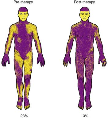
- Thermoregulatory sweat tests (TST)[14]. This involves applying a powdered substance throughout the skin and watching its changes in colour while sweat is produced on different body parts, under controlled conditions (Figure 2.2.4a).

Whether too high or too low, an abnormal body temperature causes fatalities. For instance, heat stroke can happen under prolonged periods of time in hot weather, which is a cause for heart failure[16].
References
- ↑ World Health Organisation. The Global Impact of Respiratory Disease. 2017, https://www.who.int/gard/publications/The_Global_Impact_of_Respiratory_Disease.pdf.
- ↑ World Health Organisation. Chronic Respiratory Disease. https://www.who.int/health-topics/chronic-respiratory-diseases#tab=tab_1. Accessed 14 Nov. 2020.
- ↑ ‘labeled_diagram_lungsrespirato.v2.png (1408×1125)’. https://d32ogoqmya1dw8.cloudfront.net/images/NAGTWorkshops/health/case_studies/labeled_diagram_lungsrespirato.v2.png. Accessed Dec. 09, 2020.
- ↑ World Health Organisation, “Cardiovascular diseases.” https://www.who.int/health-topics/cardiovascular-diseases/#tab=tab_1 (accessed Nov. 14, 2020).
- ↑ “Cardiovascular System Image.” https://openstax.org/books/concepts-biology/pages/16-3-circulatory-and-respiratory-systems (accessed Nov. 14, 2020).
- ↑ N. Herring and D. J. Paterson, Levick’s Introduction to Cardiovascular Physiology, 6th ed., vol. 1. Milton, 2018.
- ↑ What is blood pressure and how is it measured? - InformedHealth.org - NCBI Bookshelf. (n.d.). Retrieved December 09, 2020, from https://www.ncbi.nlm.nih.gov/books/NBK279251/.
- ↑ Arteriosclerosis / atherosclerosis - Symptoms and causes - Mayo Clinic. (n.d.). Retrieved December 09, 2020, from https://www.mayoclinic.org/diseases-conditions/arteriosclerosis-atherosclerosis/symptoms-causes/syc-20350569.
- ↑ Katakami, N. (2018). Mechanism of development of atherosclerosis and cardiovascular disease in diabetes mellitus. In Journal of Atherosclerosis and Thrombosis (Vol. 25, Issue 1, pp. 27–39). Japan Atherosclerosis Society. https://doi.org/10.5551/jat.RV17014
- ↑ Messner, B., & Bernhard, D. (2014). Smoking and cardiovascular disease: Mechanisms of endothelial dysfunction and early atherogenesis. In Arteriosclerosis, Thrombosis, and Vascular Biology (Vol. 34, Issue 3, pp. 509–515). Lippincott Williams & Wilkins Hagerstown, MD . https://doi.org/10.1161/ATVBAHA.113.300156
- ↑ Atherosclerotic renovascular disease - Kidney Research UK. (n.d.). Retrieved December 09, 2020, from https://kidneyresearchuk.org/conditions-symptoms/atherosclerotic-renovascular-disease/
- ↑ Hutchison, J. S., Ward, R. E., Lacroix, J., Hébert, P. C., Barnes, M. A., Bohn, D. J., Dirks, P. B., Doucette, S., Fergusson, D., Gottesman, R., Joffe, A. R., Kirpalani, H. M., Meyer, P. G., Morris, K. P., Moher, D., Singh, R. N., & Skippen, P. W. (2008). Hypothermia Therapy after Traumatic Brain Injury in Children. New England Journal of Medicine, 358(23), 2447–2456. https://doi.org/10.1056/nejmoa0706930
- ↑ Human thermoregulation and measurement of body temperature in exercise and clinical settings - PubMed. (n.d.). Retrieved December 09, 2020, from https://pubmed.ncbi.nlm.nih.gov/18461221/
- ↑ Thermoregulatory Sweat Test | Stanford Health Care. (n.d.). Retrieved December 09, 2020, from https://stanfordhealthcare.org/medical-tests/t/tst.html
- ↑ B9781437700015000659_gr2.jpg (352×384). (n.d.). Retrieved December 09, 2020, from https://neupsykey.com/wp-content/uploads/2016/12/B9781437700015000659_gr2.jpg
- ↑ Cui, J., & Sinoway, L. I. (2014). Cardiovascular responses to heat stress in chronic heart failure. Current Heart Failure Reports, 11(2), 139–145. https://doi.org/10.1007/s11897-014-0191-y