User:Kaitlin E. Kudlac/Notebook/Biology 210 at AU
March 22, 2014:
Zebrafish Lab Data:
Embryology and Zebrafish Development Day 5:
| Petri Dish | Number of Hatchlings | Number of Embryos | Number of Dead Embryos | Number of Dead Hatchlings |
| Control | 17 | 4 | 0 | 0 |
| Dark | 17 | 4 | 0 | 0 |
Day 7:
| Dish | Number of Hatchlings | Number of Embryos | Number of Dead Hatchlings | Number of Dead Embryos |
| Control | 21 | 0 | 0 | 0 |
| Dark | 20 | 0 | 0 | 1 |
Most of the observations that required observing the zebrafish could not be made due to the size of the fish. However one large obsrevation that was continually made throughout the bulk of the experiment was that the dark fish stayed towards the outside of the dish will the light fish or the control fish stayed torward the middle of the petri dish. The control or light fish were also lighter in color than the dark fish. Day 14: Final Day of Experiment
| Dish | Number of Hatchlings Alive | Number of Hatchlings Dead |
| Control | 0 | 20 |
| Dark | 18 | 2 |
Qualitative Observations: Day 14
| Characteristic | Light(dead) | Dark |
| Swimming | When the light fish were alive they tended to stay in the center of the plate while they were swimming and were faster than the dark fish | Not on the outside edge of water- moving a little slower than before- some are faster than others, but tend to stay near the edge of the plate |
| Eye Pigmentation | Lighter more of a clear white | Darker more distinc black color, eyes are black |
| Development of Body/ Body Segments | All at the hatchling stage, segmented bodies into three segments, all were dead however | All at hatchling stage, darker body, have not grown past hatchling stage, clearly defined segments |
| Observe Structures | Even though dead still have tais and main bodies | Long tails with shor bodies and small heads, very dark eyes, clear bodies with black outlines |
| Heart Rate | ALL DEAD | 100-120 |
| Size | 2250 micrometers | |
| Eye Diameter | 250 micrometers | |
| Body Orientation | Straight spine, eyes are big and tail is larger than head, no fins, distinct eye color and body color | |
March 22, 2014 16S Sequences for Bacteria:
Sequence 1:
GGGTTTNNGTATCAGGGTGGGGGAGGNCCNCCCCCATGACCCCTACCCATCAATGANNTGGGATGCGGGTTGGNTACCAACTANCTAATGGGACGTCTGGTCATCTTGTATCCTAGGAANTTTGATATTAAGGTGATGCGACCCGAAAATACCATGGGGAATTAATTCANATTTTACTGGGNTATTCCCCTGNNCAAGGAAGATTGCATACGCNTTACGCACCCGAGNNCCCGGCCNTTAGTCNAAAAACTTCTACACCCCCCTTGNATGTGTTTGGTCTGTNNGGTCTTTCATCTTGAGTCGGACAGCTGAANGTACTATACGAATAANCTCCGGCTANNTCCGTGCCNTCANCCCCGGC
Sequence 2:
TTCNGCTACTCTCACGAGAGTAGGTTTATCCCTATACAAAAGAAGTTTACAACCCATAGGGCCGTCGTCCTTCACGCGGGATGGCTGGATCAGGCTCTCACCCATTGTCCAATATTCCTCACTGCTGCCTCCCGTAGGAGTCTGGTCCGTGTCTCAGTACCAGTGTGGGGGATCACCCTCTCAGGCCCCCTAAAGATCGCAGACTTGGTGAGCCGTTACCTCACCAACTATCTAATCTTGCGCGTGCCCATCTCTATCCACCGGAGTTTTCAATACCGAATGATGCCATCCAGTATATTATGGGGTATTAATCTTCCTTTCGAAAGGCTATCCCCCAGATAAAGGCAGGTTGCACACGTGTTCCGCACCCGTACGCCGCTCTCAAGATTCCGAAGAATCTCTACCGCTC
Sequence 3:
TGNTCAGTTTGAACGNTGGCGGCATGCCTTACACATGCAAGTCGAACGGCAGCACGGAGCTTGCTCTGGTGGCGAGTGGCGAACGGGTGAGTAATATATCGGAACGTACCCTGGAGTGGGGGATAACGTAGCGAAAGYTACGMTAATACCGCATACGATCTAAGGATGAAAGTGGGGGATCGCAAGACCTCATGCTYSTGGAGCGGCCGATATYTGATTAGCTAGTTGGTAGGGTAAAAGCCTACCMAGGCATCGATCAGTAGCTGGTNTGAGAGGACGACCAGCCACACTGGAACTGAGACACGGTCCAGACTCCTACGGGAGGCAGCAGTGGRGAATTTTGGACAATGGGCGAAAGCCTGATCCAGCAATGCCGCSYGAGTGAAGAAGGCCTTNNGGTYGTAAAGCTCTTTTGTCANGGAAGAAACGGTGNARRGCTAATTTNCTTTGCTAATGACGGWACCWGAAGAATAANCANNNGCTAANN
February 25, 2014 Lab 5: Invertebrates Question/ Purpose/ Objective: The main purpose of this experiment is to understand the importance and various characteristics of invertebrates and to learn how simple systems, most importantly specialized cells and body plans, evolve into more complex organisms and organ systems.
Overall Procedure: At the beginning of the lab we observed various organisms and how these organisms have evolved to have more complex body plans and more specialized cells. W observed Acoelomates, Pseduocoleomates, and Coelomates to observe these various trends of evolution. After the observation of these various organisms, we collected the invertebrates that we received from our Bernese Funnel. We poured the contents into a petri dish based on the location of the funnel, there was an upper petri dish and a lower petri dish. After separating the collections from the Bernese Funnel we observed the two petri dishes underneath a microscope and attempted to identify various invertebrates using a dichotomous key.
Raw Data: We only found and observed two invertebrates in our petri dishes from our Bernese Funnels. The first organism that we observed was a flea of the phylum siphonaptera. This organisms had small wings, was rounded, tiny and black. The size of the organism was about 3 mm. The second organism that we observed in the petri dishes was a soil mite. This organism is small with lots of legs and a visibly segmented body. This organism was about .5 mm in length. The size range of the organisms that were measured was about 1- 3 millimeters. Since we only had two organisms it is hard to determine what the overall size range was in the transect but based on the two organisms that were observed 1-3 millimeters is most accurate. The largest organism was the flea and the smallest organism was a soil mite. The organisms that are most common in the leaf litter are fleas, mites, and worms of various types. Five organisms that could have been identified in our transect were earthworms either nematodes or round worms. Two types of birds that could live in the pine trees are most types of owls, various types of crows and cardinals as well. Some of the biotic characteristics that could effect our transect is that it is located on a hill and there food and other plants may have trouble growing due to the possible lack of sunlight or the chance of too much water or too little water flowing down the hill. Other possible biotic factors that could effect the transect is the location due to the fact that people might walk in and around the transect and effect the growth of food or plants that nourish other animals that live in and around the transect. Some of the abiotic factors that could effect the transect are the barriers around the transect like the church or picnic tables or the street which could prevent the necessary animals from traveling to the transect that need to feed or nourish the transect.
While I have been able to upload more pictures now then I have ever been able to before, I think I have reached the limit on open wet ware because it will not let me upload anymore images so I can not upload the food diagram that I have been trying to upload.
Discussion/Conclusion: As this was our final transect activity I am interested into see the comparison between all the various forms of the transect that we have studied. I am also interested to see how the various interactions are effected by the biotic and abiotic factors of the transect. Most of the conclusions that I have made throughout the experiment have been accurate and therefore have supported my initial hypothesis. KEK
February 16, 2014 Lab 4: Plants and Fungi
Question/ Problem/ Objective: The purpose of this experiment was to understand the various characteristics of plants and to see and observe the function and importance of Fungi. We also took various plants from our transect and characterized them in our transect.
Overall Procedure: The basic procedure of this lab involved us first going to our transect and collecting five different types of plants that we would then take back to the lab and observe and characterize according to different classifications. Throughout the classification process we followed different stations to observe various parts of plants that would help us to characterize the plants that we found in our transect. The first station was plant vascularization. Here we observed various mosses and plants and located the xylem and the phloem on the different types of plants, the two structures responsible for bringing food and water to plants. The second station involved plant specialization. Here we observed various different types of specialized cells in the plant, most significantly the stomata and the guard cells which help with gas exchange in the plant. The third station involved plant reproduction where we observed diagrams of plant reproductive organs and then classified various plants and looked at plant reproductive organs underneath the dissecting scope. We also learned about the differences between monocots and dicots and the importance and differences between the two subsets. The final section of the lab involved looking at and appreciating fungi underneath the dissecting scope. The final step of the lab was to set up the Berlese funnel in order to collect invertebrates that will be observed in the following lab.
Raw Data:
The majority of our raw data involved the characterization and classification of the five various plants that we gathered from our transect.
| Transect | Location and number in transect | Description( bush, tree, plant) also shape and size | Vascularization | Leaves and special characteristics | Seeds, evidence of flowers or other reproductive parts |
| #1 | Off of a large pine tree | Thin long needs from a pine tree- long branch but tiny needles | Grooves on the needles and the bark, can see the xylem but not the phloem as much | Long thin pine needles, seeds on the bottom as well | Gymmosperms- pine cone on the tree- reproductive seed- also tiny seeds on the needles as well |
| #2 | Floor on the transect | Circular in shape with very noticeable veins | Visible xylem and phloem also very defined veins on the leaf | Dicot- evenly distributed veins throughout the leaf- very balanced | Dicot- angiosperm- came from a bush with flowers |
| #3 | Triangular green life lying on the floor of the transect | From the floor or ground of the transect- hidden in the ivy found on the ground | Can also easily see the veins and the xylem and the phloem | Has a more purple color- triangular in shape and highlighted not as much defined veins | Dicot- angiosperm |
| No spore evidence but most likely from flowering bush or tree | |||||
| #4 | Round green leaf- found on the top of the ivy in the transect | Leaf was most likely part of the ivy or the bushes that are coating the transect floor | Visible xylem and phloem | Similar to green leaf #2- easy to follow and very defined veins, a little brown around the edges | Dicot- |
| Angiosperm | |||||
| No spore evidence but came from a flowering bush or tree | |||||
| #5 | Picked from the holly tree in the transect | Thicker leaf but smaller in size, came from the holly tree | Visible xylem and phloem but harder to see veins- not as easy to follow | Visible xylem and phloem, lighter green texture- rougher edges | Dicot |
| Some flowers and berries were found on the tree- angiosperm | |||||
The image below is an image that I drew of the bread mold fungi that was in lab on the day we observed the various plants. This image was drawn at 4x and is a typical image of bread mold. This image is a fungus due to its multicellularity, its spores that are sprouting out from the mycelium and the fuzzyness that often characterizes a fungi.
[[Image:fungi image.png
]]
Conclusions and Future Plans: One of the future plans for this experiment deals with the set up of the Berlese Funnel so that in the subsequent lab we can observe and collect the invertebrates that are found in our transect. One of the conclusions that I made from this lab is that most of the plants and trees or bushes that are found in our transect have generally the same characteristics. This is probably due to the fact that similar plants characteristics thrive in similar environments, so a dicot might be better in one environment while a monocot might be better in another environment. I am looking forward to seeing if this same conclusion can be drawn from the invertebrates.
KEK
February 15, 2014 Lab 3: Microbiology and Identifying Bacteria with DNA
Question/Problem/ Objective: Last week in lab we cultured bacteria from our hay infusions on normal agar plates and on agar plates with tetracycline. This week in lab we will be observing these agar plates and observing the various forms of bacteria and possible fungi that have grown in the last week. We will characterize the bacteria through the use of gram staining, overall colony description including color, size, shape, texture, and through a motility test using a wet mount. At the end of the lab after we have characterized the bacteria in our hay infusion, we will be isolating DNA from the bacteria in the colonies to performa PCR reaction to analyze the DNA sequences in the following labs.
Overall Procedure: Throughout this lab there were four different tasks or procedures. At the beginning of lab we counted the total number of colonies of bacteria on each plate that we cultured. We counted the bacteria on each dilution plate and on each agar and then multiplied by a conversion factor to get the number of colonies per mL. In the second part of the lab or the second procedure we observed the differences between the normal nutrient agar plates and the nutrient agar plates that also contained the antibiotic tetracycline. We noted the differences in numbers between the two plates and the differences between the types of bacteria that grew on each plate. The next part of the lab was the bacteria cell morphology observations. In this part of the lab we took the four bacteria, two from nutrient agar plates, and two from tetracycline plates, that we choose from various agar plates and created wet mounts of them. Once the wet mounts were made we observed them under the microscope to see if they moved or we could see flagella or cilia on the bacteria. After we created the wet mounts we observed the bacteria under a dissecting scope to see the overall shape, texture, color, and other defining characteristics of the bacteria that were chosen from the agar plates. After observing the overall shape of the bacteria we then performed gram stains to see if the bacteria were gram positive or negative. This process involved a variety of steps which included heat fixing the bacteria to the slide and then washing and rinsing the bacteria with various dyes in order to see if the dyes would either stick to the thick layer of peptidoglycan that gram positive bacteria had or the extra cell membrane that gram negative bacteria had. At the very end of the lab we prepared PCR reactions with one of the bacteria so that in the following lab we can observe the DNA sequence obtained from the different bacteria.
Raw Data: One of the first steps in this experiment was to observe our hay infusion for one last time. Our hay infusion actually began to smell better. I am thinking this is due to some of the bacteria and other possible little things living in there starting to die out due to the lack of nutrients, however, overall the hay infusion stayed relatively similar to what it looked like last week. In procedure I we documented the number of colonies and colonies per mL that grew on the agar plates, this information is documented in the table below.
| Dilution(Plate Label) | Agar | Colonies Counted | Conversion Factor | Colonies/ mL |
| 10^-3 | nutrient | unable to count | x 10^3 | impossible to count |
| 10^-5 | nutrient | 37 | x 10^5 | 37 x 10^5 |
| 10^-7 | nutrient | 4 | x 10^7 | 4.0 x10^7 |
| 10^-9 | nutrient | 0 | x 10^9 | 0 |
| 10^-3 | nutrient +tet | 98 | x 10^3 | 98000 |
| 10^-5 | nutrient +tet | 5 | x 10^5 | 500000 |
| 10^-7 | nutrient +tet | 0- air bubbles | x 10^7 | 0 |
In this part of the experiment we observed the differences between the antibiotic agar plates and the nutrient agar plates. Generally there were more bacteria on the antibiotic plates then there were on the regular nutrient agar plates. I think this indicates that in general the bacteria in nature is resistant to at least so variations of antibiotics and because of this can grow on either the regular nutrient agar plates or the nutrient agar plates with antibiotics. While some of the types of bacteria grew on both the nutrient agar plates and the tet plates, most of the bacteria were different. Tetracycline is a type of antibiotic that fights off various types of bacteria including gram positive and gram negative bacteria. It works by inhibit protein synthesis rather than just killing the bacteria all together. Tetracyclines fight a broad spectrum of bacteria therefore there are many types of bacteria that can survive in tetracycline and lots that are also killed. In the following portion of the experiment we choose three different types of bacteria from different plates, two from nutrient agar plates and one from a tet plate and characterized them and performed gram stains on them.

The first bacteria that we chose is from the 10^-5 nutrient agar plate. It is characterized by a thick black mound mass. While it looks black from a distance its actual color is a dark purple It is round with a slightly ovular shape and has a circular morphology. When a wet mount was created you could easily see the cell wall and the mainly coccus shape with a single cell colony. Furthermore when motility was tested of the bacteria it was very easy to see the flagella propelling the bacteria along through the water. When the gram stain was performed for this bacteria is was determined to be gram negative due to the pink color of the the extra cell membrane and the cell wall.


The second bacteria that we chose was a bacteria from the nutrient agar 10^-7 plate. It had an egg-yolk orangey color and had an overall circular shape with a raised convex elevation. When the bacteria was observed under the microscope we could not discern a cell wall and the coccus and raised elevation shape was confirmed. It was also hard to see the motility of this bacteria but it is most likely assumed to have some motility since on the plate the colonies were everywhere. After the gram stain was complete it was determined that this bacteria was also gram negative due to the pink color.

The third bacteria that we chose was from the 10^-3 tet plate. It had a similar color and shape to the bacteria above but we chose it since we had two similar bacteria on two different types of plates. This bacteria like the bacteria above is characterized by an orange color with a round convex overall shape. It also has a raised elevation and has smooth edges. When placed under the microscope the overall color was deemed a more dark yellow but the elevation and shape was about the same with no visible cell wall. After performing a gram stain this bacteria was also gram negative due to the pink color.

Conclusions and Future Plans: One of the questions at the beginning of the lab addressed whether or not we thought if archaea bacteria would grow on the nutrient agar plates and the answer is no because archaea really only grow in extreme conditions and unfortunately the agar plates are not extreme conditions. One of the conclusions that I made is that more bacteria grew on the antibiotic plates than on the regular nutrient agar plates. I was surprised to learn this because I would not think there would be many antibiotic resistant bacteria in nature but due to the influx of antibiotics being prescribed this would make sense that they are going to start appearing in nature. One of the future plans for this experiment is to determine the DNA sequence of the bacteria that we began the PCR reaction on. I am interested to see how the DNA of our bacteria and what the actual characteristics are. After sending our PCR reactions to get sequenced it was determined that the first bacteria was not related to any other known DNA sequence, at least according to the BLAST that was done on the first organism. After the second organism was blasted it was determined that the second bacteria that was tested was 100% similar to chryseobacterium. After the third organism was blasted it was determined to be 96% similar to an unknown bacteria that has not been determined as to what type of bacteria the organism actually is.
KEK
February 6, 2014 Lab 2: Identifying Algae and Protists
Question/ Problem/ Objective: The main objective or problem in this part of the lab is to observe the hay culture that we created last week and observe and identify algae and protists that are growing in our hay infusion. Furthermore, besides just the primary observations of protists and algae in our transects, the other main objectives of this lab are to learn how to use a dichotomous key and to understand the overall characteristics of the protist domain and its subsequent subsection of algae.
Overall Procedure: At the beginning of the lab, we first observed various different types of protists and algae so that when we observed these organisms from our hay infusion we would have a base to determine and identify them from. We observed various different types of algae at first including Stramenopila or reddish brown algae, green algae and then various different types of protists. When we observed the protists under the microscope we also practiced using the dichotomous key which would help us to identify protists in our hay infusion when the time came. The main objective during the first part of the experiment was focused on using the dichotomous key, though the organisms looked amazing under the microscope, the dichotomous key was important to understand since we would use it later when classifying organisms in our hay infusion and thus our transect. After observing common protsits and algae we then moved to observations involving our transect or our hay infusion. We first chose three different niches in our transect to take samples from and observe. We choose from the top bottom and middle as these were the most pronounced niches in our transect. After making wet mounts and observing the various organisms under the microscope to see the different types of algae and protsits, we then moved to preparing for next weeks lab. In preparation for next week's Microbiology lab we created serial dilutions using material from our individual cultures or hay infusions. Serial dilutions work because you start out with a large concentrated substance and then dilute it by taking a small volume from the large concentrated substance and moving it into a tube filled with water you then repeat the process until you get a very diluted version of the largely concentrated substance(this will be explained more in later sections hopefully I can upload a diagram about this).
Raw Data:
Primary Observation of Hay Transect:
At the top of the liquid in the hay infusion there is a thin film layer most likely comprised of the powdered milk and there is also some green leaves and twigs at the very top. There is a small amount of brown filament or mud like material at the top to. The hay infusion also has a very pungent smell. It smells most likely rotting leaves and trees. The color of the hay infusion is most likely a cloudy grey with a greenish brown tint towards the bottom that is settled on the floor of the tube. The middle has more of a brown color due to the leaves and twigs that mostly occupy the middle of the hay infusion. Most of the infusion seems to be dead but there could be some living organic matter at the top of the infusion due to the large concentration of powdered milk.


Characterization of Different Organisms:
Bottom Sediment Organisms:
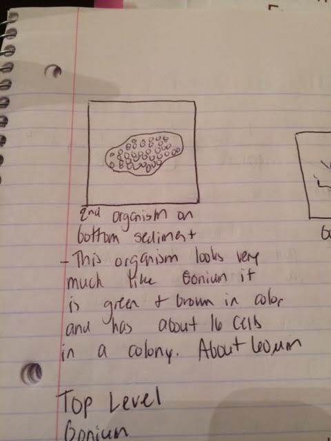
On the bottom level of the hay infusion we found two different types of organisms. The first organism that we found was a paramecium that was sitting on top of a thread like material that was found in our transect and showed up in our hay infusion. The paramecium was immobile and not moving on the thread and was about 5 micrometers big. It had a whitish red tint to it and was somewhat see through. The second organism that we found on the bottom of the transect was a protist or algae that closely resembled Gonium. It had a greenish brown tint to it and had about 16 cells per colony. In general though it was about 40 micrometers in size. It was generally an ovular shape with some rounded edges.

Middle Organisms:
On the middle level of the transect one of the first organisms that we observed was again gonium. It had the same characteristics of the gonium described above had but was a little bit larger in size, about 60 micrometers as opposed to 40, then the gonium on the bottom layer. This could be due to more sunlight being able to penetrate the middle of the transect then at the bottom of the transect and there could be more nutrients in the middle of the transect as well. The other organism that we found was chilomonas protist. This protists had an elongated cell with a narrowed center and was mostly clear in color. It seemed to have some sort of flagella on the end but it was hard to tell under the microscope. This organism was about 18 to 20 micrometers long.

Top Organisms:
Again on the top level of the hay infusion we found more gonium which was even larger than the gonium found on either the bottom or middle of the transect, at about 80 micrometers, leading to the conclusion that there is more light and nutrients for the gonium to prosper at the top of the hay infusion then in the middle or the bottom of the hay infusion. This gonium was more characteristic of the gonium that we saw last week in that it had green and brown extending arms coming from the sides of the colony. It also had about 15-18 cells within the individual colony. We also found Volvox at the top layer of the hay infusion, which is the more evolved version of Gonium. The Volvox was very large in size, about 400 micrometers with a greenish brown tint just like the gonium. It was more complex than the gonium was but still had some of the same characteristics as outlined before.

One organism that meets all the characteristics of life is gonium. The six main requirements for being alive are that the organism is made up of the most basic unit, which is the cell. Gonium fits this requirement because it is made up of cells. Even though it is only one step above being a single celled organism, Gonium is still made up of multiple cells. The second requirement for being considered alive is to use and be made up of DNA, which gonium is. The third requirement is movement. Gonium moves through the use small cilia and flagella that are on the surrounding body of the gonium. The fourth requirement for life is reproduction. Gonium uses binary fission or asexual reproduction to reproduce and continue on the gonium line. The fifth requirement for life is growth. Gonium grows much like it reproduces by creating new cells and colonies that form into one large gonium colony. The sixth and final requirment for life is some form of nutrition or stimuli which gonium does through its small appendages that uses exocitosis to absorb the nutrients in the water it lives in.
If the hay infusion culture was to be observed for another two months, I think one of the major changes that would occur is the smell and the growth of bacteria. As the organisms that lived in the hay infusion began to die the amount of bacteria would begin to increase thus causing the smell of the hay infusion to become more pungent and noticeable.I think that some of the selective pressures that affected the compositions of our samples is the varying degree of light that the hay infusion cultures got. Because the cultures were not in natural light but were in artificial light this could have caused the cultures to grow less and to cause some of the organisms that would normally be in the infusion to disappear and die to the lack of light and the increase in artificial light.

Conclusions/ Future Plans:
Some of the future plans that I have for this part of the lab is to continue to watch the hay infusion, specifically the smell. I think that if the smell starts to get worse, it could be an indication of more of the plant matter in the infusion dying. I also would like to monitor one of the observations I made earlier in the experiment which was that more complex organisms and larger organisms live towards the top of the hay infusion since there is more light and nutrients at the top then in the middle or at the bottom, specifically through the gonium colonies that were observed. The main future plan of this experiment is to observe the bacteria that were colonized and put on the agar plate that we will observe next week. I am excited to see what bacteria were discovered in our transect. KEK February 6, 2014
January 30, 2014
Question/Problem/Objective: The general purpose of this lab is to determine the general characteristics of a transect that is located on the American University campus grounds. Prior to this we also observed the origin of the Volvocine Line to help us understand how organisms have evolved and how this information can eventually lead to the how we can eventually observe other organisms in our transect.
Overall Procedure: In the first portion of the lab, we observed the origins of the Volvicine Line. We first observed the most basic version of the Volvicine Lin, Chlamydomonas. Through the observation of this most basic version of the Volvicine Line, we could observe how the Volvicine Line has evolved both in size and in reproductive tendencies. We prepared various slides of the Chlamydomonas and examined it microscopically. After observing Chlamydomonas, we then observed Gonium which is the next species in the line. This organism is much more complex then the Chlamydomonas, it has more colonies and is also a bit more complex in reproductive standards then the Chlamydomonas is. After we observed the Gonium, Volvox was then observed in the same format through a wet mount slide that was made. Volvox is generally a much more complex organism with a more intense reproductive capabilities and a more complex size and structure as well. In the second part of the lab we were assigned an individual niche on the American University campus. Our transect is located behind the seminary in an area covered and surrounded by many pine trees and many other types of treesIt is located at the bottom of a very steep hill that is very susceptible to runoff and other elements that can possibly roll down the large hill. In our area there is also a lot of various types of ivy that coat the floor underneath the many different types of trees. The transect is also located very close to Massachusetts Avenue and is this close to a lot of foot traffic, thus our transect has a very high percentage of chance of being frequented by many people that may walk in it or around it. In the back of the area there is also a tree stump possibly alluding to human interaction, like a person could have cut down the tree. On top of the ivy that coats the floor of the transect there are lots of leaves that have most likely fallen from the surrounding trees or have rolled down the hill that is located near the transect. There is also a lot of grass that surrounds the transect that is a source of food for many animals that might visit the transect. The transect does not have a lot of light that penetrates the trees due to the size of the trees in the area. After we noted the general observations of the transect we then prepared a hay infusion coming from the transect. The hay infusion culture was prepared by taking 10 to 12 grams of the soil and ground and placing it a jar with 500 mLs of water in addition to 0.1 grams of dried milk and then was mixed together.
Raw Data:
General Description of the Transect: Our transect is located behind the seminary in an area covered and surrounded by many pine trees and many other types of treesIt is located at the bottom of a very steep hill that is very susceptible to runoff and other elements that can possibly roll down the large hill. In our area there is also a lot of various types of ivy that coat the floor underneath the many different types of trees. The transect is also located very close to Massachusetts Avenue and is this close to a lot of foot traffic, thus our transect has a very high percentage of chance of being frequented by many people that may walk in it or around it. In the back of the area there is also a tree stump possibly alluding to human interaction, like a person could have cut down the tree. On top of the ivy that coats the floor of the transect there are lots of leaves that have most likely fallen from the surrounding trees or have rolled down the hill that is located near the transect. There is also a lot of grass that surrounds the transect that is a source of food for many animals that might visit the transect. The transect does not have a lot of light that penetrates the trees due to the size of the trees in the area. After we noted the general observations of the transect we then prepared a hay infusion coming from the transect. The hay infusion culture was prepared by taking 10 to 12 grams of the soil and ground and placing it a jar with 500 mLs of water in addition to 0.1 grams of dried milk and then was mixed together.
Abiotic and Biotic Factors of the Transect:
Some of the biotic factors that the were located in the transect were trees, ivy, grass, bugs, such as grasshoppers and crickets, and some small bushes underneath the ivy. Some of the abiotic factors that the transect had was small pebbles or rocks, aluminum foil and other trash. There were not many abiotic factors in our transect but due to people walking through the area frequently there could be trash in the area but it could be located underneath the ivy and not visible.
![]()
Unfortunately the picture is upside down but you can still get the overall sense of what the transect looks like.
Conclusions and Future Plans: One of our main future plans for this lab is that we are eventually going to use the hay culture infusion to observe microscopic organisms that are growing in our hay culture infusion and by association our transect. In the first part of the lab we observed the origins of the Volovcine line and how organisms in this family have evolved over time. While the Volvocine line is not directly related to our individual transects, I believe that it will help us track the evolution or relationships between organisms that we will find in our transect. Though we did not yet specifically observe anything from our transect yet, I believe that the soil and plant matter that we obtained will yield very interesting results. January 30, 2014 KEK
