User:Fabiola Garau/Notebook/Biology 210 at AU
March 22, 2014 9:52 am Introduction: In this experiment titled " Zebrafish Development" the objectives were to learn the stages of embroyonic development,compare embryonic development in different organisms and set up an experiment to study how environmental conditions affect embryonic development. The conditions for the zebrafish chosen in this experiment were specifically the dark variables. In other words, the zebrafish were put in a drawer with total darkness. The hypothesis is that the zebrafish in complete darkness will develop in a longer period of time than the one's in the control (which have normal light) and for this reason their eye size will be smaller. The prediction is that if the zebrafish in the dark conditions take a longer time to develop, then their eye size will consequently be affected.
Materials and Methods: 1. Zebrafish embryos 2. petri dishes 3. 20 mls deepark water 4. microscope 5. When the embryos are 4-5 days 10 mls of water will be remove and 25 mls of fresh water will be added. 6. Save embryos in paraformaldehyde. 7.0.02% tricaine solution
Observation and Data:
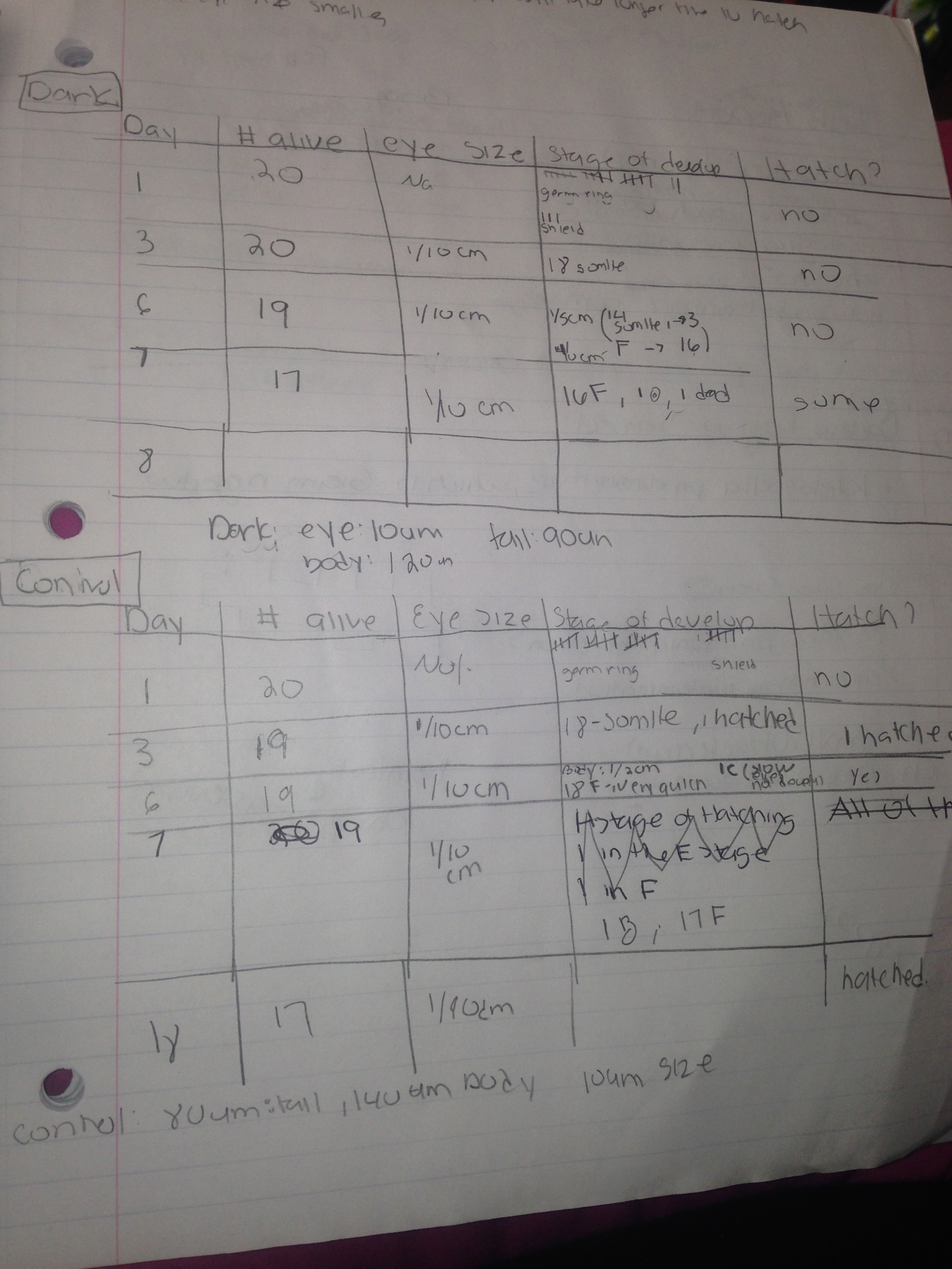
In day 1 of the experiment there was 20 zebrafish alive in the dark and control group. The eye size could no be measure because it was not develop yet. In the control group 15 zebrafish were in the germ ring stage and 5 were in the shield stage. It is important to note the the germ ring comes before the shield. In the dark group 17 were in the germ ring and 3 in the shield stage. On day 3 however, on 1 zebrafish died in the control group. The eye size in the control group was 1/10 cm and all except one were in the 18-somite phase. One zebrafish hatched!. In the dark conditions 20 zebrafish were still alive, eye size was 1/10 cm and all were in the 18-somite phase. In day 6 of the control the eye size was the same. 18 zebrafish measured 1/ 2 cm and were in the F phase , which was characterized by having a tail with pectoral fin and protruding jaw, and 1 was in the c phase, which its eye size was still developing. In the dark group, 14 zebrafish measured 1/ 5 cm and were in the 14 somite stage, which was characterized by still having a yolk sack, and 16 zebrafish measured 1/ 10 cm and were in the F phase. Finally in day 7, 17 zebrafish were in the F phase and 1 was in the b phase, which is characterized by having now two eyes with the complete elimination of the yolk sac. In the dark group, 3 zebrafish died. 16 zebrafish were in the F phase and 1 was still in the 18 somite. After this day 7, final observations regarding eye, body and tail were made. In the control group, the body size was 140 um, eye size 10 um and tail size was 80 um. In the dark group, the body size was 120 um, eye size was 10 um and tail size was 90 um. Unfortunately I was unable to take any photos but I will include photos of the stages in the power point presentation.
The table below shows the observations in stage and eye size in both dark and control groups
 Conclusions:
The hypothesis of the experiment was proven. As seen in the table of the dark group and control group, the development of the zebrafish took a longer time to develop in the dark group than the in the zebrafish in the control group. Even though the eye size in both groups measured the same, it develop at different time rates. The body and the tail was indeed affected by the dark and light conditions since the zebrafish in the control group had greater measurements than the ones in the dark group. For future experiments,I suggest that instead of letting the zebrafish died at the end of day 7, one should continue to feed the zebrafish with paramecium to observe the complete development and explore what will eventually happen with the development of the dark group.
March 1,2014 5:30 pm
Introduction: In this experiment titled "Invertebrates" the objectives were to understand the importance of invertebrates and to learn how simple systems evolved into more complex species. In this lab one observed the the soil from the AU transect to study invertebrates such as arthropods under the dissecting microscope. Under the microscope we noted the phylum and size of 6 different invertebrates .
Materials and Methods: 1.Dissecting Microscope 2. acoelomate, Planaria 3.pseudocoelomate, Nematode 4. coelomate, Annelida 5. 2 dichotomous key 6. Berlese Funnel
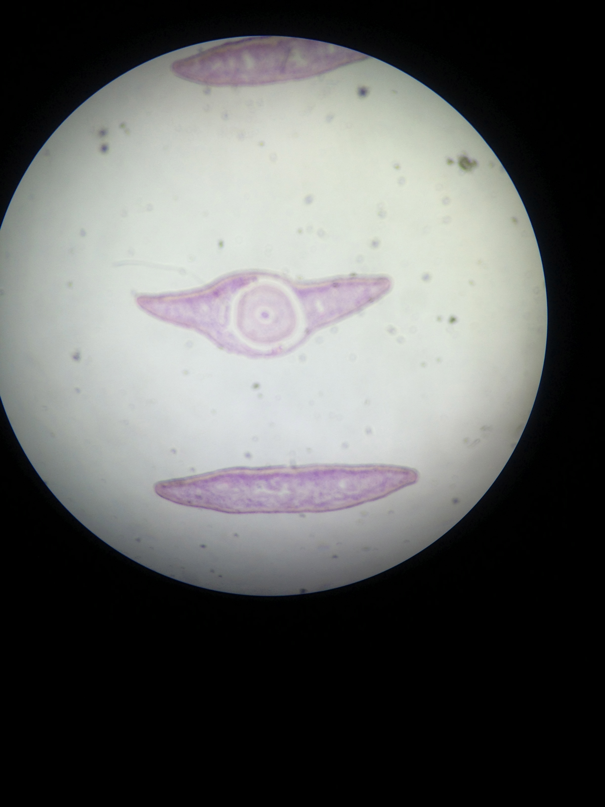
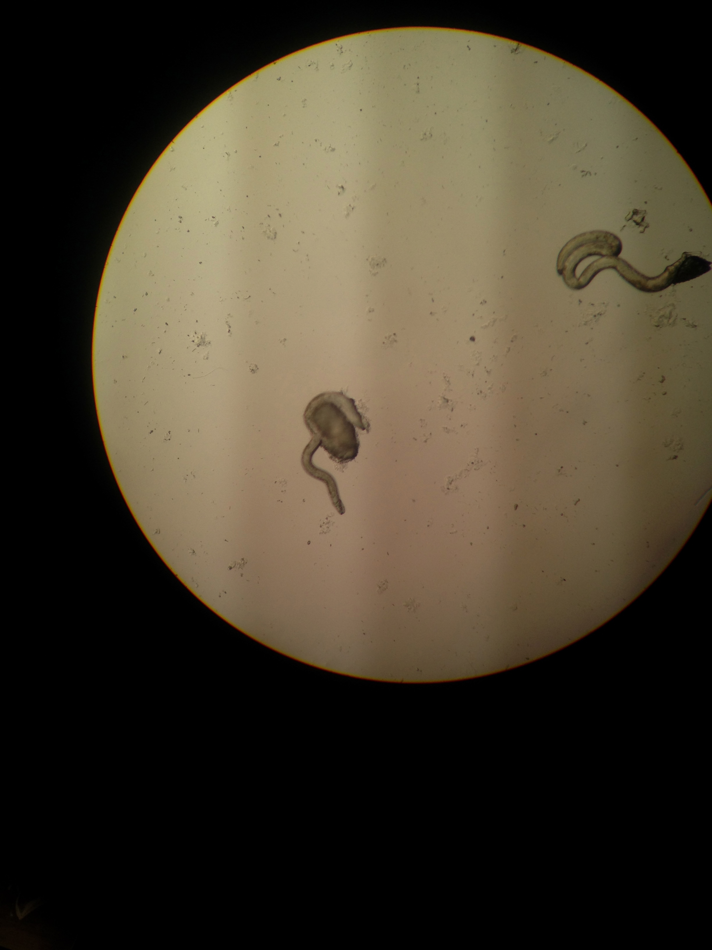
Observation and Data: In the first part of the lab, we distinguished and observed the acoelomates, pseudocoelmates and coelmates structures in three different phyla of invertebrates. The Planaria phylum was acoelomate, which means it does not has a fluid filled cavity in its digestive tract. Since this lack of fluid affects the way the muscles contract, the worm in this phylum was sliding with the use of the cilia. The Nematode phylum was pseudocoelmate, which means it had an incomplete line cavity. Without this line cavity, the worm had no circular muscle whatsoever and move in a serpentine locomotion. The Annelida phylum was coelomate, which means it had a lined, fully filled cavity. The worm in this phylum was able to contract its muscle and was extending its head and posterior body in a wave like motion.
Below there are pictures of the acoelomates, pseudocoelmates and coelmates in respective order.
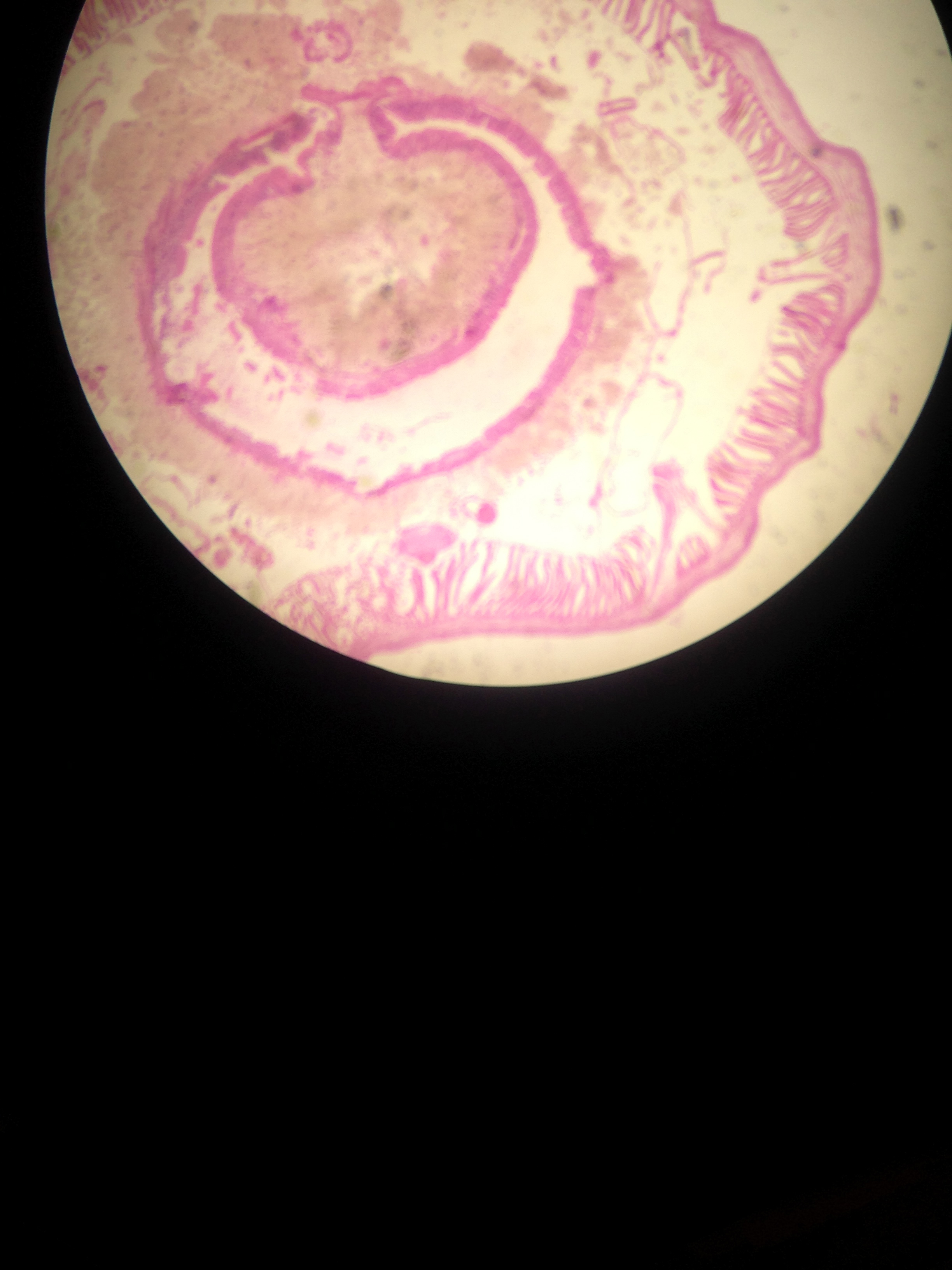
From our petri dish we identified 5 organisms and noted its size along with a description. The table below summarizes our findings.
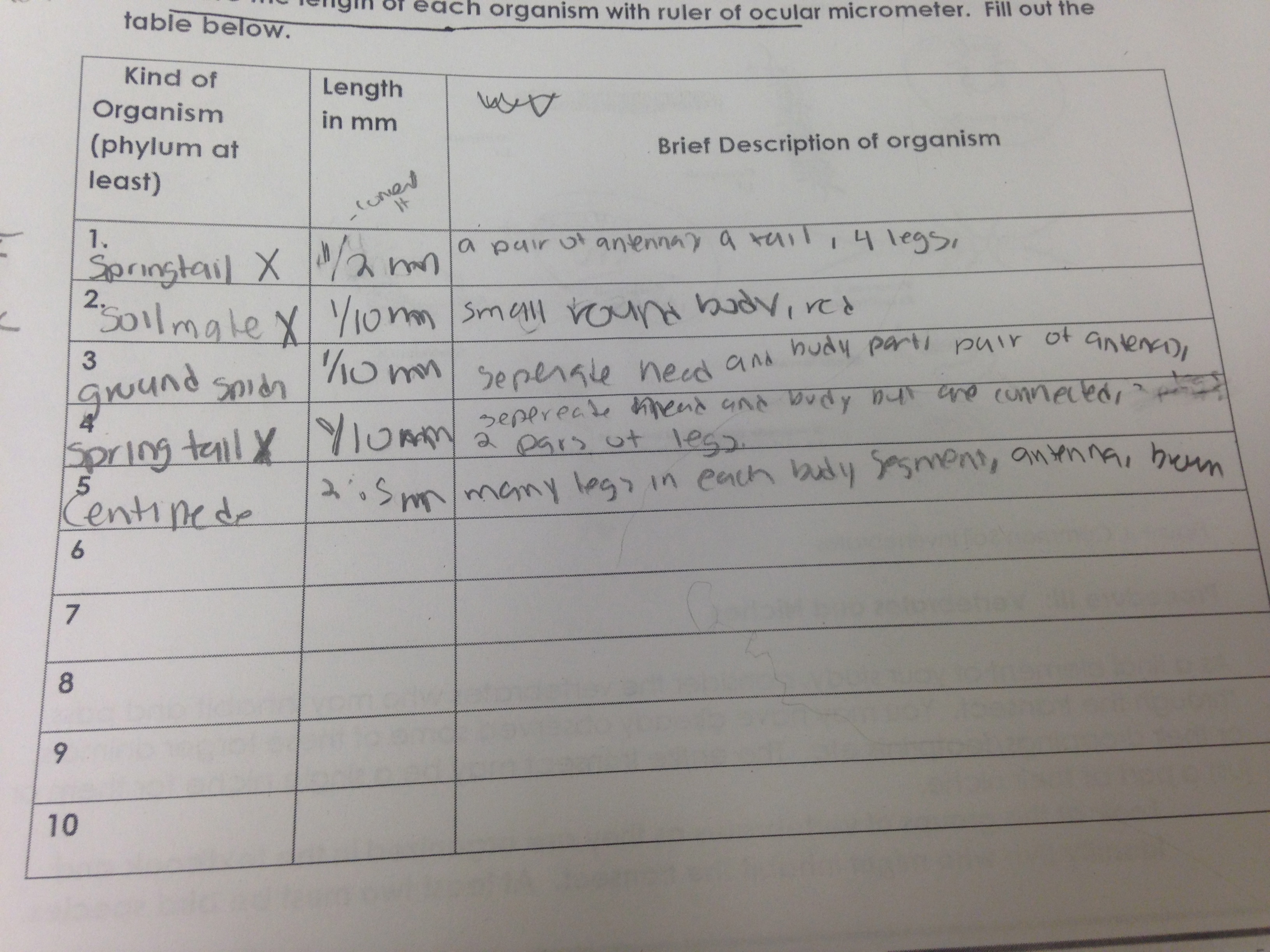
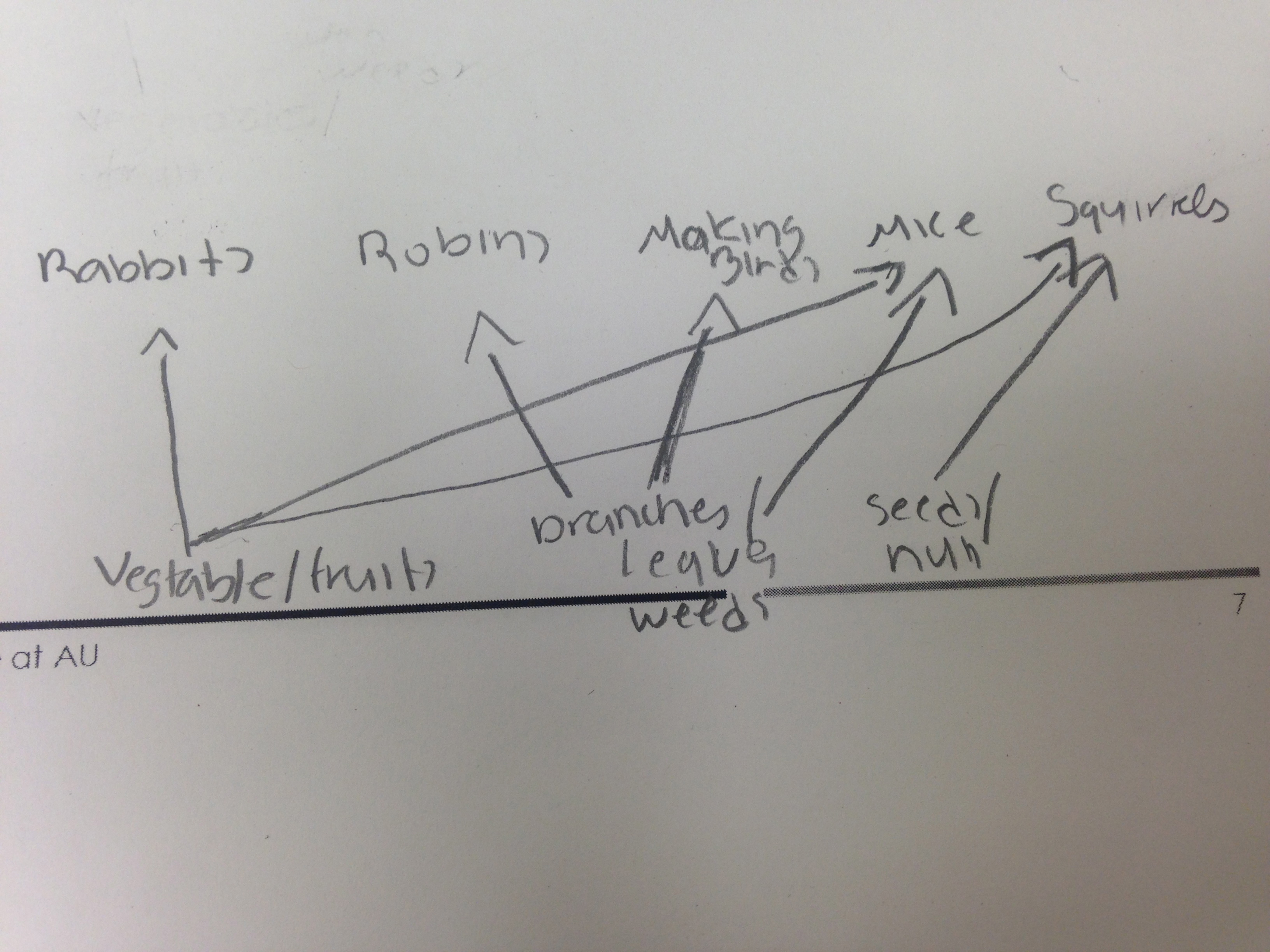
The organisms in our AU transect were really small, ranging from 1/10-2.5 mm. The largest organism was Springtail X Primitive insect and the smallest organisms were the Soil Mite X and Ground Spider. It is important to note that although the table above mentions the centipede was the largest organism, it was not from our AU transect but from Virginia. The most common organism in leaf litter are the arthropods. The five possible vertebrates from our transect are:
1.The American Robin: chordata, aves, passeriformes, turdidae, turdus, T Migratorius. -Since our transect was mostly branches and leaves, the robin can use these materials to built a nest.
2. Mocking birds: chordata, aves, passeriformes, mimidae, mimus, Mimus polyglottos. -Like American Robins, mocking birds can mostly benefit from the branches for nesting materials.
3. Squirrels: chordata, mammalia, rodentia, sciuridae, sciurus, sciurus carolinensis. -It is possible that it could benefit from the seeds in the compost area.
4. Mice: chordata, mammalia, rodentia, cricetidae, peromyscus, peromyscus boylii. -Since our transect has a compost area, the mice can benefit from the foods such as onions and tomatos as well as weeds and insects.
5. Rabbits: chordata, mammalia, lagomorpha, leporidae, nesolagus timminisi. -They would benefit from the vegetables from our compost garden.
Conclusion: We achieved the objectives in this experiment since we gained better understanding of the invertebrates along with its complexities. In the first part of the experiment we distinguished how the lining and fluid in the body cavity affect the movement of acoelomates, pseudocoelmates and coelamtes organisms. Later used our own transect to discover and named the invertebrates and possible vertebrates. For future experiments, one might carefully conduct an experiment on exactly which vertebrates are live transect.
February 23,2014 7:00am Lab 4
Introduction: In this experiment titled "Plantae and Fungi" the objectives were to understand the characteristics and diversity of plants and to appreciate the function and importance of Fungi.In the first part of the experiment one will not only observe 5 plant samples of an AU transect and observe its vascularization, specialized structures and reproduction but also observe under a microscope a Bryophyte moss, Mnium and angiosperm Lilium. Plant vascularization refers to the way plants get their nutrients and water from their environment while plant specialization refers to the cuticle, stomata cells, pailisade cells and parenchyma cells. Lastly, reproduction refers to the alternation of generation which is when plants alternates from gametophyte (haploid) and sporophyte (diploid). Reproductive features include the stigma, anther, style and wether the seed is dicot or monocot. For the second part of the experiment one will focus on the fungi, which are decomposers. In order to examine the fungi, one will observe a black bread mold in a petri dish. The hypothesis for this experiment that features in vascularization, specialized structures and reproduction directly depend on one another. The prediction is the if a plant has less vascularization features then the specialized features and reproductive features are going to be less.
Materials and Methods 1.Micrsocpoe 2. 5 samples of plant 3. Lilium, Bryophyte moss, Mnium 4. 3 bags 5. black mold petri dish 6. 25 ml of 50:50 ethanol water solution 7.flask 8.40 watt lamp 9.funnel
Observations and Data:
The five samples of plant were obtained from different parts of the AU transect near the Reeves field. The first plant sample was the "Hornbeam", which had a single red steam and three medium size hairy, green leaves. It had vascularization but it did not had a seed or any flowering/reproductive part. The second sample was the "Common Chickweed", which had many small, round, green leaves.The stems were all interconnected, it had vascularization but there was no seed/flower part. The third sample was the "American Sycamore", which had a medium size brown leaf and steam. It is important to note that this sample was from a tree. It had vascularization but no seed/flower part. The fourth sample from our transect was also the "Common Chickweed" but with the leaves now more big. Unfortunately, our transect was not sufficiently diverse to obtain a fifth plant sample. The image below describes the different parts that we obtained the plant samples as well as few images of the samples.
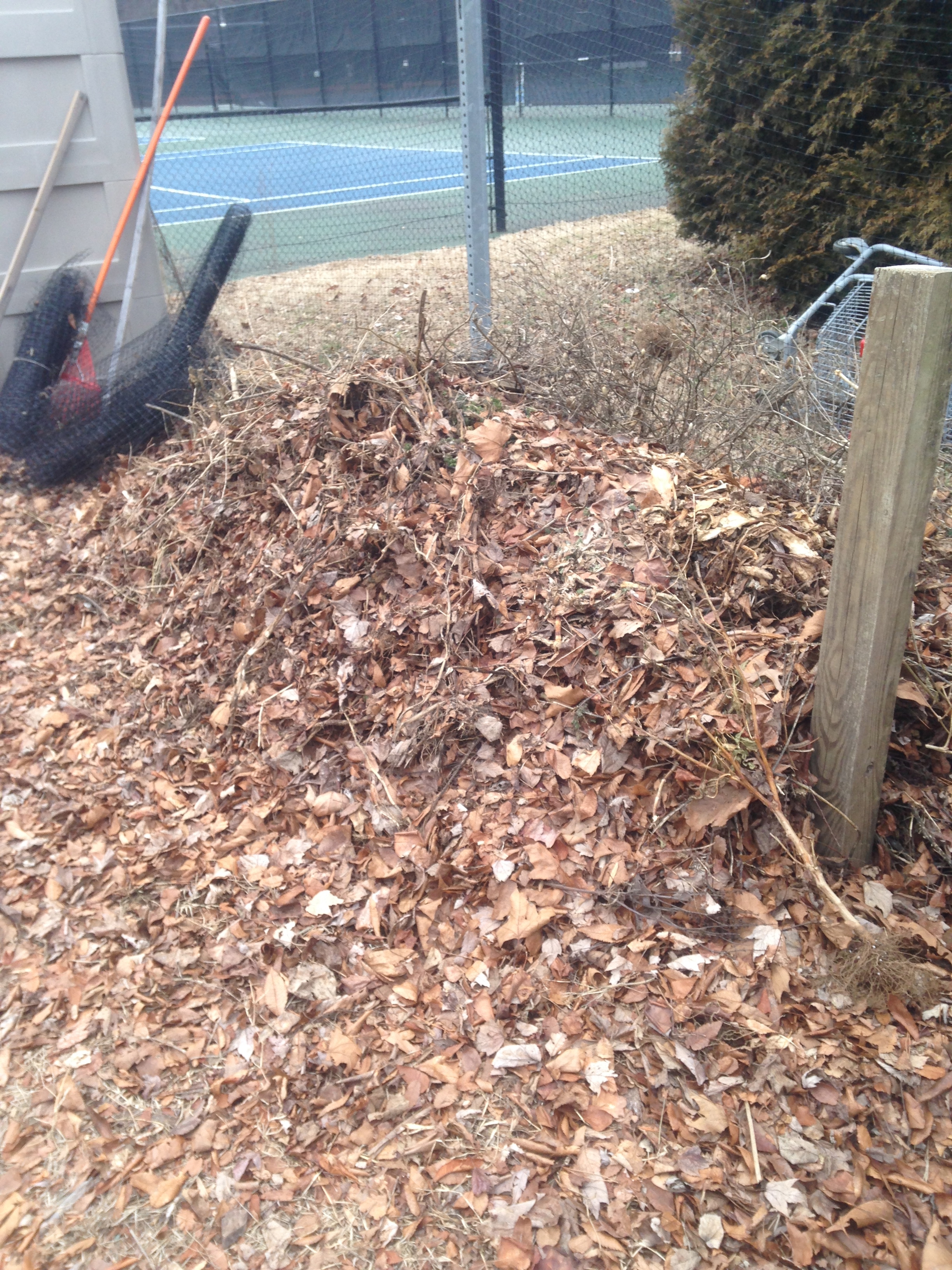
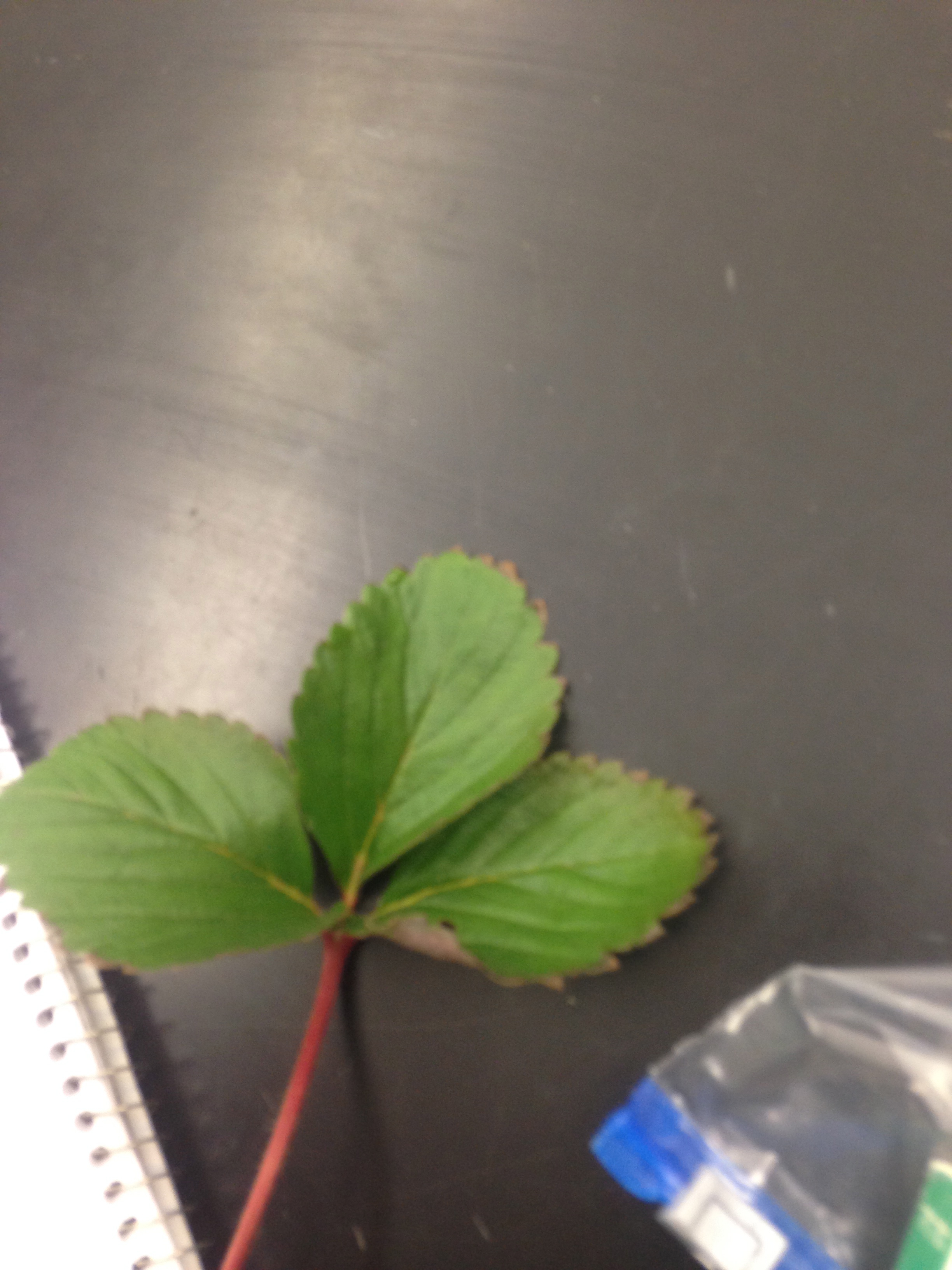
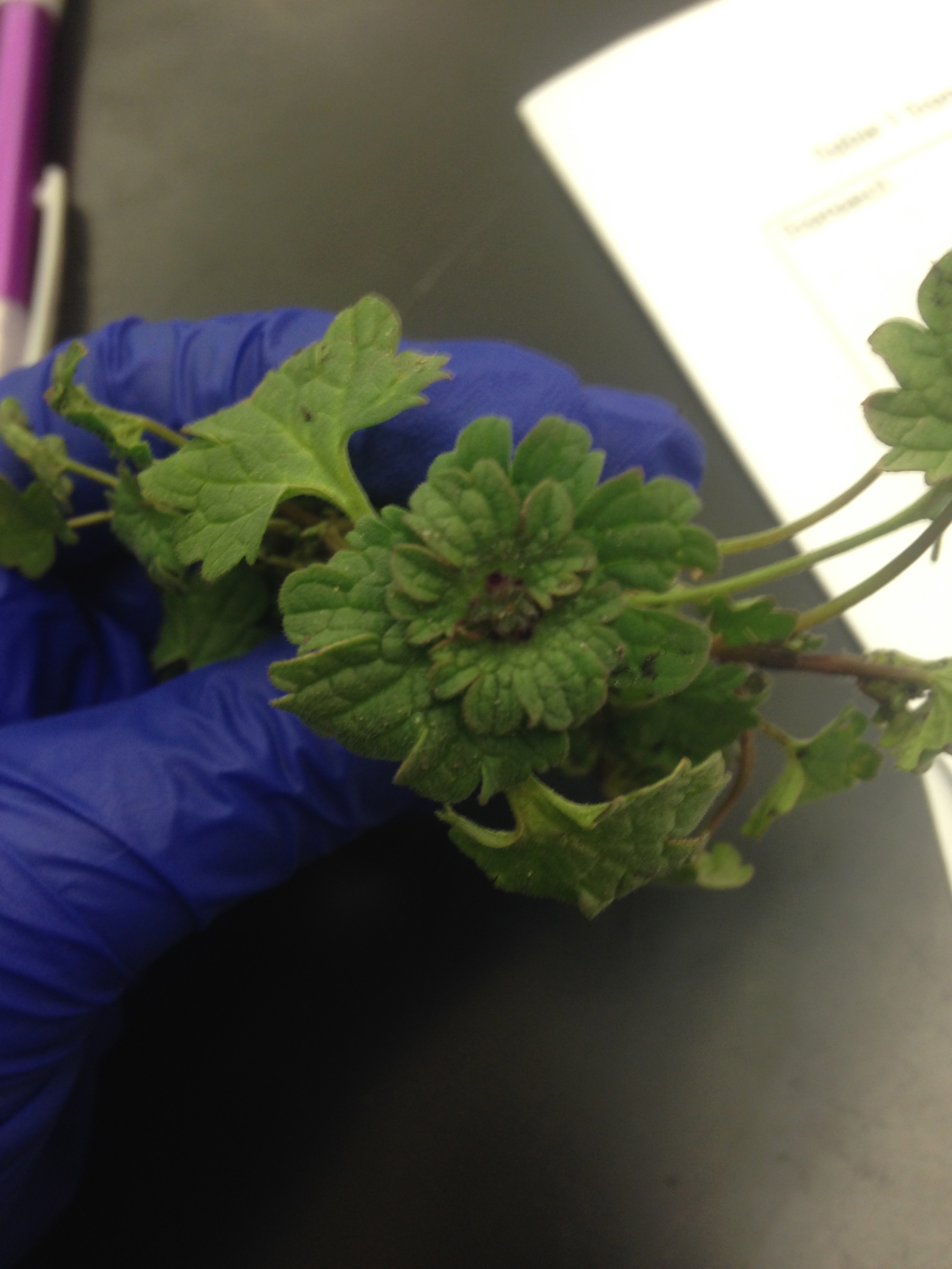
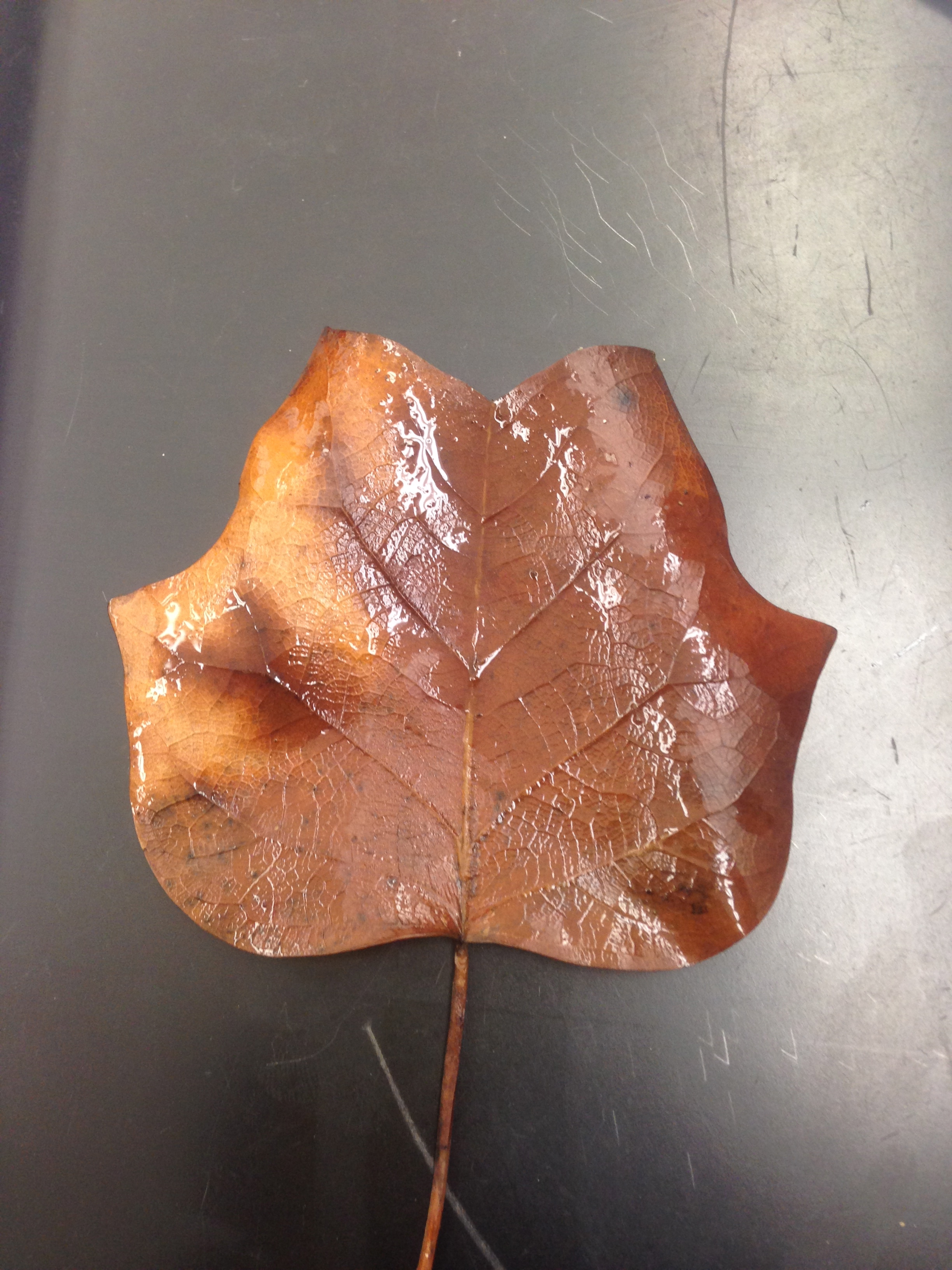
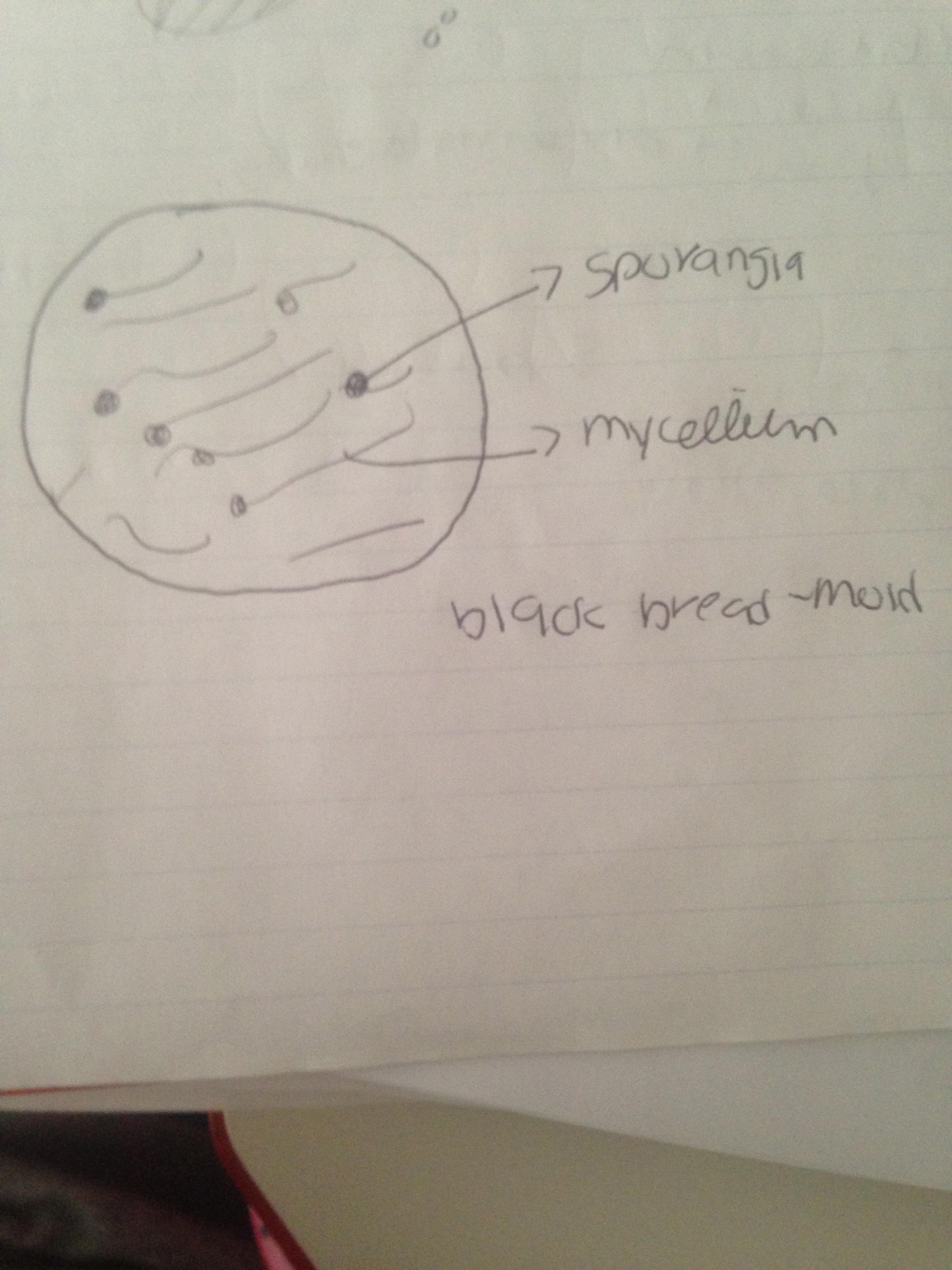
In all of our samples the vascularization was basically the same since they all had a stem to support the leaves and transport water an nutrients through the xylem and phloem. Furthermore since all of our samples had no flowering part, we could not observe any seeds in order to classify as either monocot or dicot. The Fungi sporangia is when the hyphae grows upward to form small, black, globeblack structures. The sporangia are important because they contain spores that fungus use for reproduction. Under the dissecting microscope we observed the black bread-mold/ Rhizopus stolonifer, which belongs to the Zygomycota lineage. This is indeed a fungus since it clearly has the sporangia structures as well as mycellium. Below, there is an image of the drawing of the black bread-mold.
Conclusions: The hypothesis of this experiment was not proven since even though our sample plants had indeed vascularization, there was no reproductive features. This means that that vascularization, specialized structures and reproduction do nor directly depend on one another. Even though this hypothesis was not proven, one could try another AU transect in order to discard this hypothesis completely. One thing that I found confusing in the lab was when we had to describe the vascularization of the plant samples. I did not know what we had to mention exactly. At the end of the experiment we began to set up a Berlese Funnel in order to collect Invertebrates for the next lab.
February 15,2014 4:00 pm Lab 3 Introduction: In this experiment titled "Microbiology and Identifying Bacteria with DNA" the purpose of this experiment were to: understand the characteristics of bacteria, observe antibiotic resistance and understand how DNA sequences are used to identify species. In order to understand the characteristics of bacteria and observe antibiotic resistance, one must first observe the serial dilutions of the 7 agar plates along with the handout of colony morphology, 4 out of the seven agar plates did not had tetracycline, which is has strong antibiotic resistance. With this said, the hypothesis for this experiment is that the agar plates with tetracycline will have less number of colonies than the agar plates without tetracycline. The prediction is that if the agar plates have a great number of colonies then it lacks the antibiotic resistance of tetracycline.
Materials and Methods: 1. Hay Infusion Culture 2. A microscope 3. Colony morphology handout 4. 7 agar plates 5.Crystal Violet 6. wash bottle filled with water 7. gram's iodine 8. 95%alcohol 9. safranin stain 10. centrifuge and sterile tube
Observations and Data: When I saw my Hay Infusion Culture for the second time, I noticed that the water had dropped tremendously, there was no mold on the surface and there was a peculiar smell. This change from last week is evidence that each week the appearance of the hay infusion changes due to the fact that the components in the infusion are still/ in a set place and the changes in the environment that make the infusion prone to bacteria and fungi. Once I counted the colonies in the agar plates, I noticed that the agar plate labeled 10-3 without tetracycline had the greatest number of colonies (700) and the agar plate labeled 10-9 without tetracycline had the less number of colonies due to the fact that it was the most diluted serial (120). On the other hand, the agar plate labeled 10-7 with tetracycline had the greatest number if colonies (50) while the agar plate with tet labeled 10-5 had the least number of colonies (3). In our experiment there was a mistake between the serial dilutions of 10-5 and 10-7 since the numbers are not going with the normal tendencies. The agar plates with without antibiotic had a lot more colonies than the agar plates with the tetracycline antibiotic. This difference in the number of colonies indicate that antibiotic is helpful in killing most bacteria overall. The effect of tetracycline on the total number of bacteria is that it greatly decreases its number. The species of bacteria that are unaffected by tetracycline are staphylococcal, streptococcal, and pneumococcal infections.Tetracycline works by decreasing the spread and growth of bacteria. It is effective in the treating of infections on the respiratory tract, urinary tract and the skin. The types of bacteria that are sensitive to this antibiotic are: ongonococcal urethritis, Rocky Mountain spotted fever, typhus, chancroid, cholera, brucellosis, anthrax, syphilis, and acne. When we observed the seven agar plates one could see that there were three colors that dominated the colonies: orange, white and beige. Furthermore the difference in the number of colonies in the nutrient plates and nutrient plates with tetracycline was noticeable.
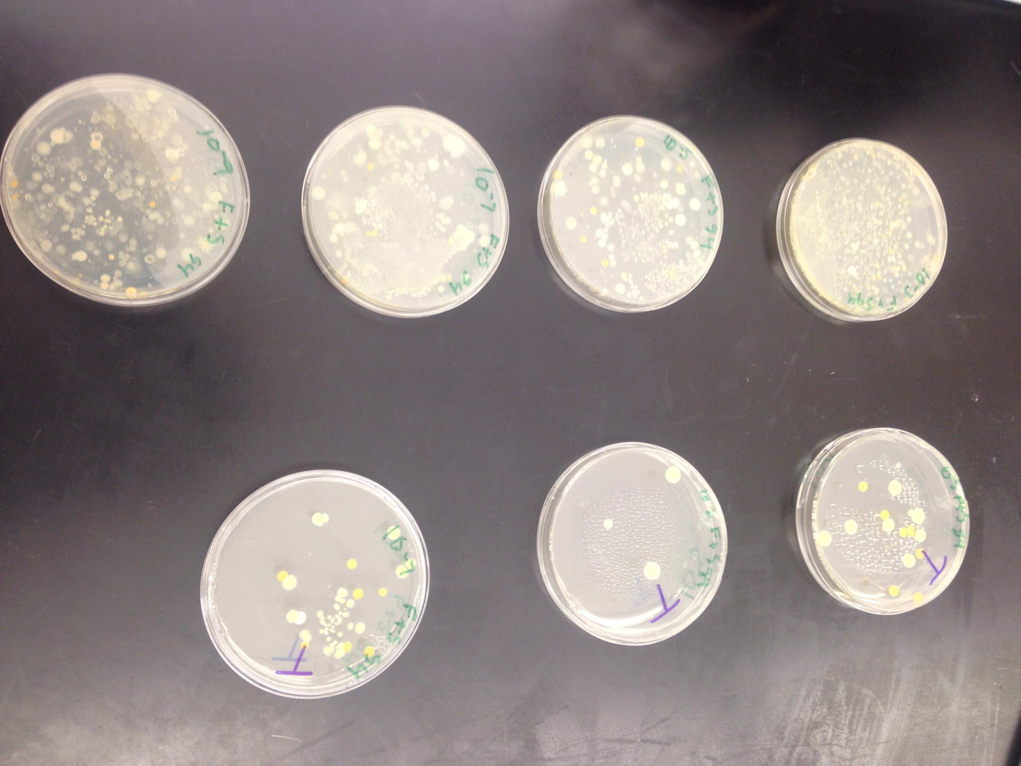
In order to examine the bacteria cell morphology, we chose the 10-5 nutrient agar, 10-3 nutrient agar, 10-5 nutrient agar plus tetracycline and 10-3 nutrient agar plus tetracycline. Once we prepared each sample of bacteria with gram stain and put them in a wet sample in the microscope, we could then observe the cell morphology.
The 10-5 nutrient agar plate (Figure 1) was identified as Diplococcus, it was gram positive and motile. The colors were beige and orange. On the other hand, the nutrient agar plate 10-5 with tet (Figure 2) was identified as Diplobacili, gram positive, motile with convex elevation and entire margins and beige colors. The nutrient agar plate 10-3 (Figure 3) was identified as Fusiform Bacili, gram negative without motility, flat elevation and entire margins, orange colors. Lastly, the nutrient agar plate with tet 10-3 (Figure 4) was also identified as Fusiform Bacili/ Streptobacili. It was gram negative without motility, flat elevation and entire margins, orange/ beige colors.
The image below further describes our drawing observations.
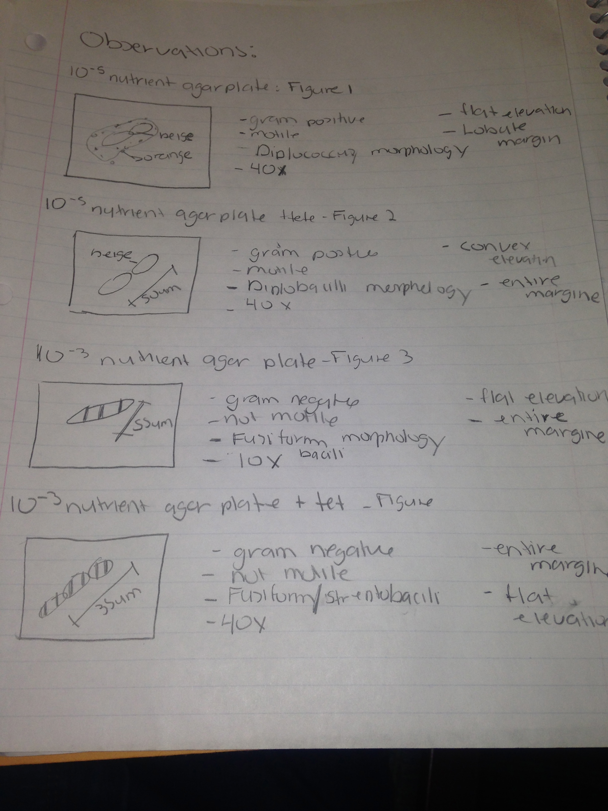
Conclusions: The hypothesis of the experiment was proven since the agar plates without the antibiotic of tetracycline presented the greatest number of colony formation. Indeed tetracycline stops the growth and spread of bacteria. One can say that the gram stain test was a success since it lead us to the identification of the cell morphology of the our four agar plates. Even though this was a success, the process was tiring since in two occasions our samples slide fell in the staining tray and we had to start again. We finished our experiment preparing colony samples for PCR in order to identify the DNA sequence for next lab.
February 9,2014 10:00 am
Introduction: In this experiment titled "Identifying Algae and Protist" the main objectives were: To understand how to use a dichotomous key and to understand the characteristics of Algae and Protist. One has to know the characteristics of such eukaryotic organisms in order to observe and identify the organisms in my Hay Infusion Culture using the dichotomous key. With this said, the hypothesis for this experiment is that the observations specifically in size, shape, and movement in the organisms in my hay infusion is critical in oder to identify them using the dichotomous key.
Materials and Methods: 1. Hay Infusion Culture 2. A microscope 3. wet mount sample of organisms in Hay Infusion 4. Ward's Dichotomous Key 5. 4 tubes of 10mls labeled with numbers 2,4,6,8 , a micropipeter set at 100 microliters and tips. 6. Four nutrient agar and four agar plus tetracycline in order to prepare 100-fold dilutions of the Hay Infusion Culture.
Observations and Data: As I carefully observed my Hay Infusion Culture, the first thing I noticed was that the water was much lower than before and how it turned yellow and brown. Even though the culture did not smelled, over the surface there was mold. Moreover, at the bottom of the culture there was dirt with a few branches and leaves.
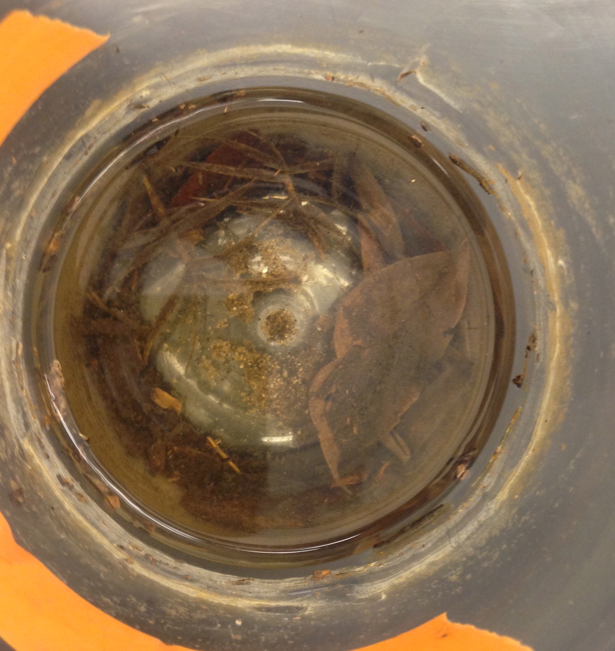
I took microscopic samples at the two ends of marked orange tape of my infusion. I took the samples at these two different ends because the organisms differ since they already established their own "niche" within the actual niche. As I observed these microscopic samples, I observed and identified 3 different organisms with the help of the dichotomous key. The first organism I observed was the Euglena, which measured around 41um. This organism can photosynthesize since it has a chloroplast and has a single flagellum that helps it move. The second organism I observed was the Paramecium Aurelia, which measured around 125um. This organism has micronucleus all throughout its "body" along with a much larger nucleus, cannot photosynthesize and has a cilia that helps it move around. Finally, the third organism I observed was the Amoeba, which measured around 550um. I noticed that the Amoeba moved constantly around using pseudopodia and had a single disc shaped nucleus.
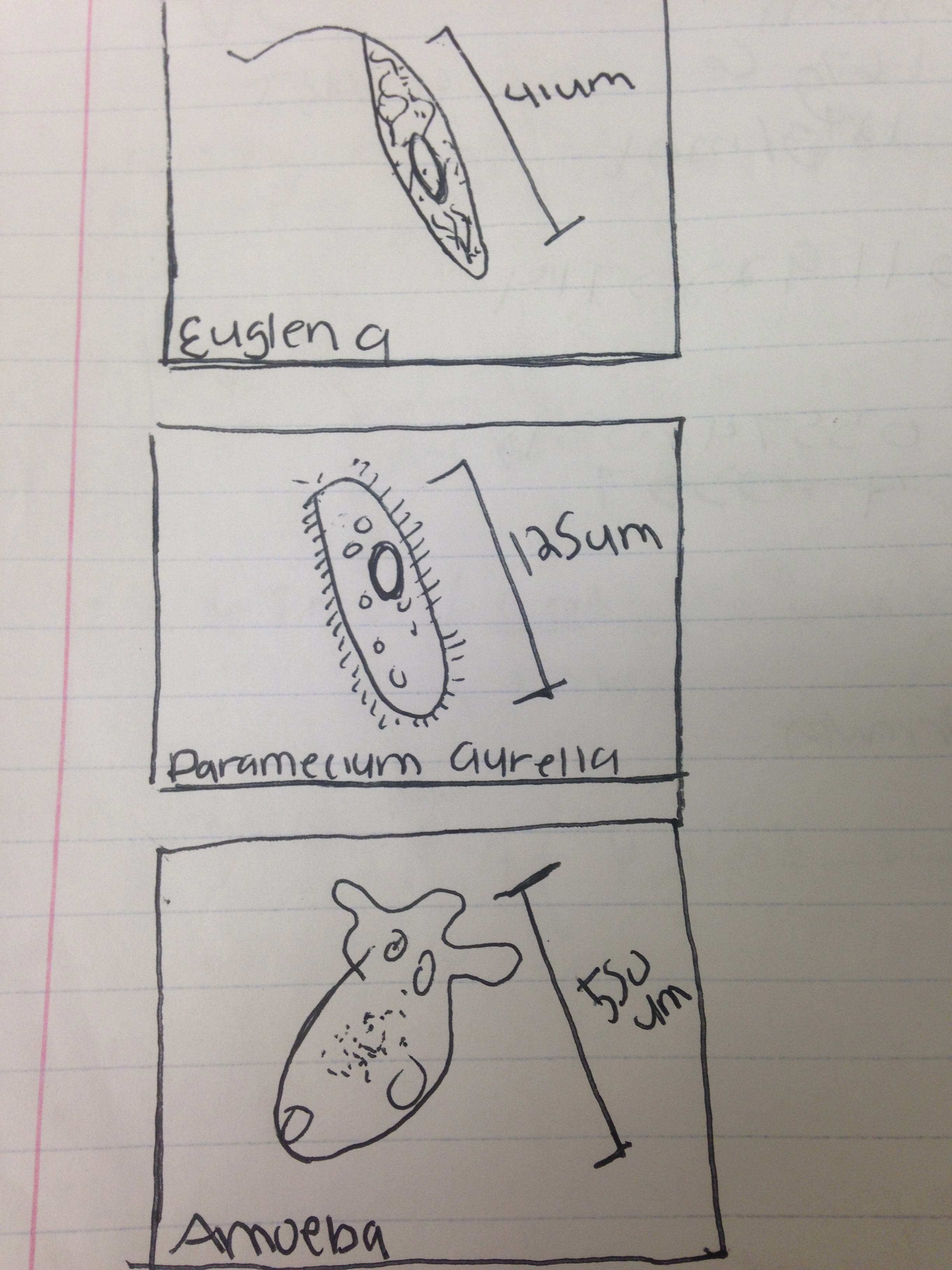
Paramecium is able to meet all of its needs for life. For instance, it can reproduce asexually by binary fission and sexually by conjugation, it uses its oral groove to acquire energy by water and nutrients and it has two nuclei: a macronucleus and micronucleus in order to keep its genetic material. Finally, there are able to move using the cilia. In other words, Paramecium meets the 5 fundamental characteristics of life: they acquire energy, are made up of cells, store information, are capable of replication and are a product of evolution.
In order to prepare for next week's lab, we prepared a serial of 100-fold dilutions with petri dishes. We first labeled 4 tubes with numbers 2, 4,6,8 and mixed the hay infusion culture. We then transferred 100 microliters of broth from each of the four tubes and then placed them on the surface of their respective agar plate. Once we completed the dilutions, the agar plates looked like the image below:

Conclusions: The hypothesis of my experiment was proved since I was able to identify the organisms in my hay infusion using the dichotomous key with the characteristics in its size, shape and movement. In order to improve my experimental design, I would have chosen next time microscopic samples at different ends of my infusion to see if there were more organisms that I could have identified using the dichotomous key. If the hay infusion had been observed for another two months, I am sure that there would be almost no water left on the infusion. Overall, the hay infusion would smell and there would be mold all throughout. I suggest that the dichotomous key instead of being black and white, was in color so that one can be more sure in the identification of the organisms, especially in the section of Paramecium.
January 29,2014 Introduction: In this first experiment titled “Biological Life at AU” the purposes were to gain a better insight at natural selection and the biotic and abiotic characteristics of a specific niche. Not only we examined samples of green algae and its Volvocine line but also examined a niche from AU.
Materials and Methods: For examining the green algae, we prepared a slide of Chlamydomonas and added protoslo in order to examine it in the microscope. We also examined a slide of Gonium to observe the Volvocine in a slide. For examining the niche, we had a map of the AU campus that helped us find a specific location for our niche. We used a 50mL sterile conical tube and a flashlight in order to collect the biotic and abiotic samples.
Observations and Data: While I examined the green algae, I recorded 600 cells for Chlamydomonas with a colony size of 0um, 400 cells for Gonium with a colony size of 10um and a total 2,000 cells for Volvox with a colony size of 10um. For my niche, I recorded leaves, little insects, grass, tree branches and worms as biotic samples while rocks, and soil for abiotic samples. The area of my niche was located in the compost garden of AU next to the Reeves Field.
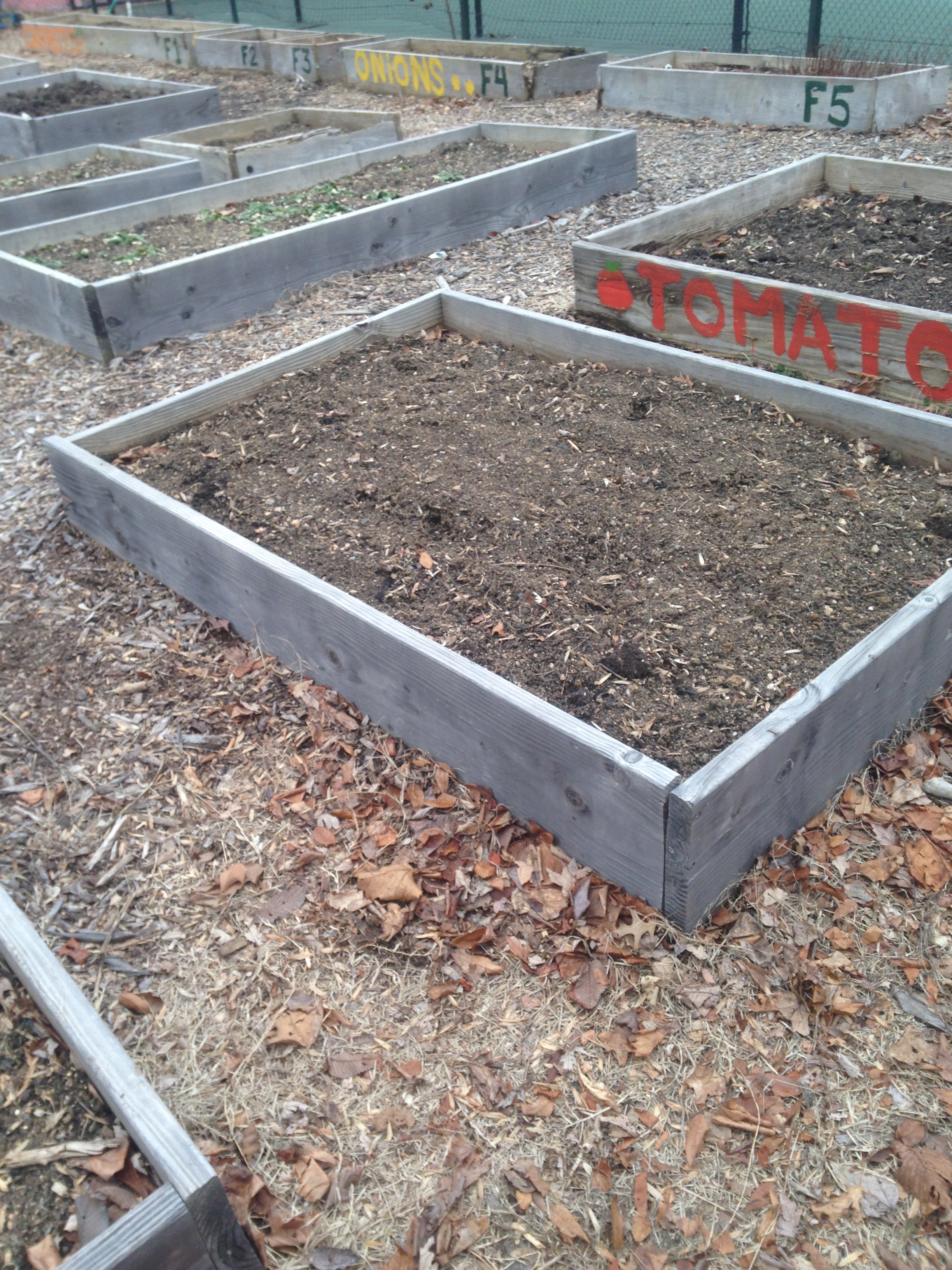
Conclusion: Overall the experiment was a success since I achieved the purposes stated in the introduction.
Good first try but a little brief. Some things could improve this text for example: You could include some more information based on the red text from the lab protocol. Separate the observation of protists from the transect work to make it more clear. Include diagrams and images of transect information. When starting the next entry please add to the top of the notebook so move older text downwards. SK
