User:Emily J. Ionescu/Notebook/Biology 210 at AU
03/19/2015 - Developmental Study: Effects of Rhodamine Exposure on Zebrafish Embryo (Week One)
Purpose
The purpose of the experiment at present is to compare the normal development of unaffected Zebrafish embryos with that of Zebrafish embryos that are being exposed to a solution containing the chemical Rhodamine.
Materials & Methods
Set up two petri dishes: one for the control group and one for the test group (containing Rhodamine). For the both groups, 20 mL of Deerpark water is to be added to the petri dish. The test group petri dish is added 2-3 drops of Rhodamine using a transfer pipette. Twenty (20) healthy, translucent embryos are to be added to each container using a dropper.
On day four, 10mL of water were removed from the dishes, and any empty egg cases and dead test subjects were disposed of. A total of 25mL of fresh water were added to both groups. Some of the dead embryos were saved and put in paraformaldehyde.
On day seven of observations, 5mL of water were removed, and replaced with 5mL of fresh water and test solution. A couple embryos from both the control groups were selected to be preserved in paraformaldehyde.
From day seven to fourteen, 5mL of water were continuously replaced with 10 mL of freshwater. Surviving Zebrafish were placed in an aquarium for safekeeping (Bentley, 2014).
Results
Observations of the development of the Zebrafish embryos were collected over a period of two weeks. The following is a summary of resulting analysis throughout the study:
02/19/2015 (Day 1)
Close attention was payed to the materials and methods setting up the experiment. Initially, each group had an equal amount of Zebrafish embryos (20 embryos).
02/20/2015 (Day 2)
On day two, the petri dishes were checked for fungi growth on the embryos. Embryos that had mold growth were replaced by non-fungal embryos. The color of the test group solution was noticed to be a very light-tinted orange color. The color of the Zebrafish was also determined to have this tint (although the solution itself might have been the reason of this appearance).
02/23/2015 (Day 5)
On day five, counts of surviving embryos in both groups were made and freshwater was added. The recorded count for the control group was 12 alive, 5 dead, while for the experimental group there were 16 alive and 2 dead embryos. The disappearance of 3 and 2 embryos, from the control and test group respectively, is unknown. However, it is possible that spillage of the petri dishes during unsupervised times of the experiment might be the cause of this error.
Qualitatively, the control group was observed to have hatched less than the Rhodamine subjects. On the other hand, no significant differences in behavior were noted. Aside from the appearance of the control group fish moving a little bit more than the Rhodamine, the two subject groups were behaving similarly. The faint color difference observed from day one was still noticable in the Rhodamine petri dish.
02/26/2015 (Day 7)
On day seven, counts of embryos were made, old water was again exchanged for fresh water and the fish were fed with one small drop of food. The control group had 16 hatched eggs, 3 dead, 16 surviving and 1 unhatched. Meanwhile, the Rhodamine group had 16 hatched embryos, 13 surviving, 2 dead and 2 unhatched. Both samples seemed to be adequately developed.
The fish were graded based on a developed swimming and movement scale ranging from 1-10 (1 being the slowest and 10 being the fastest). The control group received an overall score of 8-9 on this scale, while the Rhodamine subjects were awarded a 4 out of 10. Also, a tap and move measurement was performed for each group. The subjects would be tapped and their swimming propulsion was measured using a ruler. The control group was averaged a tap and move score of 1.5-4cm, while the Rhodamine only received a 0.2-1cm propulsion score. Thus, quantitive observations were used to compare the movement of both groups.
Qualitatively speaking, the control group subjects were more agile and moved faster and more often than the Rhodamine. Aside from being more active, the control Zebrafish embryos could also propel themselves further when swimming, being able to reach further. The Rhodamine embryos, thus, moved less and also exhibited a shaking movement, traveling minimally, moving less and unable to propel as much as their countersubjects. This was suspected to be due to underdevelopment.
03/06/2015 (Day 15)
The final observations of the Zebrafish were performed on day fifteen. Counts of embryos, as well as disposal of dead and preserving of the living embryos were taken care of. Thus, the control group had 7 living embryos, 4 dead and 9 that were unidentified. Contrary to that, the Rhodamine petri dish was found to contain only a solution, the Zebrafish having disappeared. More likely than not, this might be due to spilling of the petri dish in times were the containers were left unsupervised.
A summary of observations of the only remaining group, the control group was accomplished. The length of the two-week old embryos in the control petri dish was on average, around 3.5mm, which allowed them to be seen without a microscope. They had a white-transparent pigmentation, with yellow spots in between the eyes and along their tails and black spots along the spine (both front and back). Their fin development was small, the average fin measuring 0.2mm and was at times not visible across all subjects.
Organ development of the control group fish varied - some was complete, for example the heart, big, developed torso and the eyes were very visible, while in the majority they were not as exhibited. Motility was reduced, however, since last checked on day seven. The embryos were not very active, mostly staying still and only moving if the dish was shaken. The eye diameter was approximately 0.3mm in all of the living subjects in the control group, which is quite large.
Conclusion
Due to unknown, only suspected, causes, the experiment was not concluded as the Rhodamine-treated Zebrafish embryos had disappeared towards the end of the study. Judging from day five and seven observations, the embryos of the experimental group were affected by the chemical with which their solution was treated. The movement, development and activity was reduced compared to the normal control group. Therefore, according to this study, the Rhodamine chemical did have a negative impact on the development of Zebrafish embryos.
03/05/2015 - Transect One Bacteria 16S rRNA Gene Sequencing
Purpose
The purpose of this laboratory experiment is to adequately sample bacteria colonies from serial dilution agar plates and perform PCR on the 16S sequence from the sample. Thereafter, the aim is to use the results to adequately identify the bacterial species found to be living within the borders of Transect One.
Materials And Methods
Using samples from the serial dilution bacteria plates labeled Agar + Tetracycline 10^-6 (13A) and Agar + Tetracycline 10^-2 (13D) that were originally collected from the Hay Infusion Cultures of Transect One, DNA was isolated into a 100uL of water in a sterile tube. For ten minutes, the tube was incubted in a heat block at 100 degrees Celsius, immediately being centrifuged for five minutes at 13,400 rpm. A total of 20uL of primer/water mixture was added to a PCR tube, after which the PCR bead dissolved. Lastly, 5uL of supernatant from the centrifuge was added to the 16S rRNA reaction and the tube was placed in a PCR machine.
After one week, the PCR products were run on an agarose gel and the DNA was purified for sequencing (Bentley, et. al., 2014).
After the purification of the DNA, its sequencing was used to identify the bacterial species. First and foremost, the codes of the 16S rRNA genes labeled 13A and 13D were copied in both the 5'3 and 3'5 directions. Using the Nucleotide Blast search engine, the sequence was searched in an extensive database and the top search results were considered. The bacteria identification was concluded by confirming whether the yielded search results match the initial agar plate characterization from previous weeks.
Data & Observations
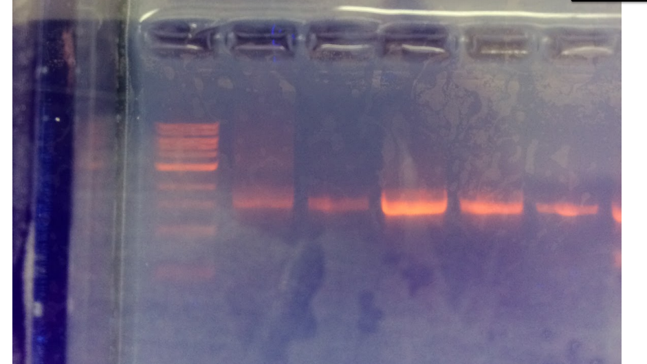
According to Figure 1, the 13A and 13D gel samples seem to have an approximate length of 1,300 base pairs.
13A And 13B Bacteria Gel Samples Collected from Transect One
Figure 1 : Bacteria gel samples appear to have a length of 1,300 bp.
After the sequence was purified and the raw sequence was generated using the Nucleotide Blast, they were analyzed and recorded, as follows:
Sample 13A: (Forward direction - 5'3)
NNNNNNNNNNNNNNNNNCNNNNNNTGCNGNNNNANGGNNGNCNGNNNNNNANCAATCCTGGCGGCGAGTGGCGAACGGGT GAGTAATACATCGGAACGTGCCCAATCGTGGGGGATAACGCAGCGAAAGCTGTGCTAATACCGCATACGATCTACGGATG AAAGCAGGGGATCGCAAGACCTTGCGCGAATGGAGCGGCCGATGGCAGATTAGGTAGTTGGTGAGGTAAAGGCTCACCAA GCCTTCGATCTGTAGCTGGTCTGAGAGGACGACCAGCCACACTGGGACTGAGACACGGCCCAGACTCCTACGGGAGGCAG CAGTGGGGAATTTTGGACAATGGGCGAAAGCCTGATCCAGCCATGCCGCGTGCAGGATGAAGGCCTTCGGGTTGTAAACT GCTTTTGTACGGAACGAAACGGCCTTTTCTAATAAAGAGGGCTAATGACGGTACCGTAAGAATAAGCACCGGCTAACTAC GTGCCAGCAGCCGCGGTAATACGTAGGGTGCAAGCGTTAATCGGAATTACTGGGCGTAAAGCGTGCGCAGGCGGTTATGT AAGACAGTTGTGAAATCCCCGGGCTCAACCTGGGAACTGCATCTGTGACTGCATAGCTAGAGTACGGTAGAGGGGGATGG AATTCCGCGTGTAGCANTGNAATGCGTAGATATGCGGAGGAACACCGATGGCGAANGCAATCCCCTGGACCTGTACTGAC GCTCATGCACGAAAGCGTGGGGAGCAAACAGGATTAGATACCCTGGTAGTCCACGCCCTAAACGATGTCAACTGGTTGTT GGGTCTTCACTGACTCANTAACGAAGCTNACNCGTGAAGTTGACCGCCTGGGGAGTACGGCCGCAANGTTGAAACTCNAA NGAATTGACNNGGACCCGCACAAGCNGTGNATGATGTGNTTTAATTCNATGCAACGCGAAAACCTTACCCACCTTTGACA TGTACNNNANTTNNNCCAGANATGGCTTANTGCTCGAAANAAAANCGTAACNCANGTGCTNCATGNCTNNCGTCNNCNTC NTGTCGTGANA
Sample 13D: (Forward direction - 5'3) NNNNNNNNNNNNNNNNNANANTGNANNCCNNAGCGGTAGCAGANGNTATCANGATGTCCGACAGCGGCTTGCNGATGAGG TACAAGTGTGGTTTATGCCTTTAGCCGGGGGAGGCACTTTCGTTGGGAAGATTACAACCCCATAATTATAATCGTGGCAT CTCTTGAAANGGACTGGTCCAGTGGAAAAAGAAGGGCCCGACCCTGATGANGCAGTTGGTACGGGGACGGTTCACCANGG CTGTGATGTTTGTGGGGCCTGANAGGGTGATCCCCCTGTGTGGTACGGAGACATTGACCCAACACCAATTGCAGGCGCCT CTGAGGAATATTGGACAATGGGTGAGAGCCTGATCNNNANTCNNCGNGAAGGATGACGGTGCTCCTGGTTGTATTCTTCT TTTGTATATTGATGGTGATTTCCTCGTGGGTGAAGCTGAATGAACTATACAAGCAGNAACCGGNGAGGCCCNTGCCTTCA GCCTCGGTNNTACNCAGGGTGTTGCCGTTTGAGAGATTTATTGNNTTNTCGAGGTTGGTTCNNGCNGANGGCNNACAATA TGCTGTANNNNTNACTNNNNGGTCAATCTGCATANGTTGGCGCGNGNCGCGACTNTTGGATATCTACCTTGCNTAAAANA NTCNNACANGGAANNCNTANATAATANCNNNNNCACCAATTGCGAANGCAGGTTACTATGTCTTAACTGACGCTGATGGA CGAAAGCGTGGGGAGCGAACAGGATTANATACCCTGGTANTCCACGCCNTNNNNNATGCTNACTCGTTTTTGGGNTCTTC NGATTCAGAGACTAAACNAAAGTGATAAGTTAGCCACCTGGGGAGTACGTTCNCAAGANTGAAACTCNAAGGAATTGACN GNNCCCGCACAANCGGNGGATTATGTGNNTTNATTCNATGATACGCNANGAANCCTTNNCCNANGCTTAANTGGGNANTN GATCGGTTTNNNANNNNACCTTNCCTTNNNCAATTTCAAGGTNCTGCATGGNTNGTCNNCNGCTNNNNCCNNNANTNNNA GNTAANTCCTGNNNNNNNGNNNCCCCNTGTCNCNNN
Sample 13D: (Reverse direction - 3'5)
NNNNNNNNNNNNTNNNTGTAGCGNACNNNNNNNGTCTNNTGGATTCGGGCCGCCNNTACTATATAGNGTNGTTGTCTGCC
TGTACCAGGAACGGGANNAAACGTCGTATTTNGGNAGATGGGCGCAACCGGAGGTTGGACGAATTTGAATTATAAGGTGN
CANTCCNATNGCAAATGAGNCCGGNACTGCAGATNNGATTAGCGCTTCACCGGGAAGTGCNCTGATGTAACTTTGTAGGA
GNGTGAGGTCCNATGATCGTTATTATGGATGGNTTGATTGGAATAATGATATGGATTTTAATGATGCANNAGAAACTATT
CTTGCTACAACTTAAAGGTTTAAACTAGTGACAGGGGTTGCGCTCGTGTACCGANNTAACCTAACNATNGAAATTACCGG
GTGAGGTTTGCATGCCAGGNTNGGGTGCTGCCGCTCCGTGGGGACATTTTCCATACGATAATTTCCTATATTATCCTTGG
TAAAGTGCCGCGCGTACCACCTAAATACACCACATAATCCACCGCTNGTGCGGGCCCTCTTCANTTCCNTTGAGTTTCAT
TCNNGCNAANGNACTCCCCNNNNNGNNNANTTATCANTTTCNCTAANTCACTGANNCCNAAGANCCNGANNGANNNNNAN
NNTNANAANNNNAGTGGACTACCANGGTATCTAATCCTGTTCGCTCCCCACGCTTTCGTCCATCAGCGTCAGTTAAGACA
TAGTAACCTGCCTTCGCAATTGGTGTTCTAAGTAATATCTATGCATTTCACCGCTACACTACTTATTCCAGCTACNTCTA
CCTTACTCAAGACCTGCAGTATCAATGGCAGTTTCACAGTTTAAGCTGTGAGATTTCACCACTGACTTACAGATCCGCCT
ACNGACCCTTTAAACCCAATAAATNCNNANAACGCTNGCACCCTCCGTATTACCGCGGCTGCTGGCACGGANTTAGCCNN
TGCTTATTCGTATAGTACCTTCAGCTTTCCACACGNGGNAAGGTTGATCCCNATANNAANANNTTTANNCCCATANGGCN
TCATCNTTCANGCNNANGGCTGGATCNGNTCTNACCCATTGNCCANTANTCCTCACTGCTGCCNCCCGTNNNANNNNG
Once the species were entered into the Nucleotide Blast Database, the species were identified based on the top search results they yielded. Thus, sample 13A was determined to be the Variovorax and sample 13D turned out to be the Chryseobacterium bacteria.
Conclusion
Sample 13A was identified to be a Variovorax bacteria, also known to be a rod-shaped, Gram-negative, motile bacteria that is isolated from soil. It is known to feed off of various substances, such as Boron, and it can also be very diverse (Willemes, 1969). This is consistent with the precious, colony description because it describes the bacteria as being motile and isolated from soil.
Sample 13D, on the other hand, was identified as a Chryseobacterium, which is a small rod-shaped, colden-yellow bacterium (Vandamme, 1994). Again, these descriptions seem consistent with the previous, colony characterization, as the colonies were initially described to have a yellow, mustard-like color.
Works Cited
Bentley, Meg, et. al., "Laboratory Manual to Accompany: General Biology II", American University (2014). 31-33.
Vandame, P., et. al., "New perspectives in the classification of the flavobacteria: description of Chryseobacterium gen. nov., Bergeyella gen. nov., and Empedobacter nom. rev.". Int. J. Syst. Bacteriol., 1994, 44, 827-831.
Willemes, A., et. al., "Comamonadaceae, a new family encompassing the acidovorans rRNA complex, including Variovorax paradoxus gen. nov., comb. nov., for Alcaligenes paradoxus" (Davis) 1969. Int. J. Syst. Bacteriol., 1991, 41, 445-450.
02/12/2015 - Identification And Characterization of Vertebrates in Transect One
Purpose
To accurately identify, classify and characterize vertebrate organisms found to inhibit or pass through Transect One. Also, to gain more understanding of the interactions with other biotic and abiotic components of the area studied.
Materials And Methods
Using deductive reasoning and careful observations of the vertebrates that inhibit Transect One, the identity and interactions of these organisms was determined. Also, using online dichromous key resources, accurate taxonomic classifications from phylum to species was made available.
Data And Observations
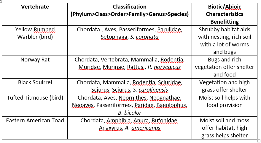
A summary of the inhabiting vertebrate species and their characteristics and classifications is offered in Table 1. Out of the animals found, two were birds (the Yellow-Rumped Warbler and Tufted Titmouse), two rodents (Black Squirrel and Norway Rat) and a toad (the Eastern American Toad).
Table 1: Summary of the identified vertebrate organisms found to inhabit Transect One.
Conclusion
As you can see, although there is not a lot of phylum diversity in terms of vertebrates in Transect One, the classes of these identified animals are definitely exhibiting a wide range. For example, as explained in Table 1, there are only two organisms with similar classes, namely the Aves class; other than that, the classes range from Mammalia to Amphibia.
Therefore, Transect One can be classified as a true community of organisms, with a stable carrying capacity due to the ecological interactions of vertebrates such as the ones studied here. Furthermore, trophic levels, or the specific positions occupied by each organism living within this niche within this food chain, are also indicative of this notion of community. If, for instance, this food chain were to be disrupted by the disappearance of the Tufted Titmouse bird, there would be an abundance of worms in the soil that could have impacts on the fertility of the soil.
02/12/2015 - Identification And Characterization of Invertebrates in Transect One
Purpose:
The goals of the research of Transect One’s invertebrates were to first observe how the movements of three different types of worms relate to their coelom structure and to understand the diversity of the five major classes of anthropods through examples of organisms. Lastly, the purpose of the study was to identify and describe five invertebrates sampled from Transect One, collected using the Berlese Funnel. The hypothesis related to the latter part of this research, therefore suspects that if there are remnants of organisms in the leaf litter gathered from the transect, the majority of the invertebrates collected will be insects.
Materials and Methods:
Part I – Movement Analysis of Acoelomates, Pseudocoelomates, and Coelomates
Using a dissecting scope, the live acoelomate, Planaria, was observed. As it was fed with egg yolk, its digestion and movement was analyzed. Also, a cross section of Planaria’s stained digestive tract was studied in detail usng a prepared wet mount. Thereafter, a cross sectional slide of various nematodes was viewed and used to describe its pseudocoelomate structure and its resulting movement. Lastly, coelomate Annelida was observed using a prepared slide. Its organ position and muscular layers were described.
Part II – Classification of Anthropods
Example organisms from the five classes of Anthropods were compared using online resources such as Museum Victoria’s website ([1]).
Part III – Identification and Description of Transect One Invertebrates
The Berlese Funnel prepared last week using leaf, vegetation and soil samples collected from Transect One was decomposed. The alcoholic solution containing the sample debris in the 50 mL cylinder was separated in two petri dishes (10-15 mL of the top was poured into one petri dish, and the remaining, bottom layer was poured into a second dish). Using the dissecting microscope, Arthropoda invertebrates were identified and classified. For any insects found, the Insecta dichromous key was used to pinpoint orders of a certain class, as well as Figure 3 (Bentley, 2014).
Data and Observations:
Part I – Movement Analysis of Acoelomates, Pseudocoelomates, and Coelomates
Based on the cross section observed of the Planaria and its stained digestive tract, the lack of a fluid filled cavity, or coelom was apparent. The simplistic structure of the digestive system is evidently lobate and it lacks separation from the outer body wall. Similarly, the head and tail of these organisms confirmed a bilateral symmetry, the mouth also being the only observed means of ingression and egression. The movement observed under the dissecting scope reflects this structural simplicity, as it is very slow, and gliding.
The cross sectional slide of the nematodes, on the other hand, revealed an only partly-lined body cavity. Its discontinuous innermost layer – the endoderm, does not come in contact with the outside, ectoderm layer. Its alimentary canal spreads from the mouth to the anus. The movement is whip-like, as the body is not as flexible, not being able to bend fully, unlike the Planaria, for example.
Lastly, the Annelida was determined to be a coelomate based on its observed, fully-lined and fluid filled coelom. The three layers: ectoderm, mesoderm, and the endoderm cause its locomotion to involve an extension of the body. This movement involving the contraction of different muscles (Reish, 2013).
Part II – Classification of Anthropods
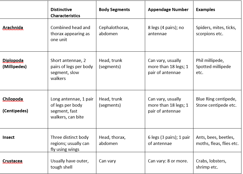
Table 1: Summary comparison of the five major classes of Anthropods, protosomal organisms with mouth forming earlier than the anus (Museum Victoria Australia, 2014).
Part III – Identification and Description of Transect One Invertebrates
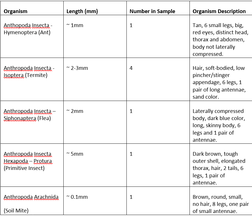
Upon close analysis of the five organisms found in the Berlese Funnel sample, four of them were identified as being part of the Insecta class of the Anthropoda phylum (ant, termite, flea, protura), and only one that was part of the Arachnida class (soil mite). Their size, number found and respective characteristics are summarized in Table 2.
Table 2: Five invertebrate organisms identified in from the Transect One Berlese Funnel sample collected one week prior and their description.
As seen, the size of the invertebrates found ranged from approximately 0.1mm to 1mm - 2mm, and even and 5mm. The Soil Mite (0.1mm) being the smallest and the Protura (5mm) being the largest of the organisms identified. Thus, the organisms most common in leaf litter appear to be insects.
Conclusion:
Following the identification of the invertebrates and their analysis, it is to be concluded that the initial hypothesis, proposing insects as the most probable organism to be found in the leaf litter collected, is therefore supported. According to Table 2, Protura, ants, fleas and termites offer a great variety of the Insecta class of the Anthropoda phylum and represent the majority of the invertebrates found on Transect One.
Also, the classes and orders of Anthropods were researched and summarized in Table 1. Furthermore, the movement of different worms, namely Planaria, nematodes and Annelida were linked to their internal structures and the presence of the coelom. The acoelomate Planaria, as a result having a simple, slow gliding motion; the pseudocoelomate nematode having a faster, whip-like locomotion and the coelomate Annelida exhibiting a less flexible, extension motion.
Works Cited
Bentley, Meg, et. al. 2014, A Laboratory Manual to Accompany: General Biology II. American University: Washington D.C., USA. 45-49
Ramel, Gordon. “The Phylum Annelida”. 2013. Earth Life (February 18, 2015) <http://www.earthlife.net/inverts/annelida.html>
“The Spider’s Parlour”. 2014. Museum Victoria Australia. (February 18, 2015) < http://museumvictoria.com.au/spidersparlour/ed1a.htm>
E.I.
02/10/2015 - Identifying Plants and Fungi From Transect One
Purpose:
The goal of the research at present was to identify and be able to describe five plants from our assigned AU transect, Transect One. Using physical characteristics and reference resources, plant life was characterized according to its genus. Vascularization, arrangement, shape and size, as well as other distinct features of the plants found were used to deduce its biological classification. Furthermore, fungi found on the area of study was dissected, observed and later decided in which of the three main groups it belongs.
Materials And Methods:
In this experiment, a total of five samples of the plant life located in Transect One were collected in plastic bag. Using a microscope, a cross-section from each plant was observed. After the collection of qualitative data was concluded, additional resources in the forms of various dichromous keys were used to identify the plant life and fungus collected.
Data And Observations:
From the transect, the five representative plant samples collected were the following: -Moss -Tall grass -Cat Tail - -
The moss was located on top of wet, rich soil on the ground, nearby an area with a multitude of rooted vegetation. It's green color helped measure its size, which was recorded to be 2-3 mm by strand.
The tall grass, on the other hand, was green, about a foot high, with stemming, long leaves that gradually thins as it grows higher. It was also found in clusters near other plant roots.
The cat tail
Here are some images of the vegetation on Transect One:
There were no seeds found within the perimeter of Transect One.
Fungi sporangia are structures characteristic of the hyphae filaments that tend to grow in an upward direction. They represent the tiny, spherical structures reaching maturity when their color deepens, containing spores that are released upon their opening. Sporangia are important because the carbon dioxide and nitrogenous wastes released from them are utilized by most plants in their carbohydrate formation. Therefore, without them plants would not be able to offer animals the nutrition they seek when consuming plants.
Some of the samples observed in the lab under the dissecting microscope included the common mushroom. These fungi are considered to be filamentous, as their cells are packed in structured shapes. Mushrooms are terrestrial and use their cap underside structure called "basidia" to reproduce sexually. Therefore the mushroom is found on the Basidiomycota lineage of Fungi.
PICTURE OF FUNGUS
Conclusion:
E.I.
02/05/2015 - Observing Antibiotic Resistance in Hay Infusion Bacteria
Purpose: The purpose of this research is to analyze, describe, and differentiate between bacteria colonies originating from Transect One Hay Infusion Culture from week one. Using Gram stains, one of the most common stains for bacteria was used to further characterize bacteria growths.
Also, changes were observed in the Hay Infusion Cultures. The lessening of water content in the jar due to evaporation, dark plant-life condensing into residue on the bottom of the container and the decreasing intensity of the putrid smell from last week's lab were the main changes analyzed. If the Hay Infusion Culture has increasing decaying of plant life in the culture, then bacteria and other microorganisms may be feeding on it.
Materials And Methods:
Bacteria Slides: The wight growth agar plates from last week's serial plating were analyzed under the microscope. Furthermore, a wet mount of four of the bacteria colonies were performed, namely the plates labeled "Agar 10^-6", "Agar 10^-2", "Agar + Tet 10^-6" and "Agar+Tet 10^-2". The procedure used included sterilizing a metal loop over a flame, taking a sample of each growth and placing it on a clean microscope slide with a drop of water. The slides were then observed under the microscope for bacterial shape, motility and color.
Gram Stain Slides: Gram Stain was performed using a sterilized loop and a sampling of each of the four plates. The bacteria were placed on a clean slide and air dried over the burner flame. Using a staining tray, crystal violet was smeared over the slide for approximately one minute and then soaked in Gram's Iodine for another sixty seconds. Thereafter, the bacteria smear was decolorized using 95% alcohol and rinsed. The gram stained sample was also observed under the microscope and characterized.
PCR of 16S Gene: Finally, the PCR replication for the 16S gene was performed. A total of 100ul of water was placed in a sterile tube with a bacteria colony sample (four sterile tubes in total, one for each chosen bacteria tray). The tubes were incbated at 100 degrees Celsius for ten minutes and then centrifuged for 5 minutes at 13,400 rpm. After, 20ul of a primer-water mixture was mixed with the PCR bead and 5ul of the centrifuged tube were added.
Data & Observations: During an entire week, the eight serial dilutions of the agar plates were left untouched. Upon observing, counting and estimating of colony concentrations, the following data was summarized in the following table (Table 1):
As shown in the above table, above the 10^-6 concentration, there is a significant reduction in bacteria colonies. This indicates that the antibiotic is effective in controlling the growth of the organisms present. Fungi, particularly mold, was present in two of the plates, namely the 10^-2 and 10^-4 dilutions of the nutrient agar plates.
Also, four of the eight agar plates were chosen for morphology observations under the microscope, specifically the plates corresponding for serial dilutions of 10^-2 and 10^-6 for both agar and agar with tetracycline. However, due to the natures of the sample, two of the chosen agar plates (agar with 10^-6 dilution and agar plus tetracycline at 10^-2) were not visible, thus not able to be described. The following data related to bacteria characterization is summarized in Table 2:
PCR replications of gene 16S was also performed. Next week, their products will be run on an agarose gel and the samples will be used for sequencing that will aid in identifying the bacteria.
Conclusion: The bacterial colony growth that resulted in the sampling Hay Infusion Culture from Transect One is therefore concluded to not have antibiotic-resistance. Results in Table 1 show the affected, declining levels of bacteria growth on agar nutrient with the added tetracycline at both 10^-6 and 10^-2 dilutions.
E.I.
01/29/2015 - Analyzing Protists And Algae In Hay Infusion Culture
Purpose: To obtain two samples from differing niches of last week's Transect #1 culture and to observe their respective wet mounts, while looking for different organisms. Using the dichotomous key, protists and algae are identified and described. Lastly, it is hypothesized that if the Hay Infusion Culture is to be left to "grow" for another two months, then many symbiotic relationships will be observed, competition within the ecosystem will take place and biotic life will develop within the culture, such as bacteria colonization.
Materials And Methods: Using the 500mL jar of the Transect #1 Hay Infusion Culture from last week's class, two samples - one from the bottom of the jar and another one from the top layer of the jar - were collected for microscopic observation. The samples representing two different niches were thereafter carefully transformed into two different wet mounts, using a disposable pipette, and each slide was observed under the microscope. Then, each organism found was measured using the ocular micrometer and then identified using the dichotomous key.
Data And Observations: Upon first observing the Hay Infusion Culture, there was a noticeable, putrid smell. The 500mL jar was filled with clear, light brown liquid, with debris on the bottom and a hardened top layer. There were a total of four organisms identified in the Transect #1 Hay Culture Infusion, namely Colpidium sp. in both samples of the culture, Pelomyxa and Bursaria truncatella on the top layer sample, and Paramecium on the bottom layer sample of the jar culture.
01/26/2015 - Observing And Sampling of AU Transect #1
Purpose: By carefully analyzing a 20 by 20 meter transect of land on American University's campus, different niche characteristics of an ecosystems were studied. Balance within a niche, interactions of biotic and abiotic components and general characteristics of topography were areas of interest in this study.
Materials & Methods: Using the designated transect (Transect #1) defined by four, marked popsicle sticks, careful analysis of topographical features and location was performed. A 50mL conical tube was used to extract a representative sample of soil and vegetation from every part of the transect. After, 11.1g of the soil and vegetation sample were added to a plastic jar filled with 500mL of deerpark water. Next, 0.1gm of dried milk was also added, and the jar was mixed for approximately 10 seconds. The jar top having been removed, the open container was thereafter left out for the duration of a week.
Data & Observations: Transect #1 is a very grassy area, filled with various types of vegetation. It is located across the AU President's house and a few feet South of the "American University" sign. Behind the transect, there is a cemented pathway that leads to Massachusetts Avenue NW. From the Western end of the transect going East (Figure 1), the topography is somewhat hilly and increases in elevation. Furthermore, most of the vegetation is concentrated on the Eastern side of the transect (Figure 1).
[[<"https://docs.google.com/a/student.american.edu/file/d/0B8Io0GUP3HWWSUJyNmc2VlpMXzg/preview" width="640" height="480"></iframe>]]
Figure 1: Labeled aerial-view of Transect #1.
Moreover, the diverse abiotic and biotic components of the area were observed and recorded. The following are five of the abiotic components found on the surface: trash, sewer cap, rocks, snow and AU flower sign. The following are the five biotic components of this ecosystem: grass, moss, Red Cardinal flower, Cat Tail flower and straw plant.
Conclusion: The careful observations conducted on Transect #1 have shown that the area is highly diverse in vegetation and in its abiotic components. The location is not expected to change drastically over the next few weeks due to non-changing whether forecasts and overall stable environmental factors.
E.I.
01/19/2014
I am extremely excited about Spring semester in Biology 210!
E.I.


