User:Dylan J. Williams/Notebook/Biology 210 at AU
Lab 1
Objectives: To understand the biotic and abiotic characteristics of a niche. A 20x20 foot transect will be measured to examine and experiment the abiotic and biotic factors in this space. Abiotic organisms will be collected and later examined under a microscope.
Steps Performed: 1. Surveyed the 20x20 feet transect that was predetermined by the instructor. 2. Examined the transect closely, looking for abiotic and biotic factors of the transect. 3. Abiotic and biotic factors were recorded. 4. A descriptive diagram of the transect was sketched on a piece of paper, including the general characteristics (location, topography, etc.). 5. A sterile 50 ml conical tube was used to take a sample of the soil and vegetation. 6. 10 to 12 grams of the sample on a scale and then placed in a plastic jar with 500 mlx of water. 7. .1 gm of dried milk was added. 8. A person closed the jar and shook it for 10 seconds. 9. The top of the jar was removed and the open jar was placed in the lab to sit for 1 week. 10. Samples of the hay infusion culture will be taken and examined in the next lab.
Data: Thursday, January 16, 2013 Biotic Factors: Some of the Biotic factors present include flowers, wood chips, leaves, Soil, and Acorns/Nuts. Abiotic Factors: The abiotic Factors present in the transect include a concrete bench, walkway stones, quadrangle sign, metal lining soil and walkway, and fertilizer. Terrain: Approx. 2/5 Grass, 2/5 Concrete, 1/5 Soil and Shrubs Grass stretches from the center of the area up into the top right and bottom right quadrants Concrete takes up the bottom left quadrant, with a concrete sign in the top left quadrant Soil and shrubbery can be found in between the concrete and grass, stretching from the top left, to the bottom right quadrants Tuesday, January 22, 2013 Change in environment: Approx. 4 inches of snowfall Small sized Footprints were left in snow of some sort of animal (Possibly a squirrel), traveled northwest bound on concrete. Leaves from bushes in snow Bushes in top left quadrant are fully submerged in snow Human footprints top right quadrant of grass Roots of shrubbery in center are submerged in snow Ice on concrete, possibly from snow truck coming through and turning snow into slush, therefore the slush later freezing.
Conclusion: The objective of the lab was specifically addressed. The transect was examined and the abiotic and biotic factors were determined by observation. In the future, tools such as a microscope and thermal vision can be used to determine more abiotic and biotic factors.
Lab 2
Objective: To understand how a dichotomous key is used and the characteristics of Algae and Protists. Slide samples of known organisms will be examined under a microscope first to practice using the dichotomous key, then samples taken from different areas/niches of the hay infusion will be examined under a microscope at 4x, 10x, 40x and identified using dichotomous key.
Steps (Continued from Lab 1): 1. The Hay infusion Culture was left unattended for a week to allow organisms to accumulate to their niches. 2. The culture was then brought to the work area carefully to not disturb the organisms formation and examined. 3. Information was recorded based off of sights and smells of the hay infusion culture. 4. Samples were take for microscopic microscopic observation. Samples from two different niches, preferably plant matter as one of the areas (Why? because different kinds of organisms feed off other living organisms-heterotrophs) and noted. 5. A dropper was used to place the liquid from the culture onto a microscope slide, and a cover slip on the top. 6. A sketch of the organisms observed under the microscope was drawn, and characterized three different organisms from each of two different of culture.
Data: Examinations- Hay Infusion Culture-
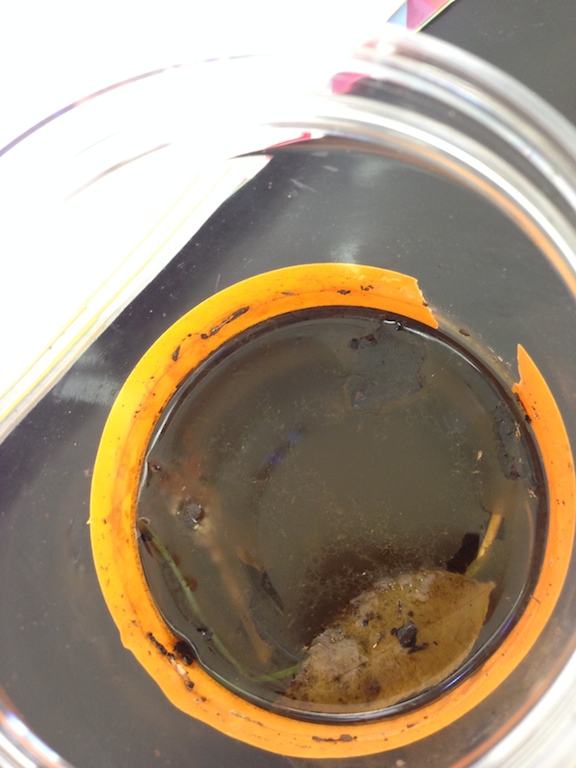
Picture of Hay Infusion Culture:
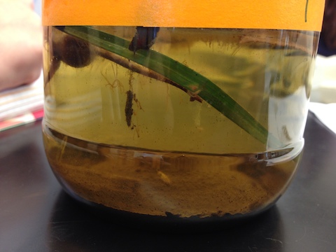
Top layer: Film on top, mold on the brown leaf, a flower bud, gray/brown opaque film Middle layer: Flower bud, piece of grass Bottom layer: Soil, Dark brown flower bud has white spider web like substance hanging from it. Misc. Observations: Grass piece is still green, the leaf is floating, there is no strong smell. Niches Observed: The bottom layer soil, and the middle layer around the piece of grass Bottom Layer: 1. Gonium (40x)- 35 micrometers, photosynthesize 2. Colpidium sp. (40x)- 55 micrometers 3. Amoeba (40x)- 35 micrometers Middle Layer: 1. Paramecium caudatum (40x)- 200 micrometers, mobile, does not photosynthesize 2. Euplotes sp.- 125 micrometers, mobile, does not photosynthesize 3. Stentor Coeruleus- 1-2mm, bacteria, mobile, does not photosynthesize 4. Endophytic Fungi- 600 mm, non-mobile, Fungus, does not photosynthesize
According to Freeman, in order for an organism to be living it has to acquire and use energy. It can be inferred that Euplotes have and energy due to the fact that they move with cilia and walks/swim with water. Another characteristic of living things is cells, and the Euplote has a oval shaped cell (Ward). Information within the cells is another characteristic of living organisms and in the Euplote, information is used to eat and make the molecules that keep it alive. Replication, the fourth out of five characteristics, happens with Euplotes. The Euplotes usually reproduce asexually, but sometimes sexual reproduction can happen via transduction (MicroscopyU). Evolution is the fifth out of five characteristics, and Euplotes are protozoa that have evolved over time.
If the hay infusion had been observed for another two months, I would predict that the living would eventually die out due to lack of nutrients in the niche. A week later the niche did not look as uniformed as it did originally when it was examined. The selective pressures that affected the compositions of my samples include the lack of nutrients in the environment in the niche. Eventually the organisms will die out due to lack of food. The organisms were taken out of their natural environment.
Source: http://www.microscopyu.com/moviegallery/pondscum/euplotes/
Conclusion: The objectives were addressed; and the protists and archeas were examined. The characteristics of the algae were determined through the dichotomous key. In the future, more kingdoms of organisms within the transect will be examined. More hands on tools can be used to convey the characteristics of the archaes and protists.
Lab 3 Objectives: To understand the characteristics of Bacteria. To observe antibiotic resistance and understand how DNA sequences are used to identify species. Slide samples of known bacteria will be examined to practice and train the eye for what is to be located. The Bacteria will also be stained to determine if one is gram positive or negative.
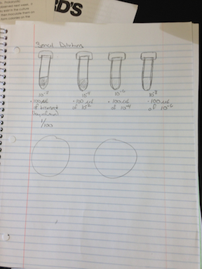
Steps: 1. Four tubes of 10 mls sterile broth were taken and labeled. 2. Four nutrient agar and four agar plus tetracycline plates were taken and labeled 10:100, 10:100,000, 1010:10,000,000, and 10:100,000,000 dilution from each of the two groups. 3. The Hay Infusion Culture was mixed, and 100 microliters was taken from the mixture, and added to the 10 mls of broth in the tube labeled 2 for 1:100 dilution 4. Took 100 microliters from tube 2 and placed into another tube for 1:10000 dilution. 5. Repeated two more times for 10:1,000,000, and 10:100,000,000 dilutions. 6. Took 100 microliters of tube 2 and placed it onto the nutrient agar plate labeled 10:1000, then carefully spreader the sample on the plate. Repeated the same procedure for the tet+ agar plate. 7. Repeated the same procedure with the number 4 tube on the 10:100,000 dilution plates, the number 6 tube on the 10:10,000,000 plates, and the number 8 tube on the 10:10,000,000,000 plates. 8. The plates were stored and sat for a week 9. The plates were taken and examined to count which ones had the most colonies of bacteria. 10. Three of the plates were selected and samples from each plate were wet mounted and examined more in depth under a microscope, looking at the cell shape and determining if there was any motility 11. Samples were then taken from each plate and heated, gram stained, then examined under a microscope at 40x to determine the cell morphology. 12. One bacteria from one tet+ plate was taken and transferred to 100 microliters of water in a sterile tube. 13. Step 12 was repeated for one bacteria from one tet- plate. 14. Both tubes were incubated at 100 Celsius fro 10 min and centrifuged. 14. 5 microliters of the supernatants were used in the PCR reaction.
Below is a drawing of the dillutions:
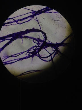
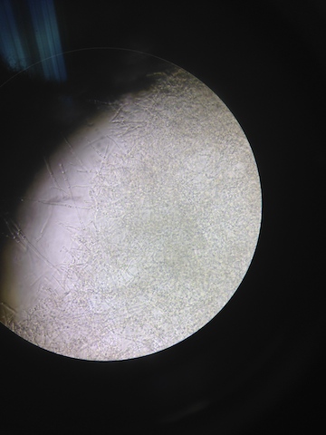
Picture of bacteria taken from the culture:
Data: Questions: Do you think any Archaea species will have grown on the agar plates? - I do not think any archaea could survive on an agar plate because it is not a suitable environment for such. Archaea live in extreme environments, like cold areas or very hot environments where bacteria cannot live. Explain why the appearance or smell might change week to week? - It might change because there will be more bacteria found on the plates, therefore an increase in their specific smell and colonies/populations.
Do you see any differences in the colony types between the plates with vs without antibiotic? - On the plates without antibiotics there are more the colonies than the plates with antibiotics. This indicates that the tetracycline did prevent growth of bacteria, and any bacteria found on the tet+ samples were resistant to the antibiotics. How many species of bacteria are unaffected by tetracycline?
- Two bacteria species appeared on the plates with tetracycline. One colony was bright orange, and the other was a clear grey with a powder like substance in the middle
According to http://www.chm.bris.ac.uk/motm/tetracycline/antimicr.htm , Tetracycline prevents the bacteria from making proteins, which are vital to a bacteria. Tetracycline can also alter the cytoplasmic membrane and cause leakages of nucleotides and other components in the cell. It affects a wide range of bacteria including richettsia, spirochetes, bronchitis etc.
The three plates examined were 10:1000 and 10:100000 nutrient plates, and the 10:1000 with tetracycline. On the 10^-3 Nutrient plate, bacteria colonies were yellow/orange, flattened with no texture, round/rod like, with flagella. There were a very colonized group of rod-shaped bacteria, compact, movement not that fast/ a lot of distance not covered, gram positive. On the 10^-5 plate the colonies were colorless and flat, rod shaped. Cell description is a streptococcus non-visible flagella and gram negative. On the 10^-3 Tet plate, the colonies were clear with powder like substance in the middle. It had a Filamentous form, Streptobacilli, non visible flagella, and gram positive.
M3-T5-1 GCTACTCTCACGAGAGTAGGTTTATCCCTATACAAAAGAAGTTTACAACCCATAGGGCCGTCGTCCTTCACGCGGGATGGCTGGATCAGGCTCTCACCCATTGTCCAATATTCCTCACTGCTGCCTCCCGTAGGAGTCTGGTCCGTGTCTCAGTACCAGTGTGGGGGATCACCCTCTCAGGCCCCCTAAAGATCGCAGACTTGGTGAGCCGTTACCTCACCAACTATCTAATCTTGCGCGTGCCCATCTCTATCCACCGGAGTTTTCAATACCGAATGATGCCATCCAGTATATTATGGGGTATTAATCTTCCTTTCGAAAGGCTATCCCCCAGATAAAGGCAGGTTGCACACGTGTTCCGCACCCGTACGCCGCTCTCAAGATTCCGAAGAATCTCTACCGCTCGG Type of Bacteria: Chryseobacterium iMTI10
AAGTCGAACGGTAGCACAGAGAGCTTGCTCTCGGGTGACGAGTGGCGGACGGGTGAGTAATGTCTGGGAAACTGCCTGATGGAGGGGGATAACTACTGGAAACGGTAGCTAATACCGCATAACGTCGCAAGACCAAAGAGGGGGACCTTCGGGCCTCTTGCCATCAGATGTGCCCAGATGGGATTAGCTAGTAGGTGGGGTAATGGCTCACCTAGGCGACGATCCCTAGCTGGTCTGAGAGGATGACCAGCCACACTGGAACTGAGACACGGTCCAGACTCCTACGGGAGGCAGCAGTGGGGAATATTGCACAATGGGCGCAAGCCTGATGCAGCCATGCCGCGTGTATGAAGAAGGCCTTCGGGTTGTAAAGTACTTTCAGCGGGGAGGAAGGTGTTG Type of bacteria: Enterobacter cloacae
GTACCTTCNGCTACCCTCACGAGGGTAGGTTTATCCCTATACAAAAGAAGTTTACAACCCATAGGGCCGTCGTCCTTCACGCGGGATGGCTGGATCAGGCTCTCACCCATTGTCCAATATTCCTCACTGCTGCCTCCCGTAGGAGTCTGGTCCGTGTCTCAGTACCAGTGTGGGGGATCACCCTCTCAGGCCCCCTAAAGATCGTTGACTTGGTGAGCCGTTACCTCACCAACTATCTAATCTTGCGCGTGCCCATCTCTATCCACCGGAGTTTTCAATTTAAAATGATGCCATTCTAAATATTATGGGGTATTAATCTCCCTTTCGAAAGGCTATCC Type of bacteria: Chryseobacterium Species: unknown.
Conclusion: The objectives were addressed. By examining the different bacteria, one can notice the different characteristics. With the tetracycline present, antibiotic resistance was present in the experiment; bacteria was on the tet+ plates. I think that all tools used were appropriate for this experiment and I would not add anything else to the experiment.
Lab 4 Objective: To understand the characteristics and diversity of plants. To appreciate the function and importance of Fungi. This will done by observing prepared slides and examples of plants and then comparing them to the five plant samples collected from the transect to understand the different types of plants.
Steps/Procedure: 1. Observed the characteristics of a Bryophyte moss, and an angiosperm. 2. Brought three bags to the transect 3. Obtained a leaf litter sample from a specific area of choice in the transect. Then found an area with soft soil and dead leaves on it. 4. Took samples from only the top "crumbly layer of the soil and plant matter above the area chosen. 5. Placed about 500g of litter into the bag (to be used for next weeks lab for invertebrates) 6. Took samples from five plants without severely damaging them, making sure they are as diverse as possible. Looked for any seeds, flowers from the plants and brought them back to the lab also. 7. Compared the moss with the stem of the angiosperm, lily. 8. Observed the moss, and noted the height of the plant. 9. Examined the cross section slide of lily stem. 10. Examined the leaves of the moss closely. 11. Studied the diagram of the bryophyte reproductive cycle, and identified the male and female gametophytes and the sporophyte. 12. Labeled the Transect Plants, including the location and number in transect, description: shape and size etc., vascularization, and if the plant had seeds or evidence of flowers
Below is a table with descriptions of the plants found in transect:
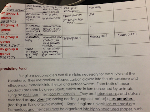
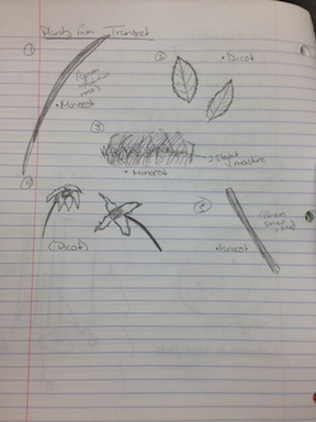
Data: Plant #1 was found under the flowering plant of the transect. It was a greenish weed, that was long and narrow. It had a monocot structure. Plant #2 was a dried leaf found from the flowering plant/bush in the transect. The leaf fell onto the soil under the bush where it was collected. The leaf was brown and short with a round oval shape. The leaf had visible veins on it, illustrating a dicot structure. Plant #3 was grass, which covered most of the transect. It was about 3 inches long and narrow. The grass was yellow-green, with a monocot structure. Plant #4 was a dead flower found on the flowering plant/bush.This flower was brown, with a brown stem. The petals of the flower were breaking with contact. There were about 4-5 petals on the flower, meaning that the flower has a dicot structure. Plant #5 were the dead roots of a bush behind the Quadrangle sign. The roots were light brown. The bottom of the roots however was grayish-white. The roots were very long and thin. The structure is monocot. Due to the absence of a body of water in the transect, all of the plants are tracheophytes, having Xylem and phloem in the stem to absorb water and nutrients underground.
Below is a drawing of the plants found:
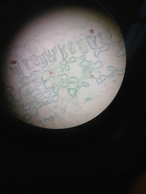
A picture showing the different structures within a plant:
Fungi sporangia are globe-like structures within the fungus that help with the reproduction/growth of the fungus. Inside of the sporangia are spores(specialized cells) which travel once the sporangia opens.
Picture of fungus found in transect:
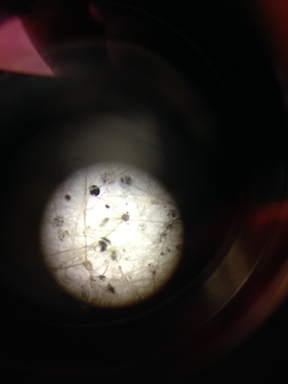
Above is a picture of fungus. The dark sphere like structures fit the description of the sporangia, and the white mass is the mycelium which is connected to the sporangium.
Conclusion: The objectives were addressed. By examining the different plants of the transect, and the prepared slides of fungus, one can notice the different characteristics of such. The proper background information was provided to indicate the different types of structures within the plants and fungus. I think that all tools used were appropriate for this experiment and I would not add anything else to the experiment.
Lab 5
Objectives: To understand the importance of invertebrates, and also learn how simple systems evolved into more complex system.
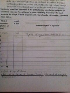
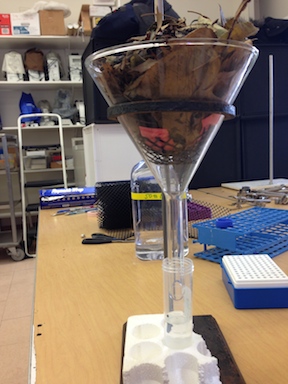
Steps/Procedure: 1. Observed the acoelomate, Planaria with the dissecting scope, then looked at a cross sectional slide of the Planaria with the microscope. 2. Observed the nematodes and a cross sectional slide of their pseudocoelomate structure. 3. Observed the coelomate, Annelida and analyzed it's structure. 4. Analyzed the invertebrates in the Berlese funnel found from the transect, and recorded the length and description of the organism
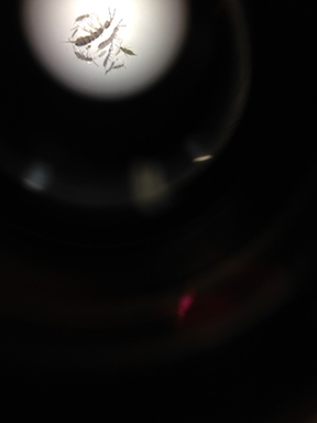
Data: The different types of prepared invertebrates were observed with the dissecting scope. These differences were not recorded due to time constraints. There were many insects of the same species found in the transect, as shown below:
These animals were 1-4 mm in length, with 3 pairs of legs, a pair of antennas, and looked similar to ant. Note: While setting up the Berlese funnel, there was a live spider found in the sample in the transect into the funnel. After a week, the spider was not located.
Lab 6
Objectives: To understand embryonic development by learning the different stages of development and comparing the stages in different organisms. To also set up an experiment to learn how environment affects embryonic development.
Steps/Procedures: Note: Due to the weather, the different organisms' development were not compared to each other. Below are the steps for the experiment on zebrafish. 1. Read and analyzed a scholarly paper on a nanoparticle dots experiment. Predicted the outcome of the experiment. 2. Observed the zebrafish embryos, and determined their developmental stage. 3. Set up the control group and the test groups in covered petri dishes. One of the petri dishes had .05 ml concentration of Quantum dots (nano particles), and another with a .5 ml concentration. 4. Use 20 mls of Deerpark water and 20 healthy translucent embryos per dish. Use a dropper to transfer the eggs to the dishes with the appropriate water. Remember if the variable tested involves an additive to the water, then every time you change water, it must have this same additive. 5. Organize the observation schedule and procedure. Make observations and carefully record those over the next two weeks in Open Wetware (see precise instructions on the next page). 6. When embryos are 4-5 days old, remove 10 mls of water with any empty egg cases and add 25 mls fresh water. (The empty egg cases will grow mold.) 7. Save any dead embryos in paraformaldehyde that will be provided. 8. One week after the experiment has begun, remove 5 mls water with any egg cases and add 5 mls fresh water or test solution. Preserve 1 to 3 embryos from the control and the experimental groups in paraformaldehyde. 9. Between 1 week and 2 weeks incubation time, remove 5 mls and add 10 mls. 10. Feed two drops of paramecium starting after one week. This can be continued just when the water is changed. 11. Made final observations and measurements at two weeks. Meaurements were completed by using anesthetics to immobilize the fish for better accuracy. 12. Remaining fish were also placed under the UV light to study the quantum dots and color of the fish.
Data: Initial amount of fish- Control: 21 fish 0.05: 20 fish 0.5: 20 fish
24 hours later- Control: 20 fish 0.05:15 fish 0.5: 17 fish
74 hours after the previous observation- Control: 16 fish- 2 taken for measurements 0.05: 13 fish- 2 taken for measurements 0.5: 15 fish- 2 taken for measurements
2 weeks from the original start date- Control: all dead fish 0.05: 1 alive fish 0.5: 2 alive fish
Measurements:
Zebrafish embryo at the beginning of the experiment: 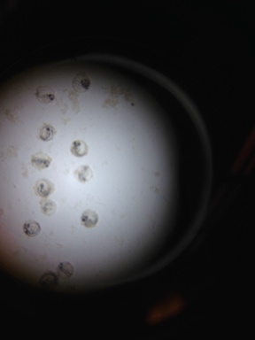
Zebrafish at 2 weeks being measured under microscope: 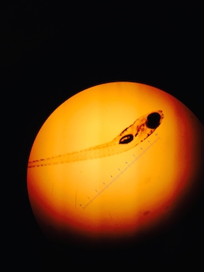
Live Zebrafish under UV light: 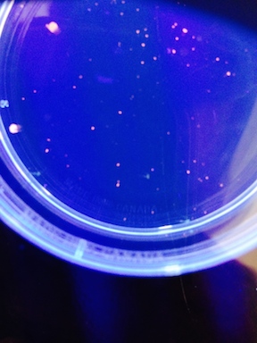
Conclusion: The objectives were addressed. By examining the development of the zebrafish, one can notice the different stages of development. The experiment showed that there were no significant effects of the quantum dots on zebrafish. I think that all tools used were appropriate for this experiment and I would not add anything else to the experiment.
