Lab 7: Vertebrate Anatomy: Difference between revisions
| Line 91: | Line 91: | ||
The urinary system (which is responsible for the excretion of urine) and the genital (reproductive) systems have little functional similarity. However, the two systems share some aspects of embryological development as well as some ducts. As a result they are often considered together. To see these structures, you will need to push aside the abdominal digestive organs, in particular the large and small intestine.<br><br> | The urinary system (which is responsible for the excretion of urine) and the genital (reproductive) systems have little functional similarity. However, the two systems share some aspects of embryological development as well as some ducts. As a result they are often considered together. To see these structures, you will need to push aside the abdominal digestive organs, in particular the large and small intestine.<br><br> | ||
'''Excretory Structures.''' The paired <u>kidneys</u> lie on either side of the vertebral column, on the dorsal wall of the abdomen. They are the site of urine production, and are supplied with blood by the renal arteries. They lie outside the body cavity (retroperitoneal) and are covered by a layer of connective tissue, which you will need to remove to see them clearly.<br> | '''Excretory Structures.''' The paired <u>kidneys</u> lie on either side of the vertebral column, on the dorsal wall of the abdomen. They are the site of urine production, and are supplied with blood by the renal arteries and drained by the renal veins. They lie outside the body cavity (retroperitoneal) and are covered by a layer of connective tissue, which you will need to remove to see them clearly.<br> | ||
Urine is transported from the kidneys by the <u>ureters</u> to the <u>urinary bladder</u> where it is stored. The ureters exit from the center of each kidney, and join the urinary bladder at its base.<br> | Urine is transported from the kidneys by the <u>ureters</u> to the <u>urinary bladder</u> where it is stored. The position of the bladder between the umbilical arteries is a good landmark to learn. The ureters exit from the center of each kidney, and join the urinary bladder at its base.<br> | ||
Urine exits from the bladder via the <u>urethra</u>. To view the next few organs you will need to remove the pubic bone; ask your instructor if you are not sure what to do. In the female the urethra lies ventral to the vagina and voids urine via the urogenital sinus to the exterior. In the male the urethra enters the penis posteriorly and voids urine via the urogenital canal and the urogenital opening at the tip of the penis. <br> | Urine exits from the bladder via the <u>urethra</u>. To view the next few organs you will need to remove the pubic bone; ask your instructor if you are not sure what to do. In the female the urethra lies ventral to the vagina and voids urine via the urogenital sinus to the exterior. In the male the urethra enters the penis posteriorly and voids urine via the urogenital canal and the urogenital opening at the tip of the penis. <br> | ||
| Line 101: | Line 101: | ||
'''Reproductive Structures.''' In the female, eggs mature in the small <u>ovaries</u>, which are located near the kidneys. Mature egg(s) enter the <u>oviducts</u> and are connected to the <u>uterus</u> by two <u>uterine horns</u>. Development of the multiple fetuses occurs in the horns rather than in the main body of the uterus formed from the union of the horns posteriorly. The <u>vagina</u> continues posteriorly from the uterus, lying dorsal to the urethra.<br><br> | '''Reproductive Structures.''' In the female, eggs mature in the small <u>ovaries</u>, which are located near the kidneys. Mature egg(s) enter the <u>oviducts</u> and are connected to the <u>uterus</u> by two <u>uterine horns</u>. Development of the multiple fetuses occurs in the horns rather than in the main body of the uterus formed from the union of the horns posteriorly. The <u>vagina</u> continues posteriorly from the uterus, lying dorsal to the urethra.<br><br> | ||
In the male, each <u>testis</u> is located within a pouch, which is enclosed in the scrotum, an exterior sac ventral to the anus. Sperm, which mature in the testis, leave via the coiled epididymus and the delicate <u>vas deferens</u> (also known as the ductus deferens). The vas deferens enters the abdominal cavity via the inguinal canal. The vas deferens from each side, then join the <u>urethra</u>, which leads to the <u>urogenital canal</u> within the <u>penis</u>. | In the male, each <u>testis</u> is located within a pouch, which is enclosed in the <u>scrotum</u>, an exterior sac ventral to the anus. Sperm, which mature in the testis, leave via the coiled epididymus and the delicate <u>vas deferens</u> (also known as the ductus deferens). The vas deferens enters the abdominal cavity via the inguinal canal. The vas deferens from each side, then join the <u>urethra</u>, which leads to the <u>urogenital canal</u> within the <u>penis</u>. | ||
<center>[[Image:111F11PigReproductive.jpg|600px]]</center><br> | <center>[[Image:111F11PigReproductive.jpg|600px]]</center><br> | ||
<center>Fig.7.2. The male (left) and female (right) reproductive system of the pig in lateral view. Image modified from: Thompson, L. H. Circular 1190 University of Illinois at Urbana-Champaign. http://www.aces.uiuc.edu/vista/html_pubs/pigs/pigs.htm#ill.</center><br> | <center>Fig.7.2. The male (left) and female (right) reproductive system of the pig in lateral view. Image modified from: Thompson, L. H. Circular 1190 University of Illinois at Urbana-Champaign. http://www.aces.uiuc.edu/vista/html_pubs/pigs/pigs.htm#ill.</center><br> | ||
Revision as of 18:13, 10 March 2015
Objectives
- To explore digestive, urogenital, circulatory and respiratory systems in a representative vertebrate, the fetal pig.
- To compare adaptations of these systems in adults of different vertebrate classes.
- To examine the adaptations of these systems during fetal development.
Lab 7 Overview
- I. Document systems anatomy in the fetal pig.
- a. Digestive system
- b. Urogenital system
- c. Circulatory system
- d. Respiratory syste
II. Compare systems anatomy in mammals with those of other vertebrate classes.
III. Contrast adaptations for fetal and adult oxygenation in mammals.
Vertebrate Anatomy: Background
The next three labs will examine the design and structure of animal anatomy through dissection, comparative anatomy, and physiological processes. The identification of anatomical structures, their function, and evidence for structural changes in organs and systems as animals diversified and adapted to different environments and habits are emphasized.
The vertebrate body can be conveniently divided into a series of organ systems, each of which contributes to a major physiological function of the body. However, all the systems interact to maintain the nearly constant environment (homeostasis) of the body. Some organs have a variety of functions and can be placed in more than one system.
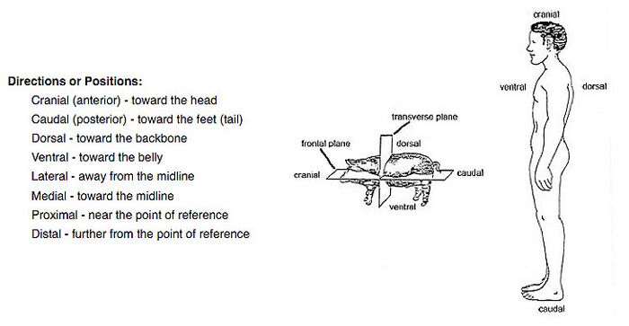
Anatomical terminology. Before beginning your exploration of vertebrate anatomy, please familiarize yourself with the following words and learn to use them in referring to the location of the body parts of your specimen.
Digestive, Urogenital, Circulatory and Respiratory Anatomy of the Fetal Pig
External Anatomy and Opening the Abdominal Cavity. A preserved pig wrapped in cheesecloth will be available for each pair of students. Three dissection manuals are available as references to aid in finding structures. DO NOT REMOVE THESE MANUALS FROM THE LABORATORY.
Bohensky, F. 1978. Photomanual and Dissection Guide of the Fetal Pig, Avery Publishing Group Inc, Wayne NJ
Smith, D. 1998. A Dissection Guide and Atlas to the fetal pig. Morton Publishing Co. Englewood, CO
Walker, W. F. 1988. Dissection of the Fetal Pig. W H Freeman and Co, New York, NY
The body of the pig is divided into three major regions: the head, neck and trunk. At the caudal (posterior) end of the trunk is the tail. Note that the trunk is divided into two regions: the thorax and the abdomen and that the limbs of the pig are directed ventrally. Determine the sex of the pig by examining the external genitalia, see p. 9 in Bohensky (1978).
To open the abdominal cavity and the posterior part of the thoracic cavity, begin your incision at the level of the forelimbs and make a vertical cut down to the level of the umbilicus. Initially cut only the skin and leave the abdominal muscles intact. When you reach the umbilicus, cut around it. Stop at the hind limbs and then make two horizontal cuts: one below the forelimbs and the other above the hind limbs, see p. 61 in Bohensky (1978).
Following the same incision lines made for the skin, cut through the muscle layers to expose the contents of the abdomen. In order to access the abdominal cavity you will need to transect (sever) the umbilical vein; make the cut midway so you can easily find both ends of the vein later. Also break the connection of the diaphragm with the ventral body wall. Pin down the lateral flaps to examine the contents of the abdomen.
The Digestive System of the Fetal Pig (see pp 31-41 in Smith, 1998).
You may need to drain and/or rinse extraneous fluids from the abdominal cavity.
In the abdominal cavity the liver is the large, dark organ that dominates the anterior abdomen. It is divided into five lobes, and is the primary site of conversion of glucose into glycogen (a carbohydrate storage molecule) and the reverse of this process, the breakdown of glycogen into glucose. It is also the site of the synthesis of bile, a product that aids in the digestion of fats by emulsifying them. Bile is stored in the gall bladder, a sac located beneath the right central lobe of the liver, from which it exits via a series of ducts to the small intestine.
The stomach lies on the left side of the upper abdomen. Food enters the mouth, travels through the pharynx, and when swallowed enters the esophagus. The esophagus is an easily compressible tube that lies immediately dorsal to the trachea. The esophagus traverses the thoracic cavity, and then passes through an opening in the diaphragm to enter the abdominal cavity. You will find these organs later in the dissection.
The esophagus transports food to the stomach, which is a major site of protein digestion. Caudally, the stomach empties the semi-digested chyme into the small intestine. The most anterior part of the small intestine, the duodenum, receives bile synthesized by the liver and digestive enzymes from the pancreas. The pancreas is a lobulated structure somewhat lighter in color than the neighboring intestines. The main body of the pancreas lies in the loop of the duodenum. Carbohydrates, fats, and proteins are all digested in the small intestine. Although the long narrow spleen is not a digestive organ, its position, wedged between the stomach and the diaphragm, make discussion of it appropriate. In the pig and many other mammals, the spleen is quite muscular and can eject large quantities of blood into the circulation to correct sudden blood loss. The human spleen is not muscular so it cannot contract nor store a large quantity of blood. It functions as an important part of the immune system, but is not an essential organ, meaning humans can live without one if it must be removed due to trauma or disease.
Follow the course of the small intestine, which is supported by a membrane, the mesentery. The end of the small intestine joins the large intestine (colon), which is noticeably larger in cross section. At the juncture a short blind sac, the caecum, is formed. The large intestine is a site of water re-absorption. Follow its course through the posterior abdomen to its terminal portion, the rectum. You will not be able to see the rectum until you remove the pubic bone later in the dissection. Wastes leave the digestive system via the anus directly below the rectum.
The Urogenital System of the Fetal Pig (see pp. 71-78 in Smith, 1998)
The urinary system (which is responsible for the excretion of urine) and the genital (reproductive) systems have little functional similarity. However, the two systems share some aspects of embryological development as well as some ducts. As a result they are often considered together. To see these structures, you will need to push aside the abdominal digestive organs, in particular the large and small intestine.
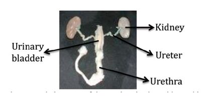
Excretory Structures. The paired kidneys lie on either side of the vertebral column, on the dorsal wall of the abdomen. They are the site of urine production, and are supplied with blood by the renal arteries and drained by the renal veins. They lie outside the body cavity (retroperitoneal) and are covered by a layer of connective tissue, which you will need to remove to see them clearly.
Urine is transported from the kidneys by the ureters to the urinary bladder where it is stored. The position of the bladder between the umbilical arteries is a good landmark to learn. The ureters exit from the center of each kidney, and join the urinary bladder at its base.
Urine exits from the bladder via the urethra. To view the next few organs you will need to remove the pubic bone; ask your instructor if you are not sure what to do. In the female the urethra lies ventral to the vagina and voids urine via the urogenital sinus to the exterior. In the male the urethra enters the penis posteriorly and voids urine via the urogenital canal and the urogenital opening at the tip of the penis.
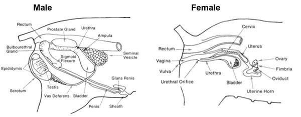
Reproductive Structures. In the female, eggs mature in the small ovaries, which are located near the kidneys. Mature egg(s) enter the oviducts and are connected to the uterus by two uterine horns. Development of the multiple fetuses occurs in the horns rather than in the main body of the uterus formed from the union of the horns posteriorly. The vagina continues posteriorly from the uterus, lying dorsal to the urethra.
In the male, each testis is located within a pouch, which is enclosed in the scrotum, an exterior sac ventral to the anus. Sperm, which mature in the testis, leave via the coiled epididymus and the delicate vas deferens (also known as the ductus deferens). The vas deferens enters the abdominal cavity via the inguinal canal. The vas deferens from each side, then join the urethra, which leads to the urogenital canal within the penis.
The Respiratory System of the Fetal Pig (see pp 65-67 in Smith, 1998).
Look at the diaphragm, the musculotendinous sheet that separates the thoracic cavity, containing the heart and lungs, from the abdominal cavity that contains the abdominal viscera. There are three tubes that pass through the diaphragm. Once again, find the esophagus, and then identify the other two: the posterior vena cava and the dorsal aorta.
Cut through the ribs close to the upper limbs, with your scissors. Cut across the distal end of the ribs, and through the clavicles and remove parts of the ribs that can readily be cut out. Don't remove the sternum connecting the two sets of ribs in the midline lying above the top of the heart since there are blood vessels right below it that we want to preserve. Look at the position of the heart within the thorax and note its relationship to the lung.
Cut in a cranial direction up to the tuft of hairs under the chin and locate the larynx (voice box). You will need to remove some thymus gland, which is very extensive in young animals, to see it clearly. Note the tough cartilaginous larynx. The trachea (wind pipe) is a tube extending from the larynx into the thoracic cavity. The air passage is kept open by C-shaped rings of cartilage along the entire length of the trachea. Note that the rings do not completely encircle the trachea, but are incomplete on the dorsal surface. Note the dark brown thyroid gland on the ventral surface of the trachea. The trachea branches to form two bronchi (you won't see these since they can only be seen if you remove the heart).
Look at the lung and observe its division into several lobes. Remember that fetal pigs have not used their lungs to breathe and that there is no air present in the alveoli (air sacs in the lungs). Look at the lung in relation to the thoracic cage. In an adult this space would be totally occupied by the inflated lungs and heart.
The Circulatory System of the Fetal Pig (see pp. 43-60 in Smith, 1998)
Heart. Carefully cut the through the pericardial sac and look at the heart and the large vessels leaving the heart. The major features of the vertebrate heart will be identified on the larger calf heart in Lab 8. Observe these features as far as possible on the smaller fetal pig heart today.
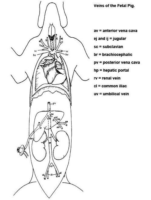
Veins. Identify the following veins: If needed gradually and carefully cut away at the remaining sternum and remove the thymus gland and the pericardial sac.
Link to a downloadable version of Fig. 7.3 labeled: File:Pig veins.docx
anterior (superior or cranial) vena cava -major vein draining anterior portion of body
brachiocephalic - formed from the union of:
subclavian - from the forelimbs
external and internal jugulars - from the head
posterior (inferior or caudal) vena cava - major vein draining posterior portion of body
hepatic portal veins - from the digestive organs to the liver
renal vein – drains kidney
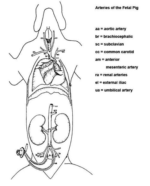
Arteries. To find the arteries at the anterior end of the heart, you will likely have to remove the overlying veins. Also if needed gradually and carefully cut away at the remaining sternum and remove the thymus gland and the pericardial sac.
Link to a downloadable version of Fig. 7.4 labeled: File:Pig arteries.docx
Identify the following arteries:
pulmonary artery - brings blood to the lungs
aorta - the largest systemic artery
brachiocephalic (innominate) - divides into:
right subclavian - to the right forearm
right and left common carotids - to the head
left subclavian - to the left forearm anterior mesenteric - to the small intestine renal - to the kidneys external iliac - to the hindlimbs umbilical - to the placenta
Circulatory System Adaptations for Oxygenation in the Fetus
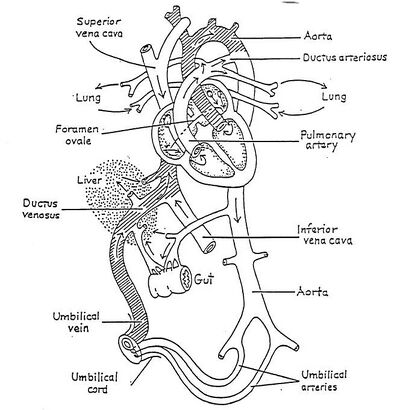
While a fetus is in utero the exchange of oxygen, nutrients,and waste products occurs across the placenta. Because the digestive tract, lungs and kidneys of the fetus will not begin to function until after birth, the fetal circulation has been modified to adjust blood flow to these organs. Oxygenated blood is brought to the fetal heart through the umbilical cord via the umbilical vein, and then the inferior vena cava.
Follow the umbilical vein, transected earlier in the dissection, as it seems to disappear in the liver. In Figure 7.5 notice how the umbilical vein divides into two branches before reaching the liver. One branch heads to the liver while the other (called the ductus venosis) shunts about 50% of the oxygenated blood directly to the inferior/anterior vena cava and thus to the fetal heart. The blood entering the liver is eventually directed to the vena cava through the hepatic veins. After birth the umbilical vein and umbilical arteries atrophy and the ductus venosus gradually fills with connective tissue and closes (ligamentum venosum). This is one of three modifications to the fetal circulatory system.
Because the lungs are not functioning in the fetus, two modifications to the circulation are found within the heart: The ductus arteriosus and the foramen ovale. We will look at these two modifications more closely when dissecting the calf heart.
Link to a downloadable version of Fig. 7.5 labeled: File:FetalPigCirculation.docx
Comparative Adult Anatomy of Systems in Vertebrates
A central argument for the common ancestry of vertebrates is the presence of the same body systems in all subgroups. While the body systems do have extensive similarities, they also show adaptations to various habitats over evolutionary time.
Dissections of four vertebrates have been prepared before class for comparative study. These vertebrates are: Perch, genus Perca - a member of the Class Osteichthyes (bony fish) Mudpuppy, genus Necturus - a member of the Class Amphibia Rabbit, genus Sylvilagus - a member of the Class Mammalia Rattlesnake, genus Crotalus - a member of the Class Reptilia Each group of four should choose one of the four systems below and consider the questions posed as you compare the anatomy of the various specimens. Be prepared to briefly present your ideas and discoveries to the rest of the class before you leave today. You may not know the most accurate answer to these questions right now and that is okay! We will discuss the answers together.
Digestive System - labeled by the Orange flags
Using the numbered key and the flags, compare the following structures in all of the specimens, unless otherwise noted, and answer the questions below.
1. Teeth
2. Esophagus (mudpuppy, rattlesnake and rabbit only)
3. Stomach
4. Caecum (rabbit only)
5. Small intestine
6. Large intestine
7. Cloaca (perch, mudpuppy, and rattlesnake - Note that these animals have an anal opening within the cloaca.)
8. Anus (rabbit)
9. Liver
Observe the skulls and teeth of the animals. What role do the teeth or jaw play in feeding? Do any of the organisms with teeth suggest specific teeth functions such as grinding, tearing, chewing, cutting etc?
As you compare the digestive tract, what do you notice about the number or placement of the organs involved in digestion for each animal
Note the large relative size/length of the esophagus of the snake compared to the other animals. Why might these animals need a large elastic esophagus?
Note the large sac at the beginning of the large intestine of the rabbit. It is called the caecum. Do you see a similar structure in the other vertebrates available for comparison, including your fetal pig? What is the main difference in diet between these animals and the rabbit? Suggest a likely function for the larger caecum in the rabbit.
Note the clear, membranous balloon-like structure in the body cavity of the perch. It is not pinned since it would break if a sharp structure is stuck into it. This structure is called the swim bladder, and it is formed by an outpocketing of the digestive system. However, it does not serve a role in the digestive tract. What do you think is the function of the swim bladder? (Hint: Think of the environment in which you would find a perch!)
Overall, how do you think length/complexity of the digestive tract is related to diet?
Respiratory System - labeled by the white flags in each of the specimens
Using the key below, compare the flagged structures in the pig, Perch, Necturus, rattlesnake, and rabbit and answer the questions below.
1. Gills (perch and Mudpuppy)
2. Lungs (mudpuppy, rattlesnake and rabbit)
3. Trachea (mudpuppy, rattlesnake and rabbit)
4. Diaphragm (rabbit only)
Observe the gills and/or lungs in the specimens. What traits are typical of respiratory structures (gills/lungs) in general? For example, do they appear to have thick membranes? Do they have many blood vessels running over/through them? What medium (water, air, or both) is used by the organism to obtain oxygen?
How does each animal make contact between the respiratory medium (air/water or both) and their respiratory structures? Identify the specific parts of the respiratory system that participate in gas exchange between the external environment and the circulatory system?
Compare the lungs of the snake to the lungs of the rabbit. Are both lobes of the lung of equal relative size in both organisms? Observe the area immediately under the lungs in the rabbit and you will see the muscular diaphragm similar to the one found in the fetal pig. Movement of this muscle helps to take in air during inhalation and expire air from the lungs during exhalation. Does the snake have a similar structure? If not, why do you think this structure is not present and how does the snake inhale and exhale?
What structure(s) in addition to gills and lungs might be considered gas exchange tissues? Do you think any of the organisms might use such alternative methods of respiration?
Urogenital System - labeled with Yellow pins (flags) in each of the specimens
Using the numbered key below compare the following flagged structures in all of the specimens, unless otherwise noted, and answer the questions below. We have mostly female specimens so we will focus on the female urogenital system.
1. Ovary (perch, mudpuppy, rattlesnake)
1a. Testicle (perch, rabbit)
2. Oviduct (Necturus, rattlesnake, [present but not visible in the perch])
2a. Ductus deferens (rabbit present but not visible in the perch)
3. Epididymis (rabbit)
4. Penis (rabbit)
5. Urinary bladder (perch, Necturus, rabbit)
6. Urethra (rabbit) or urogenital opening (perch)
7. Cloaca (Necturus, rattlesnake)
8. Kidney (one of two pinned in the perch, Necturus, rattlesnake, rabbit)
Reproductive system: Most snakes lay eggs but pit vipers, including the rattlesnake, keep the eggs inside the body releasing live young. How might egg laying compared to bearing live young be reflected in the reproductive system? If animals bear live young, where are they "housed" while they undergo embryological development? What is the source of nutrition during development for each of the organisms? How is mode of fertilization tied to the site of embryological development? Is the number of offspring affected by the method of reproduction?
Urinary system: Organisms must remove nitrogenous wastes in the form of uric acid (solid), ammonia (gas), or urea (liquid). Humans and other mammals excrete urea, a non-toxic waste product, while fish secrete ammonia and many reptiles and birds excrete uric acid. Follow the path of liquid or solid waste production (kidney to urinary bladder (if present) to the urethra or cloaca). What is the difference between a urogenital canal and a cloaca?
Circulatory System - labeled with Red pins in all of the specimens. Please also see the diagrams of blood flow to and from the heart.
Compare the following structures in all of the specimens and answer the questions below.
1. Single atrium (Perch)
2. Single ventricle (Perch, mudpuppy, rattlesnake)
3. Left atrium (mudpuppy, rattlesnake, rabbit)
4. Right atrium (mudpuppy, rattlesnake, rabbit)
5. Left ventricle (rabbit)
6. Right ventricle (rabbit)
7. Aortic arch (rabbit)
8. Anterior vena cava (rabbit)
9. Posterior vena cava (rabbit)
Observe the diagrams that display the path of blood flow in each of the specimens. How many chambers of the heart (atria and ventricles) does each animal possess? What can you suggest about the functional implications of division of the heart into chambers?
In each organism, compare and contrast blood flow from the heart to the respiratory structures? How does the presence of gills or lungs alter the circulation patterns? Does the oxygenated blood re-enter the heart after entering the blood vessels of the respiratory structures? How is blood pumped to the rest of the body? What might this mean in terms of efficiency of the circulatory system?
Assignment
1. Material in this lab will be part of the Lab Practical. Your lab instructor will describe the format of the lab practical, which will require detailed identifications and functions. Make sure that you can find and identify each of the structures underlined in Lab 7, learn their function(s) and understand the adaptations in the comparative organisms. Follow the instructions of your instructor for organizing the comparative anatomy material.
3. Complete Pre-Lab 8 quiz.
Other Labs in This Section
Lab 8: Vertebrate Circulation and Respiration
Lab 9: Conduction Velocity of Nerves
