Uploads by Ruixin Zhu
From OpenWetWare
Jump to navigationJump to search
This special page shows all uploaded files.
| Date | Name | Thumbnail | Size | Description |
|---|---|---|---|---|
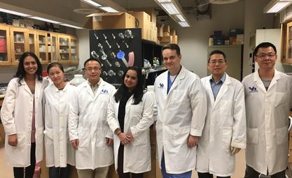
| 03:41, 14 February 2017 | GILab2016.jpg (file) |  |
34 KB | |
| 03:29, 14 February 2017 | Tavleen Bhatia.jpg (file) |  |
36 KB | Tavleen Bhatia |
| 03:29, 14 February 2017 | Shelly Choudhury.jpg (file) |  |
27 KB | Shelly Choudhury |
| 03:29, 14 February 2017 | Sebastian Zavoian.jpg (file) |  |
25 KB | Sebastian Zavoian |
| 03:29, 14 February 2017 | Reham Abdou.jpg (file) |  |
7 KB | Reham Abdou |
| 03:10, 14 February 2017 | Shelly Choudhury.jpg.jpg (file) |  |
26 KB | Shelly Choudhury |
| 03:10, 14 February 2017 | Sebastian Zavoian.jpg.jpg (file) |  |
25 KB | Sebastian Zavoian |
| 03:10, 14 February 2017 | Reham Abdou.jpg.png (file) |  |
54 KB | Reham Abdou |
| 03:10, 14 February 2017 | Tavleen Bhatia.jpg.jpg (file) |  |
35 KB | Tavleen Bhatia |
| 10:57, 2 May 2014 | Saritasinghal.jpg (file) |  |
6 KB | |
| 10:55, 2 May 2014 | MAlkhatib.jpg (file) |  |
6 KB | |
| 10:55, 2 May 2014 | CNugent.jpg (file) |  |
8 KB | |
| 10:54, 2 May 2014 | AdrianChapa.jpg (file) |  |
7 KB | |
| 07:38, 6 August 2012 | RicardoArbizu.jpg (file) |  |
7 KB | |
| 07:38, 6 August 2012 | Aljomah.jpg (file) |  |
8 KB | |
| 04:28, 1 August 2011 | ZebunnissaMemon.jpg (file) |  |
7 KB | |
| 04:27, 1 August 2011 | DianaAlexandraMoya.jpg (file) |  |
6 KB | |
| 00:49, 19 October 2010 | Sonal Desai.jpg (file) |  |
23 KB | Sonal_Desai |
| 00:48, 19 October 2010 | Razan H Alkhouri.jpg (file) |  |
22 KB | Razan_H_Alkhouri |
| 02:46, 2 March 2010 | Patelraza.jpg (file) |  |
24 KB | Raza Patel, M.D., |
| 01:22, 2 March 2010 | Lixinzhu.jpg (file) |  |
20 KB | Dr. Lixin ZHU Assistant professor, Director of GI Lab, SUNY Buffalo |
| 01:16, 2 March 2010 | Wenshengliu.jpg (file) |  |
22 KB | Dr. Wensheng Liu at Lixin's Lab |
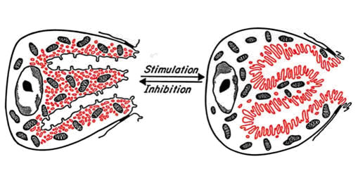
| 01:03, 2 March 2010 | Zhulab-resfig1.jpg (file) |  |
72 KB | Schematic representation of the parietal cell in resting and stimulated states. Drastic morphological change occurs with stimulation. In the resting state the apical canaliculi extend into the cell presenting short microvilli. Tubulovesicles containing ca |
| 02:55, 1 December 2009 | DSCF3817.png (file) |  |
758 KB |
