Uploads by Reuben Cutfield
From OpenWetWare
Jump to navigationJump to search
This special page shows all uploaded files.
| Date | Name | Thumbnail | Size | Description |
|---|---|---|---|---|
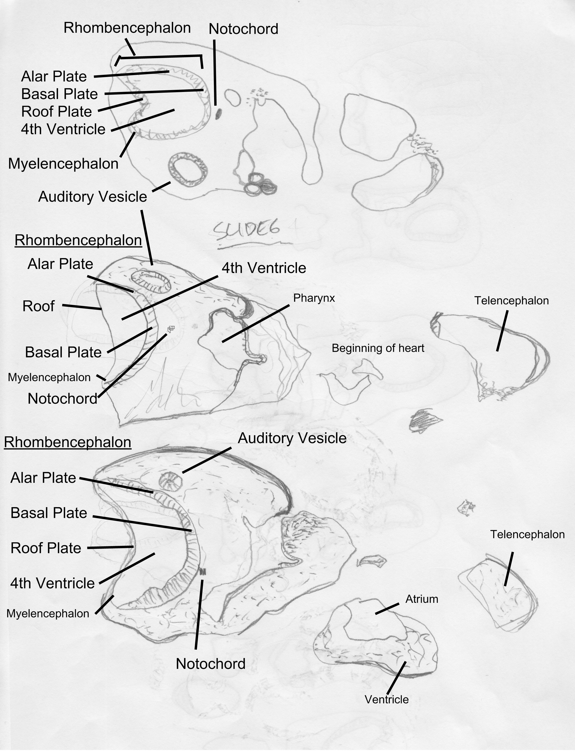
| 22:18, 20 February 2011 | MH008 section from slide6 labeled.jpg (file) |  |
780 KB | These diagrams were drawn from embryo MH008. The labels are part of a training exercise to label the different features of the developing embryo and may not be accurate. |
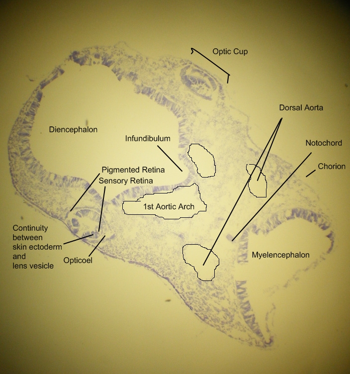
| 02:10, 9 February 2011 | MH008 labeled section (practice).jpg (file) | .jpg) |
347 KB | These may not be correct annotations, practice embryo only. |
| 01:32, 7 February 2011 | MH007 in trimmed block.JPG (file) |  |
584 KB | |
| 01:31, 7 February 2011 | MH007 in block ready to cut.JPG (file) |  |
590 KB | |
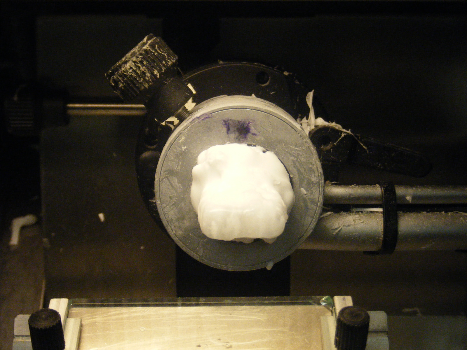
| 01:30, 7 February 2011 | MH007 in mould large.JPG (file) |  |
638 KB | |
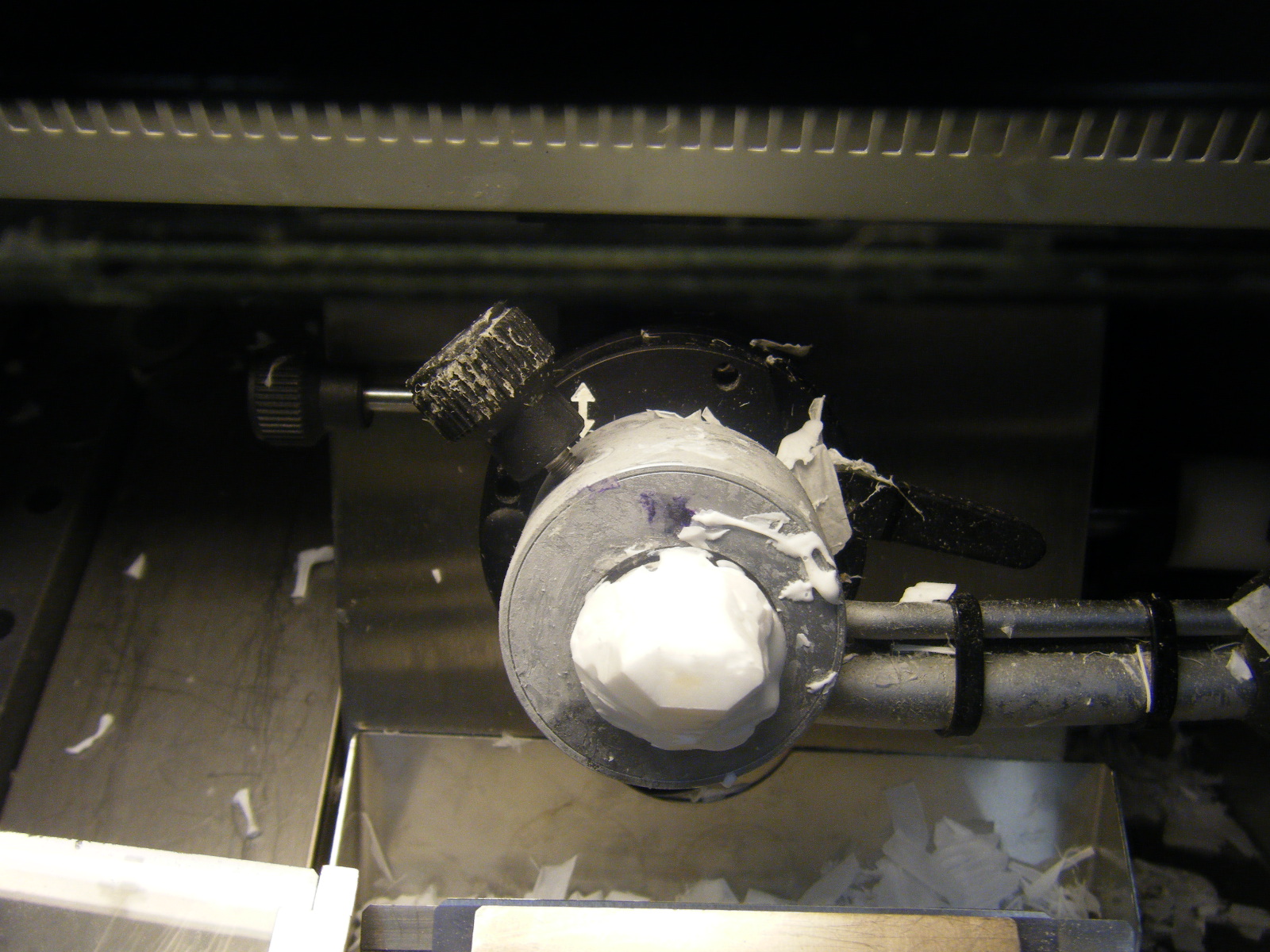
| 01:27, 7 February 2011 | MH007 in mould.JPG (file) |  |
527 KB | |
| 01:25, 7 February 2011 | MH007 Cut at wingbud.JPG (file) |  |
615 KB | |
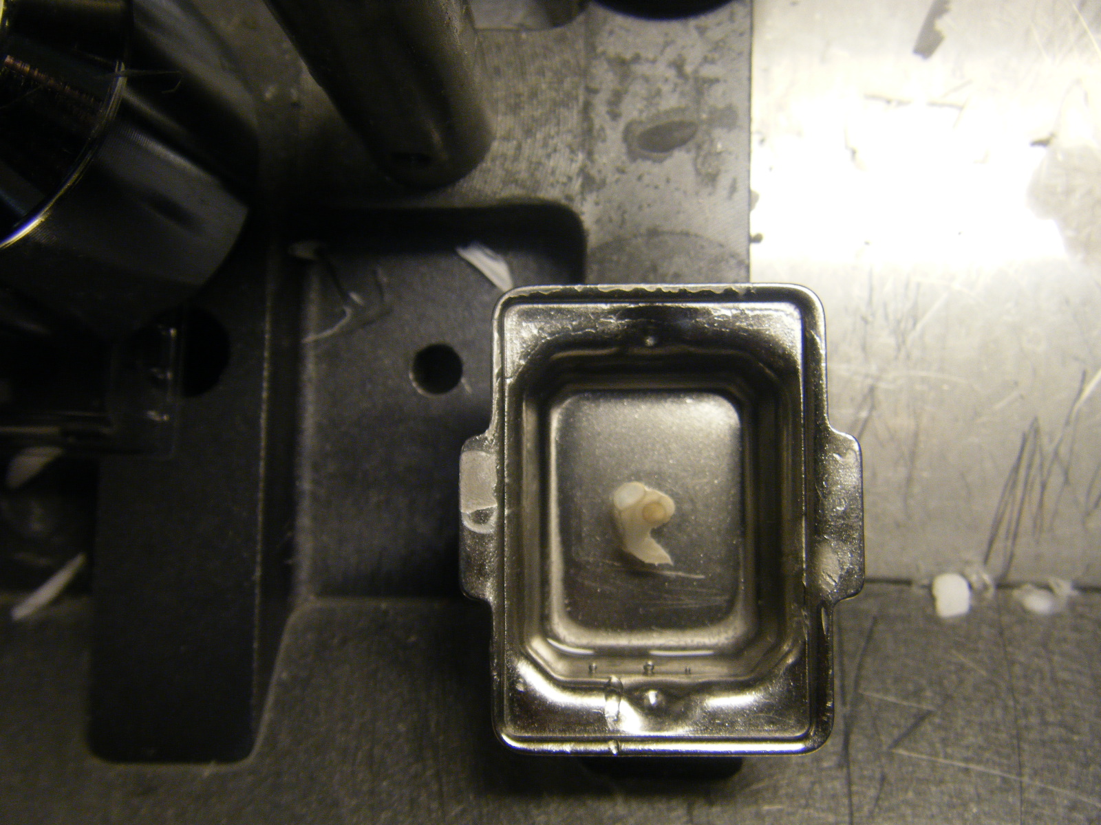
| 01:24, 7 February 2011 | MH007 ST25.JPG (file) |  |
541 KB | |
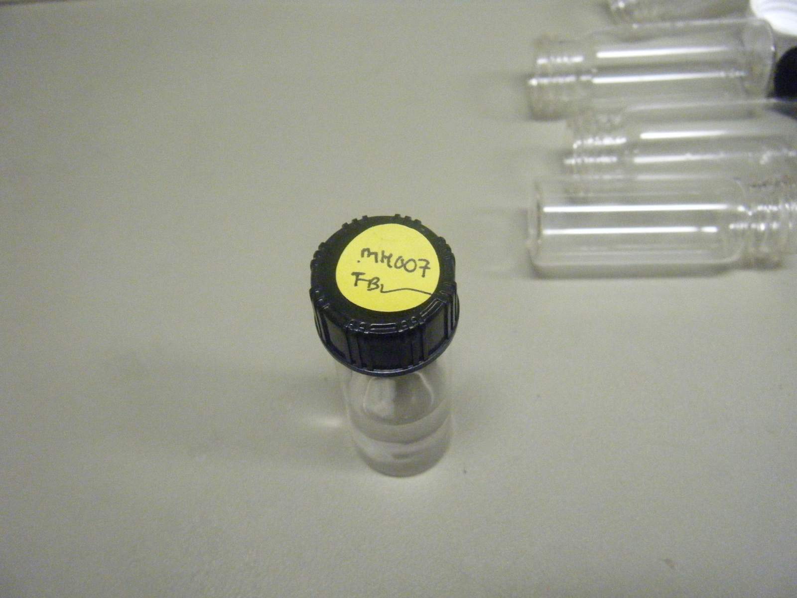
| 01:18, 7 February 2011 | MH007 Fixed and Stored in a Vial.JPG (file) |  |
605 KB | |
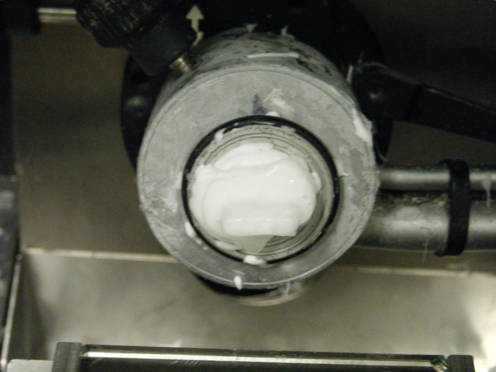
| 03:56, 21 January 2011 | Embryo RC015(20+) embedded in OCT and incubating in the cryostat.jpg (file) | _embedded_in_OCT_and_incubating_in_the_cryostat.jpg) |
623 KB | Embryo RC015(20+) embedded in OCT and incubating in the cryostat's metallic chuck holder. |
| 03:53, 21 January 2011 | Embryo RC015 submerged in OCT aligned in plastic mould.jpg (file) |  |
600 KB | Chick embryo RC015 (ST20+) submerged in OCT aligned in plastic mould prior to freezing.jpg |
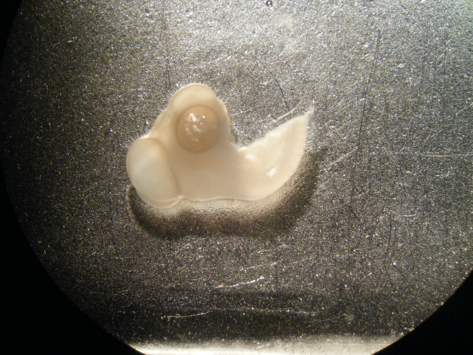
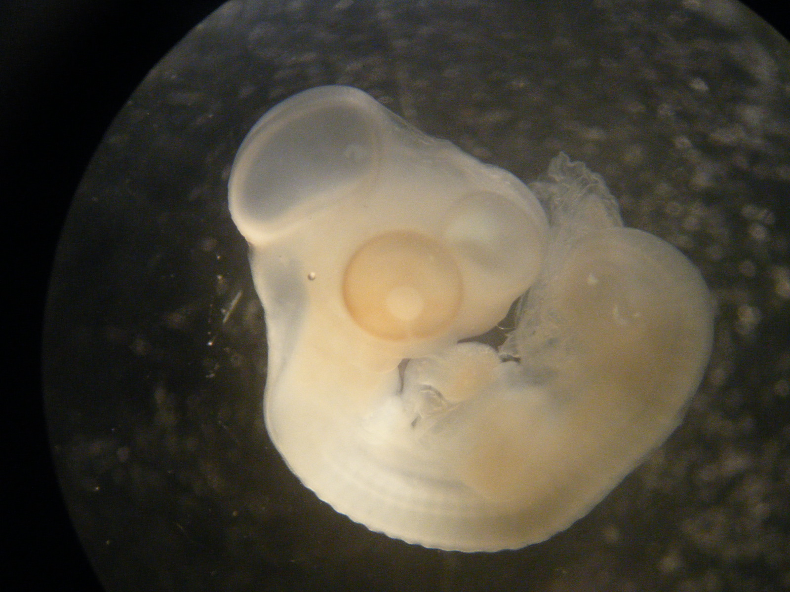
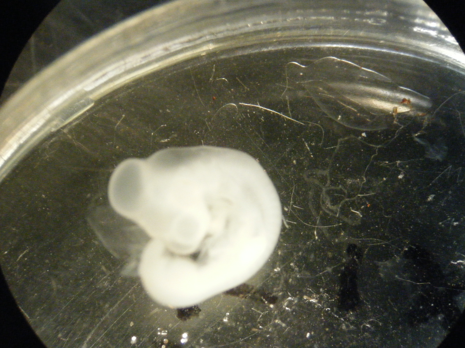
| 03:51, 21 January 2011 | RC015 whole embryo.jpg (file) |  |
552 KB | This is a photograph taken of the chick embryo RC015(ST20+) under a dissection microscope. |
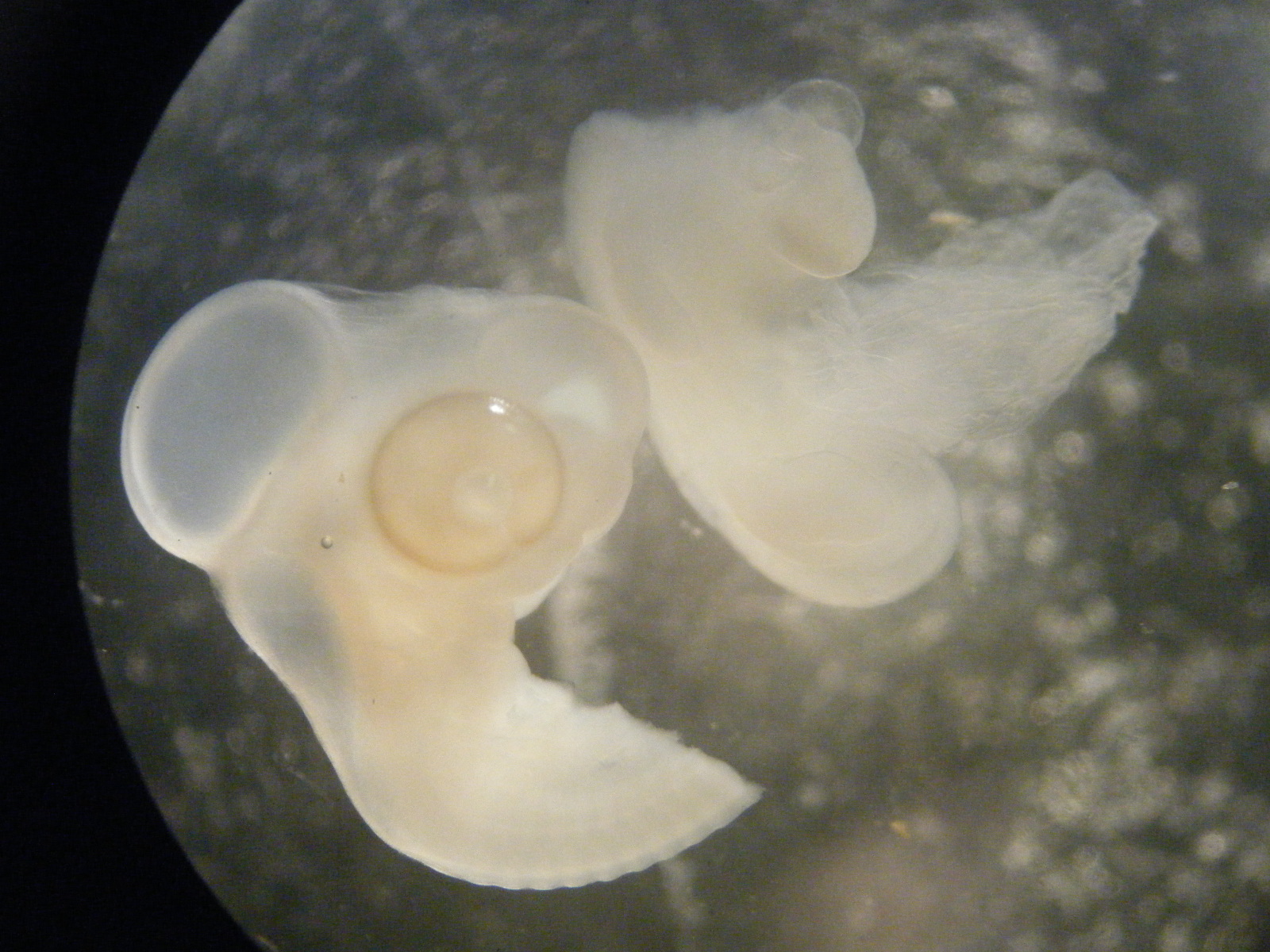
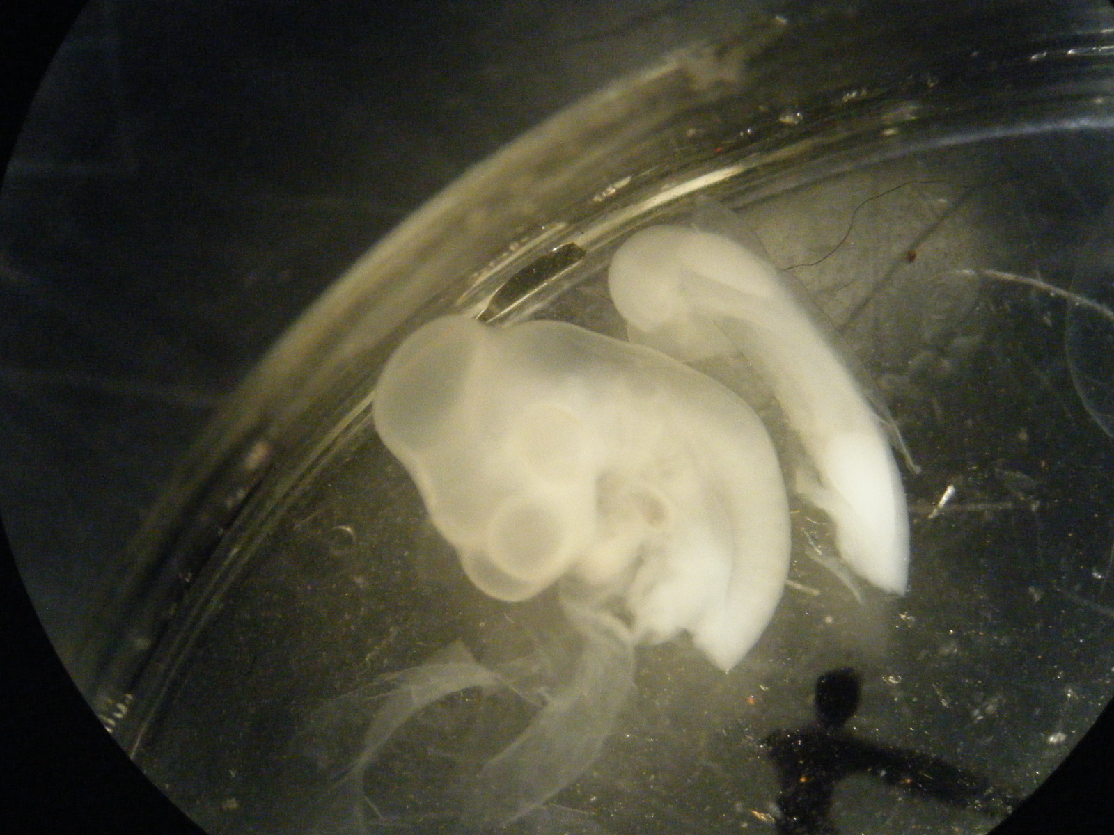
| 03:00, 21 January 2011 | RC015 following cutting at wing bud.jpg (file) |  |
635 KB | This is a photograph taken using a dissection microscope. It shows a ST20+ chick embryo which has been cut coronally at the level of the wing bud. The tail region is seen to the right of the head region. *'''~~~~''': |
| 00:42, 20 November 2010 | Oldlabphoto.JPG (file) |  |
1.8 MB |
