Uploads by Peter M. Carlton
From OpenWetWare
Jump to navigationJump to search
This special page shows all uploaded files.
| Date | Name | Thumbnail | Size | Description |
|---|---|---|---|---|
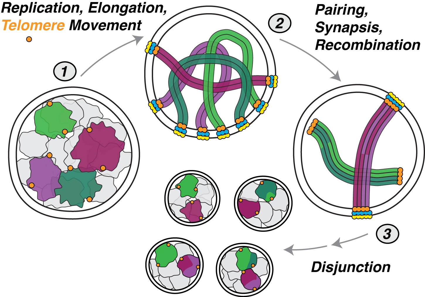
| 02:16, 11 December 2009 | Carlton About meiosis.png (file) |  |
307 KB | An overview of the changes in chromosomes that occur during meiotic prophase. |
| 23:26, 3 December 2009 | Carltonlab-logo-1.png (file) | 2.99 MB | The Carlton lab at iCeMS, Kyoto University | |
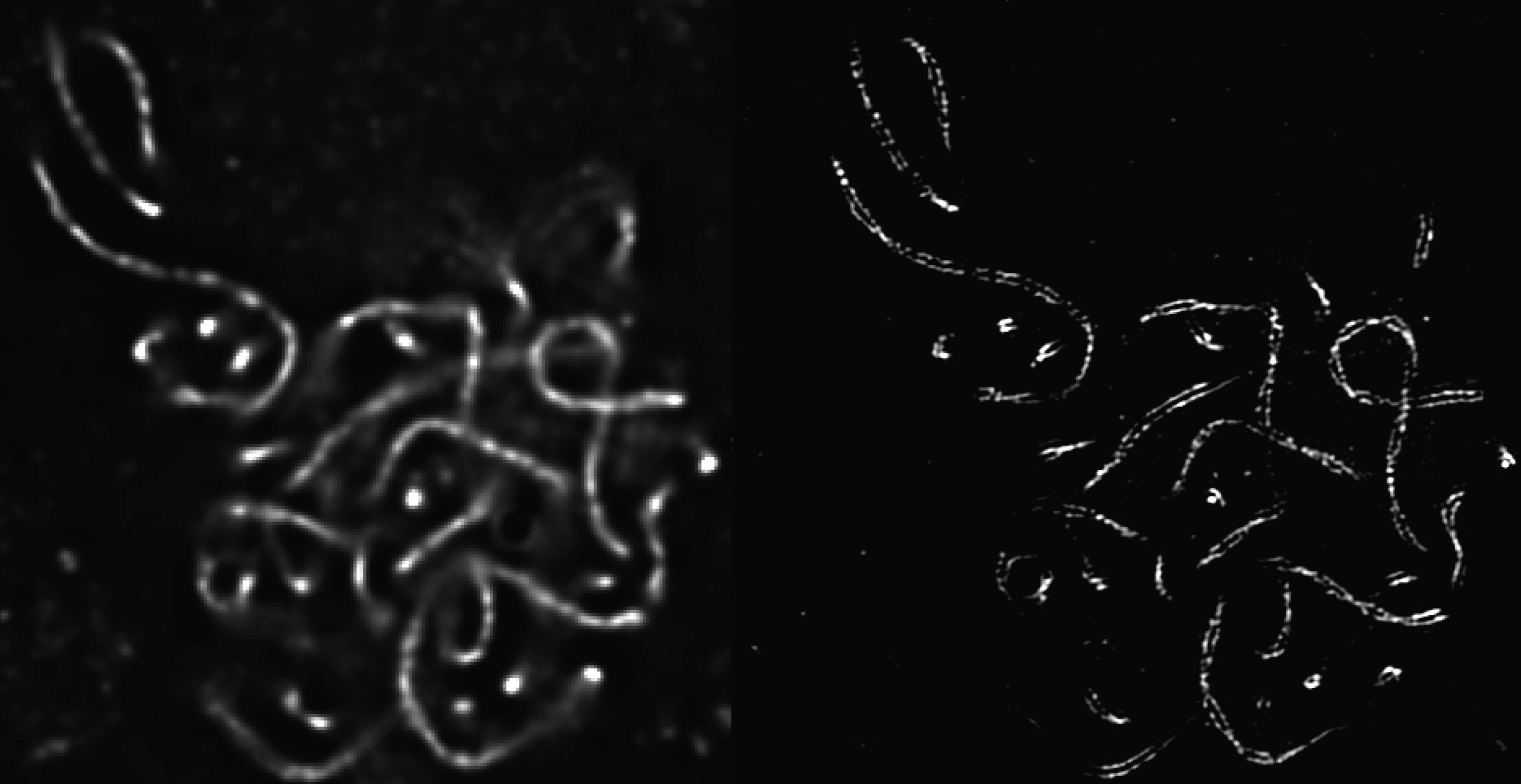
| 19:20, 3 December 2009 | MaizeSCs.png (file) |  |
457 KB | The resolution doubling of 3D-SIM is shown on an optical section of a corn (Zea mays) meiotic nucleus. The synaptonemal complexes are stained with antibodies against AFD1, a homolog of the meiosis-specific sister chromatid cohesion protein Rec8. At a sepa |
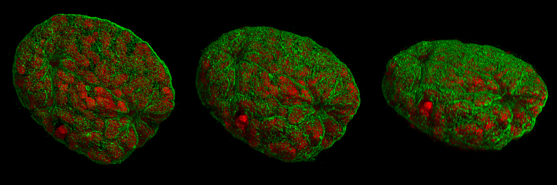
| 18:20, 3 December 2009 | 3D-SIM-2 Nucleus prophase 3d rotated.jpg (file) |  |
377 KB | Rotated projection views of a mouse nucleus at prophase, recorded with 3D-SIM. The chromosomes, stained with DAPI, are shown in red; the nuclear lamina is shown in green. Protrusions of the nascent spindle into the lamina are visible at both poles. |
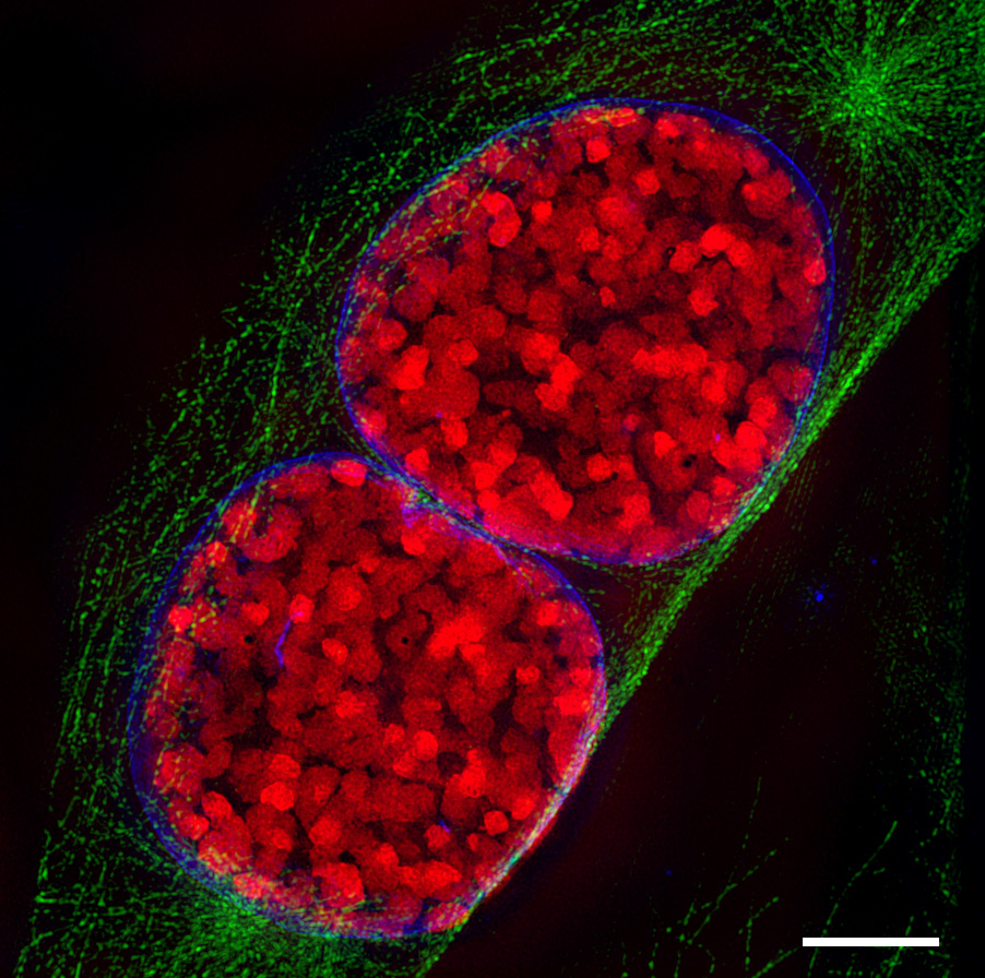
| 07:46, 3 December 2009 | 3D-SIM-3 Prophase 3 color.jpg (file) |  |
386 KB | Light-optical section through two mouse cell nuclei in prophase, recorded with 3D Structured Illumination Microscopy (3D-SIM-microscopy). condensed chromosomes are red, the nuclear envelope blue and microtubuli, which belong to the cytoskeleton, are green |
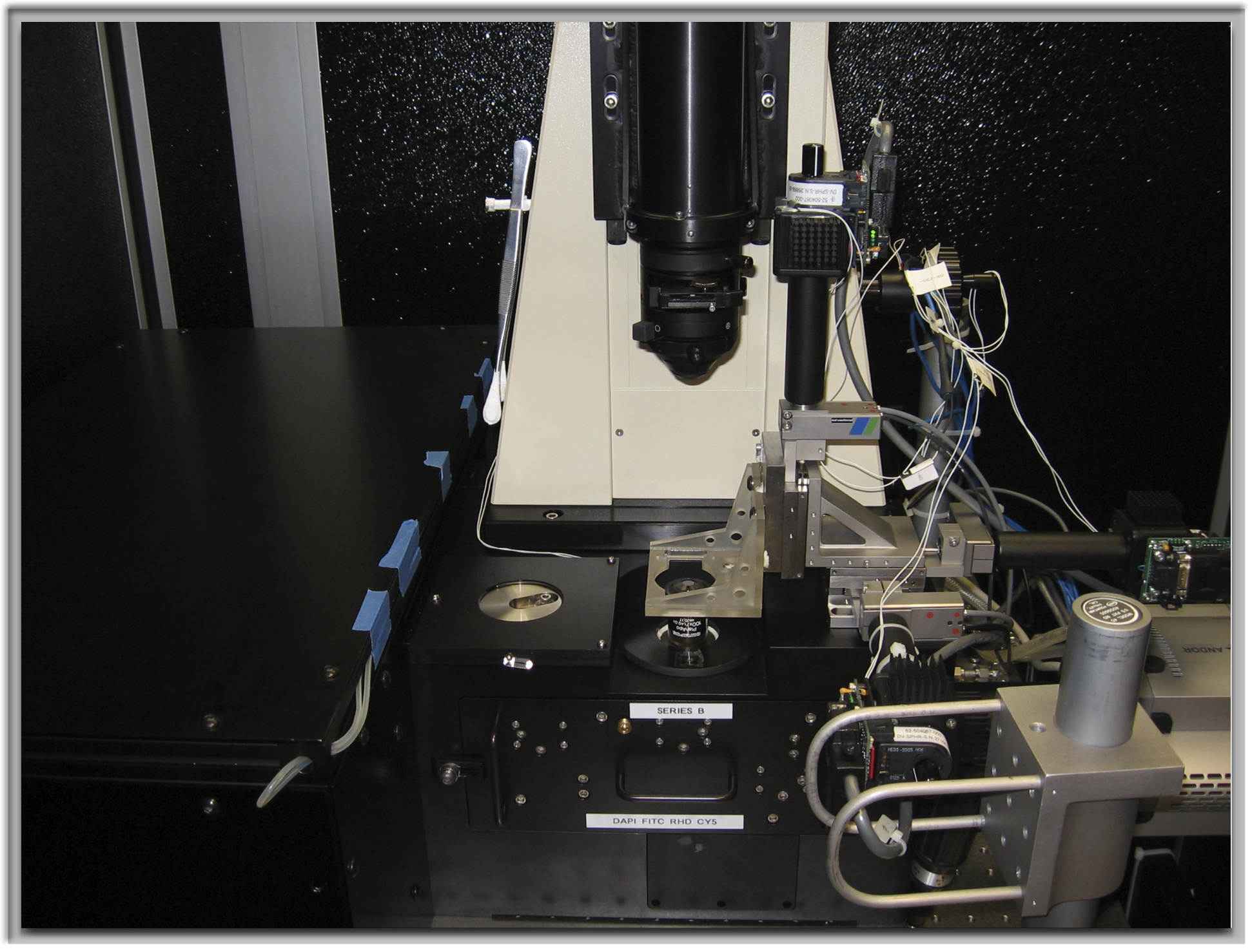
| 07:36, 3 December 2009 | Omx-image-1a.png (file) |  |
3.25 MB | The OMX Microscope, an experimental platform for <br> 3D Structured Illumination and Fast Live Imaging |
| 07:22, 3 December 2009 | PeteCarlton.jpg (file) |  |
17 KB |
