Uploads by Luke Blauch
From OpenWetWare
Jump to navigationJump to search
This special page shows all uploaded files.
| Date | Name | Thumbnail | Size | Description |
|---|---|---|---|---|
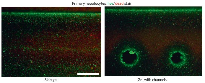
| 18:14, 8 April 2014 | Miller live cells.JPG (file) |  |
46 KB | |
| 18:10, 8 April 2014 | Sprouting miller.JPG (file) |  |
27 KB | |
| 18:09, 8 April 2014 | Sprouting.JPG (file) |  |
27 KB | |
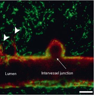
| 17:53, 8 April 2014 | Miller vasculature.jpg (file) |  |
48 KB | |
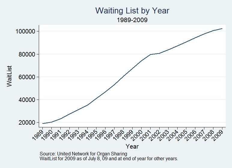
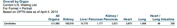
| 15:04, 8 April 2014 | Waiting list by year.jpg (file) |  |
44 KB | |
| 14:56, 8 April 2014 | Waiting list.JPG (file) | 20 KB | ||
| 05:28, 8 April 2014 | Skull in patient.JPG (file) |  |
31 KB | a- low-profile titanium microfixation plates. b,c- small gaps between the patient's skull and implant. d- a nonexistant artifact from the CT scan |
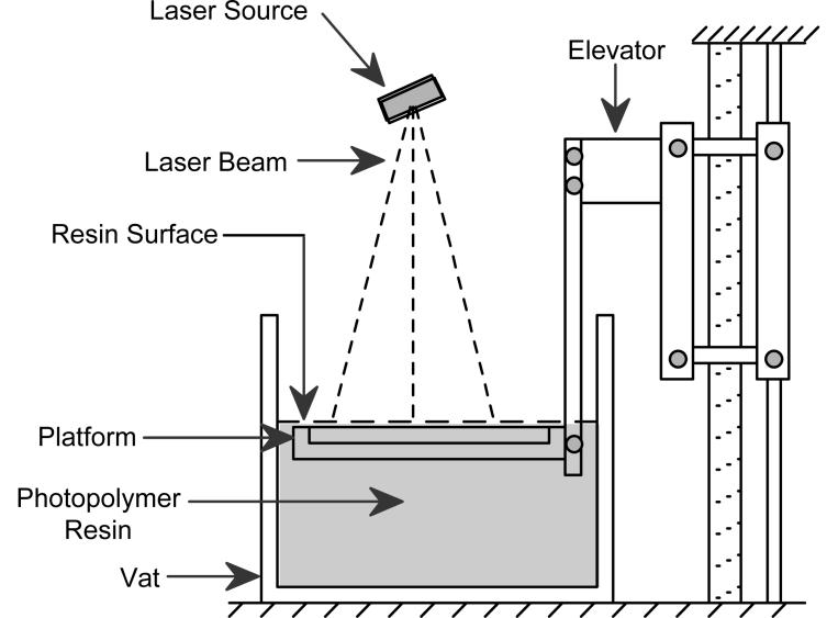
| 04:43, 8 April 2014 | Stereolithography.jpg (file) |  |
45 KB | An image of a typical 3D printer. This particular kind serves many uses outside of tissue engineering as well. |
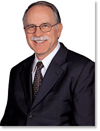
| 04:08, 8 April 2014 | Hull.jpg (file) |  |
11 KB | A picture of Charles Hull, the inventor of the 3D printer. |
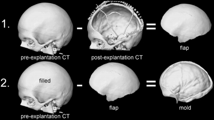
| 04:07, 8 April 2014 | Skull.jpg (file) |  |
66 KB | The doctor uses a CT scan of the patient’s skull before surgery (often taken upon onset of cranial trauma), then takes another after the flap of skull has been removed. Through digital subtraction of the two images, the flap’s shape can be determined, |
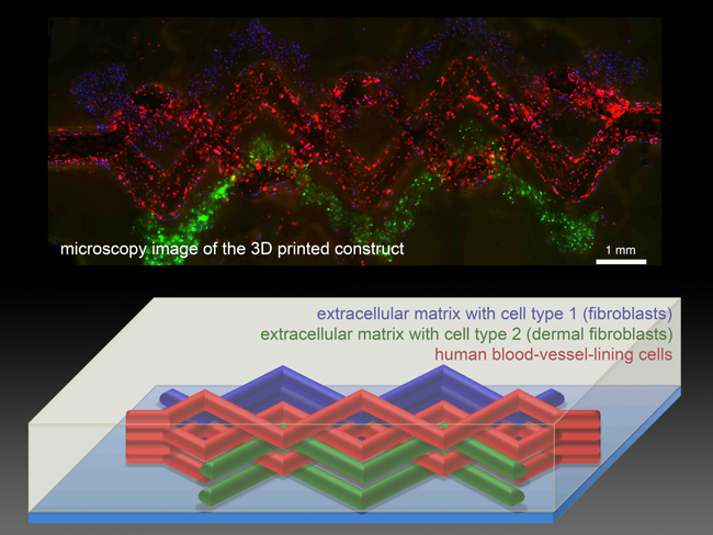
| 04:04, 8 April 2014 | Bioprinting.jpg (file) |  |
262 KB | From the Wyss Institute |
