Planck's Constant Lab Notes
Objective
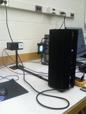
This lab is an investigation into the energy levels of electrons emitted in the photoelectric effect and the relationship between these energy levels and the frequency and intensity of the light source. This experiment uses a mercury vapor light source and an h/e
Apparatus consisting of a vacuum tube wth a cathode emitter with a low work function and an anode receptor which develop a potential difference as electrons build up on the anode. When the stopping potential of the electrons is reached, the voltage difference, which can be read with a voltmeter, is stabiilized and can be correlated to the kinetic energy of the electrons.
The relationships between the stopping potentials and the frequencies and intensities of the incident light are then analyzed with respect to both classical and quantum theories of light.
Planck's constant can be found through the equation:
E = h * frequency = KE(max) + Wo which can be rewritten in the form V(stopping) = h/e * f + Wo/e ,
the equation of a line with slope equal to h/e. Multiplying by the value for e, one may calculate the value of h, Planck's constant.
Notes
In this lab, David Weiss and I worked together again. We set up the equipment and then spoke with Professor Koch about safety concerns.
We connected the Mercury lamp to warm it for twenty minutes, and checked the batteries of the h/e Apparatus which read at 15.1 volts on the digital multimeter. Connecting to output, the reading went to 1 volt just from ambient light. I turned the grating assembly to see if one way or the other gave a brighter light, but could not tell the difference at this point.
We adjusted the angle between the lamp and the receiver to cast different spectral lines along the aperture slit of the h/e apparatus. We are looking for five spectral lines: yellow 578nm, green 546.074nm, blue 435.835, violet 404.656nm , and ultraviolet 365.483nm (wavelengths noted from Dr. Gold's Lab Manual, Prof. Gold's Lab Manual.)
There is also a line straight on that appears a dimmer yellow green, but may appear white when other lights are off. This is the zeroth order where the colors are not separated by diffraction.
We had some confusion about how to determine the stopping voltage, whether it should be at a final stable point, or at the same point for each intensity of a frequency. We aimed at the latter, but found that at some intensities, we could not reach the target voltage. This did not happen in a consistent way across the changes in intensities.
Watching the multimeter more closely, I noticed that the green at 40% immediately jumped to 0.8+ volts, dropped to 0.7+ volts, then took its time to reach 0.803 volts, in 96 seconds.
The measurements seemed to vary alot. We need to decide on a feasible voltage level to aim for that is both reasonably attainable and still allows for a measurable difference in times between different intensity levels.
Day two : Spent some time to research the target voltages that other people had used in this lab. Found yellow at 0.847, green at 0.7--, violet at 1.49. Still things seemed slow. changed all batteries and much better response times. chose to start with 1.48 for violet. David and Pranav had a long discussion trying to figure out causes for the discrepancies in voltage readings from last week. They changed out the batteries on the h/e Apparatus since it was not reaching the same voltage as from the day before, and a new voltage of 18.9 V was read on the multimeter. The new batteries only helped for the first trial run of one or two readings. Even with a lower target voltage, it continued to take more time to attain the stopping voltage even on the 100% transmission grating. The times seemed erratic.
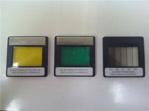
We changed the focus plate slightly, but it was clear we did not understand the proper operation of the equipment. Green band with green filter and no relative transmission filter gave 0.814v Yellow band with yellow filter and no relative transmission filter gave 0.680v
We continued on to the second experiment to determine stopping potentials of various wavelengths.
Violet: 1.642, 1.641, 1.643 Blue: 1.432 1.431 1.43
David typed the data for Yellow and Green into a spreadsheet, I handwrote it and set it up in a Google Doc later on.
Out of the lab, I read more carefully about the procedure, and will try to adjust the set up for Wednesday, with hopes that we can retake the data more accurately. This I did, as noted in the section on Day Three Data.
Safety
- Electrical Shock: The main safety concern of this lab is with respect to the Vapor Lamp which operates on 5000 volts, enough to cause serious injury, and important to be grounded. As always, check cords and connections for wear.
- Toxicity: The Mercury Vapor Tube contains toxic gas, so it is especially important to be careful with the lamp to avoid breakage.
- Equipment: The Mercury vapor tube inside the lamp is a high pressure vapor tube in order to provide more intensity and therefore more collisions and more electron emmissions. As a result, it is more fragile, and so it is important to avoid turning the light on and off more than necessary. It is also important to not look directly into the light as it can harm vision. The focus lens and the h/e apparatus, which has a static sensitivity, need to be treated with care to avoid damaging them. The colored filters and the variable transmission filter need to be handled only by their edges.
Equipment
- Mercury Vapor Light Source and Light Block(Model OS-9286)
- Voltage Range: 108-132 VAC
- Power: 125 W MAX
- Frequency: 47-63 HZ
- 115 Volts
- Light Aperture (Model AP-9369)
- Coupling Bar (Model AP-9369)
- h/e Apparatus (Model AP-9369)
- 3 Filters
- Yellow Line
- Green Line
- Relative Transmission Grating
- Digital Voltmeter (model FLUKE 111)
- 2 Connection Cables (Model 8-24)
- Two 9 Volt Batteries
- Duracell-Procell
- David's cell phone timer
Set Up


- We connected the Mercury lamp to warm it for twenty minutes, and checked the batteries of the h/e Apparatus which read at 15.1 volts on the digital multimeter. Connecting the multimeter to the output of the h/e Apparatus, the reading went to 1 volt just from ambient light.
- It is important to understand the working of the h/e apparatus and its alignment. The vapor lamp emits light that is diffracted by the Lens/Grating Assembly mounted on the vapor lamp housing. As a result, the light is displayed in first and second orders of spectral lines to either side of the zeroth order which appears as a white line. The vapor lamp can be rotated to align the various orders and individual spectral lines with the slit on the reflective mask of the h/e Apparatus (h/e). Then the h/e needs to be rotated on its stand so that the spectral line, that is shining through the slit, falls upon the opening in the h/e tube inside the black box. This can be observed by turning the cylindrical light shield to the open position which allows viewing of the inside tube. The spectral line can be focused at that point by adjusting the position of the lens/Grating Assembly on its support bars.
- We are looking for five spectral lines: yellow 578nm, green 546.1nm, blue 435.8, violet 404.7nm, and ultraviolet 365.48nm.
There is also a line straight on, the zeroth order, that appears a dimmer yellow green, but may appear more white when other lights are off, which it does, rather a very pale yellow.
Data Day One
The following data was recorded and formatted by David while I adjusted and read the instruments.
This data I formatted later on to show the relationship between the light intensities and the inverse of times to reach a set potential for the different spectral lines.
Data Day Two
Experiment 2
- We began with the first order colors and took 3 measurements of each, starting with violet and then going to blue, green and yellow spectral lines. Both the green and yellow were measured with their corresponding filters.
- We then took measurements of the 2nd order readings, starting again with the violet and moving through the same colors, adding the filters to the green and yellow spectral lines. In the second order, the lines were more diffuse. The reading for the green line was way off, and we found that there was a crossover of light from the third order ultraviolet spectral line which raised the kinetic energy of the emitted electrons and therefore also raised the stopping potential, reflected in the higher voltage reading.
My own first order data sheet is shown below, with averages in the fourth column. David's more complete records are included in the Day One data form above, as he was typing while I was adjusting the equipment and taking the readings.
Data Day Three
Pranav cleaned the filters and lens for me. I explored more about the set up and adjustment of the equipment, and found how to set the spectral lines to fall more precisely on the openings inside the h/e Apparatus.
We set up to retake the data for Experiment 1. I adjusted the h/e apparatus and the focus on the aperture inside for each spectral line. This was especially important between orders, and between the blue and green lines in the second order. I put some of the averages of this data into excel spreadsheets, which I unfortunately cannot retrieve at this time due to my limited experience with this interface. I also plotted some graphs by hand of the relationships between the transmission filter percentages and the inverse stopping times, which gave a series of diagonal lines with positive slopes.
I plotted average stopping potentials versus frequencies on paper and found a slope of 4.18 x 10^-15 eVs. This is compared to the accepted value of 4.14 x 10^-15 eVs, from Wikipedia.
The data is included in Day Three of David's spreadsheet from the Day One entry.
I was later able to plot the data in Excel, and the results are given in the data analysis.
Data Analysis
Experiment I was undertaken to determine the relationship between the intensity of given frequencies of light and the energy of the emitted photoelectrons. For this part, we used only the first order of the spectral lines. Different intensities of the same frequency light gave similar stopping potentials, and therefore similar maximum energies of the photoelectrons. This was slightly modified at lower intensities (stopping potentials were slightly less) due to the drain on the stopping potential from the connection to the voltmeter.
Experiment II was undertaken to verify the relationship between the intensity of given frequencies of light and the energy of the emitted photoelectrons. We took data from both first and second orders of the spectral lines, but since the second order data was skewed by cross-over from the third order spectral lines, I used only the first order data to calculate Planck's constant. Different frequencies of light gave different stopping potentials which reflected different maximum energies of the photoelectrons. When the stopping potential of each frequency was plotted as a function of frequency, the slope of the resulting best linear fit gave an approximation to Planck's constant.
Day one and two we used the spectral line orders to the left side of the zeroth order. Day three we used the orders to the right side of the zeroth order.
I put some of the averages of the data into Excel spreadsheets, which I unfortunately cannot retrieve at this time due to my limited experience with this interface, so then plotted some graphs by hand of the relationships between the transmission filter percentages and the inverse stopping times, which gave a series of diagonal lines with positive slopes.
I plotted the average stopping potential for each frequency versus the frequency and found a rough calculation of the slope from the endpoints gave:
Slope = `(1.642-0.7130)/(7.413-5.1903) = 4.18 x 10^-15
A more careful calculation through Excel LINEST gave:
Slope = 4.306 x 10^-15 with a Standard Error of the slope = +/-0.04597
y-intercept = 1.382 which represents the work function of the cathode.
Comparing this slope to the accepted value of 4.135 x 10^-15 eVs for Planck's constant, from Wikipedia Wikipedia, gives a percent error of:
(4.306 - 4.135)/4.135 = 4.1% error
Considerations
In this lab I learned additional skills in setting up equipment and developing awareness about the fine-tuning adjustments necessary to obtain accurate data. I also learned about deciphering and organizing data to be able to analyze it and present it in an understandable format. In this process, some of the concepts underlying the photoelctric effect and Planck's constant became clearer.
In additional work with this lab, I would take more measurements with the first order lines on both sides of the zeroth order and research more of the questions and concepts that arose as a result of the original experiment that formed the basis of this lab. I would also like to include photographs of the spectral lines and schema of the equipment set-up and the photoelectron emission process.
Concerns and Sources of Error
- Systematic Error: Since measurements are affected by the voltmeter such that the voltage is constantly lowered by some amount, the voltage reading attained is less than the actual stopping potential, especially at the lower intensities where the voltage is sustained at a lowere rate. In addition, the time to attain the stopping voltage is greater because of this drain through the voltmeter. The static electricity of the observer in touching the h/e Apparatus to reset the voltage reading can also affect the reading. I attempted to contact only the reset button (and not the metal casing) when it was being released. Crossover of light from third order spectral lines to the second order affected the stopping potentials for that order.
- Random Error: A major component of random error was the variance in human response times between reaadings, and communication times between the observer and the recorder. Another source of random error was the adjustment of spectral lines on the aperture inside the h/e Apparatus, which we were not properly aware of until the third day.
- The data consistency is also affected by the connection to a voltmeter. The time to attain the stopping voltage is affected more strongly by the drain of voltage through the voltmeter at lower intensities because the charging rate is slower.