File:RK-SennaCa204.JPG
From OpenWetWare
Jump to navigationJump to search

Size of this preview: 800 × 600 pixels. Other resolutions: 2,560 × 1,920 pixels | 4,000 × 3,000 pixels.
Original file (4,000 × 3,000 pixels, file size: 2.15 MB, MIME type: image/jpeg)
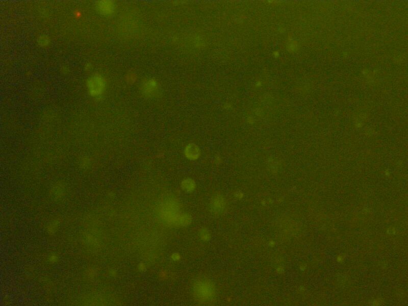
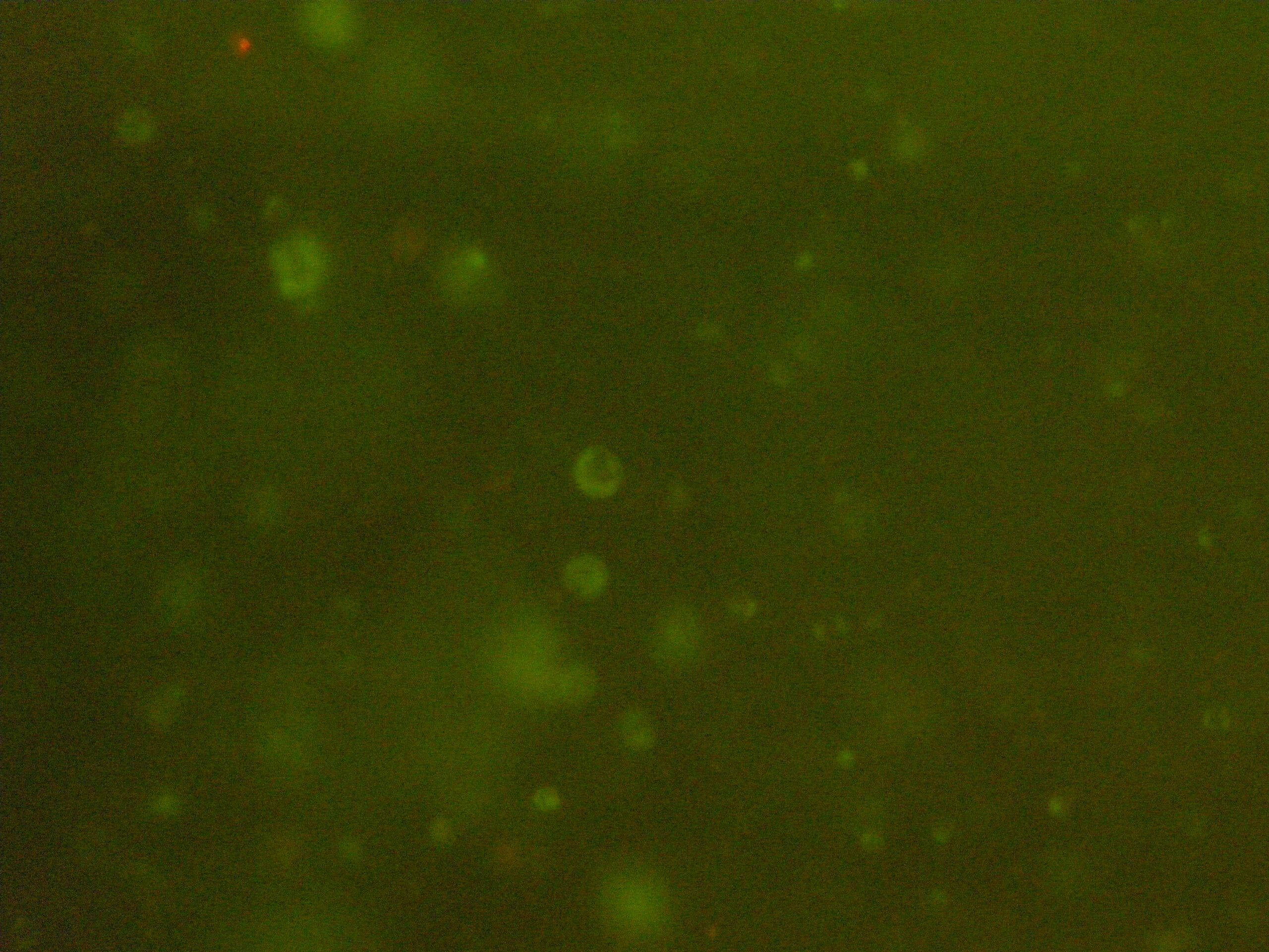
Fluorescent microscopy of alginate bead cross-linked with 204 mM CaCl2 containing chonodrocytes. Objective magnification is 10X. Green cells are living cells that were stained with SYTO 10, while red cells are dead cells stained by Ethidium.
File history
Click on a date/time to view the file as it appeared at that time.
| Date/Time | Thumbnail | Dimensions | User | Comment | |
|---|---|---|---|---|---|
| current | 19:21, 23 April 2010 | | 4,000 × 3,000 (2.15 MB) | Ragheb El Khaja (talk | contribs) | Fluorescent microscopy of alginate bead cross-linked with 204 mM CaCl2 containing chonodrocytes. Objective magnification is 10X. Green cells are living cells that were stained with SYTO 10, while red cells,not observed in this image, are dead cells staine |
You cannot overwrite this file.
File usage
The following page uses this file:
