Cytokinesis Lab
Dr. Katie Shannon's Cytokinesis Lab at Missouri S&T
 |
 |
 |
My major research interest is the assembly and contraction of the actomyosin ring in budding yeast.
Other interests are microscopy, synthetic biology and iGEM, and cellular uptake and transport of nanoparticles.
Contact and Biographical information for Dr. Shannon user:Katie_B_Shannon
Assembly and Contraction of the Actomyosin Ring in Budding Yeast
What is the actomyosin ring?
    The actomyosin ring is a structure composed of actin filaments, type II myosin, and other proteins.  Myosin is a molecular motor that “walks”
    along actin filaments.  Myosin movement along actin filaments causes a contraction or constriction of the ring.  As the actomyosin ring
    contracts, it pulls the plasma membrane of the cell.  This process is called cytokinesis, which is the physical division of one cell into two.
    Click [1] to see a movie of myosin-GFP contraction in a living yeast cell.
Why study budding yeast?
    Budding yeast, Saccharomyces cerevisiae, are also commonly known as baker's or brewer's yeast.  These yeast cells are eukaryotic,
    and many genes are conserved between yeast and human cells.  Both yeast and human cells utilize a contractile ring made of actin filaments
    and type II myosin (the actomyosin ring) in order to divide.  We hope that by studying the complex process of cytokinesis in the simpler
    yeast cells, we will learn fundamental concepts that will be applicable to  cytokinesis in other cells.
Why is regulation of actomyosin ring assembly and contraction important?
    When a cell divides, it is important that each cell receives the correct number of chromosomes and therefore the complete genetic blueprint.
    Cancer cells almost always have an abnormal number of chromosomes.  Recent studies have shown that an early step in tumor formation may be
    cytokinesis failure, which leads to tetraploidy (four copies of each chromosome).  Cytokinesis must be coordinated with chromosome
    segregation (mitosis) to ensure that each cell receives the right number of chromosomes.  Determining how cytokinesis is normally regulated
    will help us to understand how this process can fail and contribute to tumor progression.
What are our current lab projects?
    We are focusing on two proteins involved in cytokinesis, Iqg1 and Hof1.  Iqg1 is required for actin ring formation, and Hof1 is required for
    normal contraction of the ring.  We are studying the regulation of these proteins by phosphorylation.  We are also looking at the role of a
    signaling pathway, the mitotic exit network (MEN) in regulating actomyosin ring contraction.
Microscopy
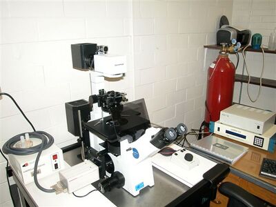
Dr. Shannon has established the Cellular Imaging Facility at Missouri S&T. An Olympus inverted epifluorescent microscope optimized for both fixed and live cell imaging was set up in August 2006. Both Dr. Shannon’s lab and collaborators in Biological Sciences, Chemistry, Chemical and Biological Engineering, and Environmental Engineering have heavily used this imaging equipment. Dr. Shannon has trained 25 students to use the microscope, and almost 600 hours of microscope use have been logged in the past year. Using her expertise in microscopy, Dr. Shannon is working with Dr. Huang to investigate the import and transport of nanoparticles in cells. Collaboration between Dr. Shannon and Dr. Ercal using the microscope to study toxic effects of radiation and ethanol on cells lead to a publication in the Journal of Applied Toxicology.
The set- up is an Olympus IX51 inverted microscope with 40X and 60X phase objectives and a 100X DIC objective. Brightline triple band filter set for multiple color fluorescence using FITC or GFP, TexasRed, and DAPI. A Longpass filter optimized for Quantum Dot excitation and emission is also available. Images are captured with a Hamamatsu ORCA285 CCD camera. Shutters, filters, and camera are controlled using SlideBook software. Z axis auto-focusing is driven using Prior stepper motor.
Synthetic Biology and iGEM
Synthetic Biology is a new field that asks if biological functions can be engineered from DNA "parts" [2]
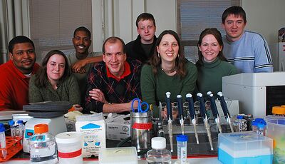
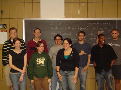
I helped start a student Synthetic Biology design team here at Missouri S&T with the help of my colleague Dave Westenberg. Our students have competed in the annual iGEM Jamboree at MIT for the past two years and will again this fall. You can learn more about iGEM and our team by following the links below.
iGEM team Missouri S&T iGEM2007 iGEM2008 2008 Missouri Miners team Wiki iGEM2009 2009 Missouri Miners team Wiki
 |
 |
Nanoparticle Research
I am collaborating with Drs. Huang and Winiarz on a drug-delivery system using Protein Transduction Domains coupled to nanoparticles called Quantum Dots. You can read about the project [http://news.mst.edu/2009/07/quantum_dot_research_could_lea.html here.