CT Imaging, by Elizabeth Swanson
Background
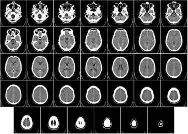
Computer Tomography, or CT, images are a form of x-ray scan which creates a 3D image of the scanned object. CT images are a compilation of computer processed x-ray images taken at a range of angles around the object to produce a single cross sectional image. Then the object is moved forward within the machine to scan the next cross section using the same technique. All of the cross sectional images can then either be viewed side by side or stacked on top of one another to create a 3 dimensional scan of the object.1 An example of a CT scan with the 2D images lined up next to one another can be seen to the left in Figure 1.
The most common CT scan referred to is the X-ray CT scan yet there exist other forms of CT scanning methods. Those include the PET scan, or positron emission tomography, and the SPECT scan, or single photon emission tomography. The PET scan is most often used to monitor metabolic processes within the body while SPECT scans are used more for functional cardiac or brain imaging.2,3
History
The development of CT scanners relied on two backgrounds: x-rays and tomography (a cross sectional view of a scanned object).
- 1895 – X-Rays are discovered by Wilhelm Rontgen4
- 1930s – Tomography beginning to be developed
- 1971 – 1st CT scanner invented by engineer Godfrey Hounsfield and physicist Allan Cormack
- 1974-1976 – 1st CT scanners are installed into hospitals
- 1980 – CT scanners are widely available
- 2008 – Siemens introduces new CT scanner
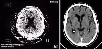
The first CT scanners developed had very low resolution with a resolution of 80 x 80 images and took about 5 minutes to take each scan at every angle, then an additional 2.5 hours to process the images into a single computer processed CT image .5 Modern day CT scanners have a resolution ranging from 1024 x 1024 to 2048 x 2048 and have the ability to take scans in less than a second, allowing from CT images of organs in motion, such as a beating heart.6 The improvement of over time of CT image resolution can be seen in Figure 2 to the right.
How CT Scanners Work
X-Ray
CT scanners rely on x-ray technology in order to create images from scans. X-rays are high energy electromagnetic radiation, with wavelength from 0.01 nm to 10 nm, below that of visible light (380 nm to 740 nm). Due to their low wavelength, X-rays have the ability to pass through most objects. Different tissues have varying ability to block these rays. For example, a dense tissue like bone is good at stopping the x-rays allowing for high contrast images. Yet softer tissues block less creating faint images and can be hard to see.7
A commercial X-ray machine works by having a fixed x-ray source emit x-rays at the target tissue. The x-rays pass through the tissue and the remaining radiation is recorded on a radiograph behind the tissue. Denser tissues, like bone, appear much darker while other tissues appear lighter caused by their varying ability to block the x-rays.
CT Scanner
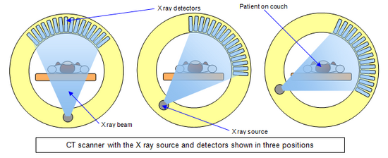
Similar to a an X-ray machine, CT scanners have a x-ray source, yet instead of being fixed, the source rotates around the object being scanned. The CT scanner takes a series of images as the x-ray rotates shooting narrow x-ray beams which are then picked up by a detector directly across from the source, seen in Figure 3. The x-ray detector measures the x-ray it receives and monitors any attenuation in its wavelength. Once the CT scanner completes a full rotational scan for a specific cross section, all of the information is compiled using complex mathematical calculations to create a single detailed CT image. The next "slice" is then scanned, and when all cross sections have been scanned, further processing can be performed to create a 3-dimensional model of the scanned object.8 Additionally, since many tissues have a low abillity in blocking x-rays, contrast agents are often used to make certain tissues stand out more in a CT scan. One example being, injecting an iodine based radio-contrast agent into the pulmonary arteries for diagnosis a pulmonary embolism.
Types of CT Scanners
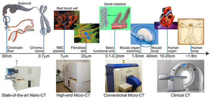
As seen above in Figure 4, a range of CT images can be created on a large range of scales. The most common and most widely used being the commercial CT machine used in hospitals. Micro CT scanners are more often used for research, seen in the next section, and are able to capture images in the micro scale with excellent detail. Lastly, nano-CT scanners are rare with only a few available currently, with the ability to image cellular components and are exclusively used for research.
Applications
Purpose in the Medical Field
CT scanners are most widely know for their use in the medical field. A CT scan can be ordered for a variety of conditions in order to scan people with tumors, identify a disease, or visualize an injury. Specific examples can be seen below:
- Head CTs: Can be used to detect tumors, blood clots, hemorrage, injuries, etc.
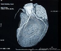
- Heart: Useful in diagnosing various types of heart disease. An example of an excellent 3-dimensional CT scan of a human heart can be seen in Figure 5.
- Lungs: Useful for detecting tumors, blood clots, excess fluid and conditions such as pneumonia and empysema, etc.
- Bone: very useful for viewing complex bone fractures or tumors since CT scans have more detail than a conventional X-ray
Tissue Engineering
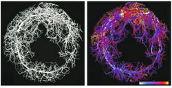
Mirco-CT scanners can be useful in tissue engineering in order to view the structure of tissue engineered constructs such as scaffolds or cells. Physical properties such as pore size, porosity, interconnectivity, and vasculature can be analyzed from a micro-CT scan. 10 The images have fairly high resolution ranging from 10 μm to 1 μm allowing for example, identifying individual capillaries. Yet, when measurements on the diameter of capillaries from an image were taken they were often over estimated most likely due to adjacent capillaries blurring together in the image.11
Another example of micro-CT technology being used in research includes one study using CT images to monitor the optimal conditions for bone mineralization on various scaffolds by comparing spacial distribution and the interconnectivity of newly developed bone.12
One study utilized micro-CT imaging to track stem cells treated with a iron oxide contrast agent. The research group was experimenting with a method of treating Duchenne muscular dystrophy by injecting stem cells to the target site. By injecting CD133+ stem cells into homozygous scid/mdx mice (mice who exhibit traits similar to that of DMD) the researchers monitored how the stem cells migrated through the body till eventually, after 2 hours, 90% resided at the target site.13
Downsides to CT Scans
While a CT scan creates an excellent 3-dimensional image of the body, it also increases a persons risk for cancer. Since CT scans utilize x-ray technology some risk is associated with the exposure to radiation. X-rays are damaging to tissues since they are a form of ionizing radiation, or have the ability to ionize molecules which can be damaging to cells and lead to cancer.14 While increased exposure, or multiple CT scans, are harmful , isolated CT scans increase cancer risk by a minimal amount and are more helpful than harmful. Yet a scan should only be undergone when necessary to limit the amount of radiation a patient is exposed to. Several factors influence the risk associated with a CT scan such as the dosage and patient age. For example, an abdominal CT has a radiation dosage comparable to that of ambient comic radiation accumulated over 3 years, or 10 millisievert. 15
Another source of risk when undergoing a CT scan is the contrast agent. 1-3% of patients given non-ionic contrast have an adverse reaction to a contrast agent, while 4-12% of patients given ionic contrast have an adverse reaction. Most reactions are mild including nausea, vomiting, and/or an ichy rash. More severe reactions are much less common and associated more with older radiocontrast agents which were known to cause anaphylaxis in 1% of patients, while present day agents have a risk of 0.01-0.04% of causing anaphylaxis.16 People at higher risk for an adverse reaction include females, the elderly, and patients in poor health.17
References
[1] Herman, G. T., Fundamentals of computerized tomography: Image reconstruction from projection, 2nd edition, Springer, 2009
[2] Elhendy, A; Bax, JJ; Poldermans, D (2002). "Dobutamine stress myocardial perfusion imaging in coronary artery disease.". Journal of Nuclear Medicine. 43 (12): 1634–46. PMID 12468513.
[3] Bailey, D.L; D.W. Townsend; P.E. Valk; M.N. Maisey (2005). Positron Emission Tomography: Basic Sciences. Secaucus, NJ: Springer-Verlag. ISBN 1-85233-798-2
[4] Markel, Howard. "Science Diction: How 'X-Ray' Got Its 'X'" NPR. New England Public Radio, 18 June 2010. Web. 13 Apr. 2017.
[5] Beckmann EC (January 2006). "CT scanning the early days" (PDF). TheBritish Journal of Radiology. 79 (937): 5–8. doi:10.1259/bjr/29444122. PMID 16421398.
[6] "40 years of Siemens CT - Siemens Healthineers Global." Semens Healineers. Semens, n.d. Web. 13 Apr. 2017.
[7] "X-ray". Oxford English Dictionary (3rd ed.). Oxford University Press. September 2005.
[8] "Computed Tomography (CT)." National Institutes of Health. U.S. Department of Health and Human Services, 02 Feb. 2017. Web. 13 Apr. 2017.
[9]Hodgkins, Leila. "Computerised axial tomography (CAT or CT)." Schoolphysics. N.p., n.d. Web. 13 Apr. 2017.
[10] Wang Y., Bella E., Lee C.S., Migliaresi C., Pelcastre L., Schwartz Z., Boyan B.D., and Motta A. The synergistic effects of 3-D porous silk fibroin matrix scaffold properties and hydrodynamic environment in cartilage tissue regeneration. Biomaterials 31,4672, 2010
[11] Nam, Seung Yun et al. “Imaging Strategies for Tissue Engineering Applications.” Tissue Engineering. Part B, Reviews 21.1 (2015): 88–102. PMC. Web. 13 Apr. 2017.
[12] Campi G., Ricci A., Guagliardi A., Giannini C., Lagomarsino S., Cancedda R., Mastrogiacomo M., and Cedola A. Early stage mineralization in tissue engineering mapped by high resolution X-ray microdiffraction. Acta Biomater 8,3411, 2012
[13] Farini, Andrea et al. “Novel Insight into Stem Cell Trafficking in Dystrophic Muscles.” International Journal of Nanomedicine 7 (2012): 3059–3067. PMC. Web. 13 Apr. 2017.
[14] Brenner DJ, Hall EJ (November 2007). "Computed tomography – an increasing source of radiation exposure" (PDF). N. Engl. J. Med. 357 (22): 2277–84. doi:10.1056/NEJMra072149. PMID 18046031.
[15] Radiological Society of North America (RSNA) and American College of Radiology (ACR). "Patient Safety - Radiation Dose in X-Ray and CT Exams." RadiologyInfo.org. N.p., n.d. Web. 13 Apr. 2017.
[16] Christiansen C (2005-04-15). "X-ray contrast media – an overview.". Toxicology. 209 (2): 185–7. doi:10.1016/j.tox.2004.12.020. PMID 15767033.
[17] Namasivayam S, Kalra MK, Torres WE, Small WC (Jul 2006). "Adverse reactions to intravenous iodinated contrast media: a primer for radiologists.". Emergency radiology. 12 (5): 210–5. doi:10.1007/s10140-006-0488-6. PMID 16688432.
[18] "SBES ADVANCED MULTI-SCALE CT (SAM-CT)." Biomedical Imaging Division, VT / WFU School of Biomedical Engineering and Sciences. N.p., n.d. Web. 13 Apr. 2017.
