Biomod/2014/AFM
<html>
<head>
 <title> Mainpage </title>
  <style>
    body{
    align: left;
    width: 1200px;
    height: auto;
    margin: 0 auto;
    background-color:#be1e3c;
    border:#be1e3c thin solid;
    }
    #goTopBtn {POSITION: fixed;TEXT-ALIGN: center;LINE-HEIGHT: 30px;WIDTH: 100px;BOTTOM: 35px;HEIGHT: 100px;FONT-SIZE: 12px;RIGHT: 30px;}
    #content{margin:0;padding:0;border:0px;}
      /*hidden section*/
      .firstHeading{display:none;}
      #sidebar-main{display:none;}
      #p-cactions{display:none;}
      #p-personal{display:none;}
   </style>
</head>
<body>
<a href="http://www.tu-braunschweig.de/index.html"><img src="http://openwetware.org/images/thumb/2/27/Nanoscooter_TUBS-siegel.jpg/800px-Nanoscooter_TUBS-siegel.jpg" width="383" height="142" alt="Logo TU Braunschweig"></a> |
<img src="http://openwetware.org/images/thumb/c/c3/Nanoscooter_Gruppenfoto-Banner.jpg/800px-Nanoscooter_Gruppenfoto-Banner.jpg" width="463" height="142" alt="our group" title="our group (Nanoscooter) for Biomod competition"> |
<img src="http://openwetware.org/images/2/24/Nanoscooter.jpg" width="165" height="142" alt="Logo Nanoscooter"> |
|---|
Team Nanoscooter Braunschweig
<style type="text/css"> body { height:500px; } div { }
</style> <body> <a href="Braunschweig"><img src="http://openwetware.org/images/b/bf/Zur%C3%BCckpfeil.png"
width="103" height="88" alt="Back"align="left"></a>
</body> |
AFM
The atomic force microscopy (AFM) uses a small (almost atomic size) metal tip to sense a surface to determine attributes of the surface like the topography. In these work two different modes, the AC-Mode and the Hyperdrive, of the AFM are used to verify that the DNA origami is folded as expected.
<img src="http://openwetware.org/images/thumb/0/03/AFM.png/800px-AFM.png" width="50%" height="50%" >
Figure 1: Laser distraction of an AFM.
Mica is a sheet silicate mineral with a wide range of structural characteristics. On the basis of these structural differences, mica can be divided in classes for example called muscovit, lepidolite or biotit. As a natural occurring mineral mica can be found in sedimentary rock or graniticpegmatites and is mostly mined in the USA, Russia, Finland and China.
<img src="http://openwetware.org/images/thumb/c/cd/Mica-Pl%C3%A4ttchen.jpg/734px-Mica-Pl%C3%A4ttchen.jpg" width="30%" height="30%" >
Figure 2: Mica plate for sample preparation in atomic force microscopy.
The ultraflat surface also allows the movement of DNA origami structures, through diffusion processes. Positive ions like Mg2+ act as a salt bridge between the negativly charged mica surface and the negatively charged DNA origami. Electrostatic interactions lead to the adsorption of the DNA origami on the mica surface. The intensity of this adsorption can be controlled by the dosage of Mg2+ ions in the buffer. The addition of monovalent ions like Na+ can reduce these electrostatic interactions by replacing the salt bridge or the Mg2+ ions, respectively. This enables a (controllable) movement of the DNA origami on the mica surface.
<img src="http://openwetware.org/images/a/a9/Muskovit.png" width="" height="" >
Figure 3: Nearly transparent sheets of muscovit.[1]
For AFM imaging we used a NanoWizard® 3 ultra AFM (JPK Instruments AG, Berlin, Germany). As the mica surface (Quality V1, Plano GmbH, Wetzlar, Germany) has to be completely (atomically) flat in order to achieve best results, the first step in an AFM experiment is the cleavage of the surface. The negatively charged mica sheet gets then loaded with Mg2+-ions by incubating with a solution of 10 mM MgCl2 for 2 minutes.
By AFM, nanoscale structures can be made visible. This makes it an ideal tool to verify the correct folding of our Nanoscooter. In the following images, 6 µL of the DNA origami solution as received from purification are mixed with 2 µL of buffer and incubated as described above.
<img src="http://openwetware.org/images/thumb/1/17/AFM-Scooter_neu.png/800px-AFM-Scooter_neu.png" width="" height="" >
Figure 4: AFM image of the Nanoscooter DNA origami. a) 3D image of several Nanoscooters. b) AFM image of a single Nanoscooter. c) Sketch of Nanoscooter in similar orientation as 1b).
Our AFM scans clearly verify the correct folding: We measure a length of about 70 nm, a width of 40 nm and a height of 10 nm in the front and 20 nm in the back of the DNA origami. This is in very good agreement with the structure designed so we are positive about successful creation of the first DNA origami Nanoscooter!
<img src="http://openwetware.org/images/a/a3/AFM-NROl.png" width="" height="" >
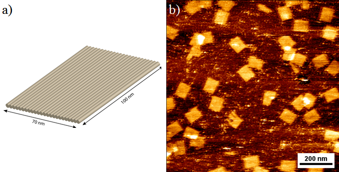
Figure 5: Rectangular DNA origami. a) Sketch of the rectangular DNA origami: It has a size of 70 x 100 nm and consists of 24 helices. b) AFM image of the rectangular DNA origamis.
We took AFM images of the rectangular DNA origami before and after 4 hours incubation in a 30% H2O2 solution. Sample preparation was carried out as described before: 2 µL of the purified DNA origami sample were diluted with 8 µL of buffer (TAE with 12.5 mM MgCl2) and incubated for 2 minutes.
<img src="http://openwetware.org/images/7/7f/AFM-H2O2.png" width="" height="" >
Figure 6: AFM images of rectangular DNA origamis. a) Under normal conditions. b) After 4 hours of incubation in H2O2.
The comparison between the two AFM images clearly shows that during the incubation time of 4 hours, the DNA origamis stayed intact. We therefore state that our proposed fuel is compatible with our Nanoscooter.
<img src="http://openwetware.org/images/7/7b/AFM-Steady.gif" width="" height="" >
Figure 7: A time series of 5 AFM images (intevals of 10 min) shows that the Nanoscooters are firmly bound to the mica surface. Anyhow the AFM shows a visible drift to lower left corner.
As can clearly be seen, the Nanoscooters are firmly bound to the surface. Anyhow, this experiment demonstrates a movement to the lower left corner, which is caused by the drift of the AFM.
<img src="http://openwetware.org/images/thumb/6/68/AFM-Floaten.png/651px-AFM-Floaten.png" width="" height="" >
Figure 8: AFM-scans with rectangular DNA origami of the same region within 30 minutes. a) Picture without NaCl-solution. b) Image after 15 minutes of incubation with NaCl. c) After additional 15 minutes.
The series shows a movement of the rectangular DNA origamis. Unfortunately, we cannot follow the pathways of single DNA origamis here because of the slow frame rate of about 15 minutes. However, it shows clearly that upon changing the buffer, the DNA origamis can now freely diffuse on the surface.
<img src="http://openwetware.org/images/0/08/Gasblasen.png" width="" height="" >
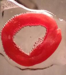
Figure 9: Emerging oxygen gas of the H2O2 decomposition.
|
</body>
</html>
