Biomod/2012/UTokyo/KaseiRunners/Result
<html> <head> <style>
- column-one { display:none; width:0px;}
.container{background-color: #ffffff; margin-top:0px} .OWWNBcpCurrentDateFilled {display: none;}
- content { width: 0px; margin: 0 auto auto 0; padding: 0em 0em 0em 0em; align: center;}
- column-content {width: 0px; float: left; margin: 0 0 0 0;padding: 0;}
.firstHeading {display:none; width:0px;}
- globalWrapper{width:848px; background-color: #ffffff; margin-left: auto; margin-right: auto}
- column-one {display:none; width:0px;background-color: #ffffff;}
- content{ margin: 0 0 0 0; align: center; padding: 12px 12px 12px 12px; width: 824px;background-color: #ffff; border: 0;}
- bodyContent{ width: 800px; align: center; background-color: #ffffff;}
- column-content{width: 824px;background-color: #ffff;}
- footer{position: center; width: 848px}
@media screen {
body { background: #F5F5F5 0 0 no-repeat; /* changed default background */ }
} /*************************
テンプレ
- /
ul.menu, ul.menu li, ul.menu li ul, ul.menu li ul li .others{ margin: 0; padding: 0; background: none; font-style: normal; font-weight:100; font-size: 180%; text-align: left; list-style: none; zoom: 1; } ul.menu li, ul.menu li ul, ul.menu li ul li { font-size: 100%; } ul.menu a{ color: #FFF; text-decoration: none; } ul.menu a:link, ul.menu a:visited { background: #002040; } ul.menu a:hover, ul.menu a:active { background: #204060; }
ul.menu li { float: left; position: relative; margin: 0; } ul.menu li a { display: block; width: 160px; text-align: center; line-height: 170%; } ul.menu li ul.result li a{ text-align: left; } ul.menu li ul.result li{ } ul.menu li ul li { float: none; margin: 0;
}
ul.menu li ul { display: none; }
ul.menu li:hover ul { display: block; position: absolute; z-index: 100; } ul.menu a.others{
background: #002040;
} .maintitle{
font-size:250%;
line-height:150%;
font-weight:900;
margin:0;
} .resultttt{ white-space: pre-wrap; } </style> </head> <body>
<img src="http://openwetware.org/images/d/df/Top_rogo_kasei.jpg" width=800px height=100px>
</body> </html>
<html> <head> <style> .resultback{
background-image: url('http://openwetware.org/images/2/27/Schemen_kasei.png');
width:800px;
height:532px;
margin:60px 0 0 0;
padding:0;
list-style: none;
}
.nav1{position: relative; top: 50px; left: 120px; }
.nav2{position: relative; top: 160px; left: 250px;}
.nav3{position: relative; top: 160px; left: 70px; }
.nav4{position: relative; top: 250px; left: 540px; }
.nav5{position: relative; top: 280px; left: 270px; }
/*************************
↑achieve ↓本文
- /
p.paragraph{ font-size :130%; line-height:140%; font-weight:normal; margin:15px 20px; } .method{ font-size :200%; line-heoght:100%; font-weight:bold; margin:0 20px; } a.tttop, h1.title a{
 display: block;
 text-decoration: none;
 color: #000;
} h2.title, h1.title{
clear:right;
clear:left;
} h2, h1{ font-weight: bold; margin-top:40px; } ul.goal{
list-style: none;
}
</style>
</head>
<body>
<a class="tttop" name="tttop">Result<a>
<a class="nav1" href="#r1"><img src="http://openwetware.org/images/4/4a/121026_idea_title1.png" alt="OPEN index" border="0" width=210px  height=41px ></a>
<a class="nav2" href="#r2"><img src="http://openwetware.org/images/8/8a/121026_idea_title2.png" alt="OPEN index" border="0" width=215px  height=43px></a>

<a class="nav3" href="#r3"><img src="http://openwetware.org/images/c/c3/121026_idea_title3.png" alt="OPEN index" border="0" width=155px  height=59px></a>

<a class="nav4" href="#r4"><img src="http://openwetware.org/images/8/85/121026_idea_title4.png" alt="OPEN index" border="0" width=182px  height=49px></a>

<a class="nav5" href="#r5"><img src="http://openwetware.org/images/0/02/121026_idea_title5.png" alt="OPEN index" border="0" width=310px  height=48px></a>
<a name="r1">1. Triggers opened the DNA tube.</a>
<img src="http://openwetware.org/images/e/e6/Result_scheme1.png" style="float:left">
<img src="http://openwetware.org/images/6/64/White.PNG"  width=40 height=400 style="float:left">
This section describes the DNA tube with toehold is opened by key strands.
1-1. Structure change of the tubular DNA nanostructure
1-2. Morphological change of the tube-dsDNA template conjugate
1-3. RNA polymerase binding to the integrated template dsDNA in the closed and opened tube.
Method
<img src="http://openwetware.org/images/8/8e/Kaseimethod1.png" width=323 height=350 style="float:left">
<img src="http://openwetware.org/images/6/64/White.PNG"  width=40 height=350 style="float:left">
 <img src="http://openwetware.org/images/f/f0/Method1_2kasei.png" width=397 height=350>
We made six-helix bundled DNA tube using DNA nano technology (Endo et
al., 2012). Using the built in toehold system, specific key (DNA
strands) induced the opening of the tubular structure. Left figure shows
the schematic drawing of the tube construction. The right figure shows
the modified design of our DNA Runner. Two staples were conjugated with
biotin for cargo binding (mark "b") and other two staples were
conjugated with Halo- or SNAP- Tag Ligand for kinesin binding (mark
"K"). Red flags indicate the toehold part (upper right figure). After
addition of the complement strands to toehold sequence, helix-helix
interaction along the dotted line cleaved. Therefore the tube opened.
Endo M et al., Transcription Regulation System Mediated by Mechanical
Operation of a DNA Nanostructure JACS 134, 2852-2855 (2012)
1-1. Structure change of the tubular DNA nanostructure
<img src="http://openwetware.org/images/3/37/Figure_1_1_new-01.png">
Figure 1-1. We made six-helix bundled DNA tube using DNA nano
technology (Endo et al., 2012). Using the built in toehold system,
specific key (DNA strands) induced the opening of the tubular
structure.
We also succeeded in integrating gfp gene into toehold- attached tube.
Addition of the key induced the opening of the tubular structure.
Analysis using 4% native polyacrylamide gel electrophoresis (PAGE).
Endo M et al., Transcription Regulation System Mediated by Mechanical
Operation of a DNA Nanostructure JACS 134, 2852-2855 (2012)
1-2. RNA polymerase binding to the integrated template dsDNA in the closed and opened tube.
<img src="http://openwetware.org/images/8/8f/Figure_1_3.jpg">
Figure 1-2. We tested if tube wraps the head of template DNA (40bp). The DNA in this experiment has T7 promoter sequence in its front. Theoretically, if it’s open, T7 RNAP binds to the DNA, but it’s closed, that doesn’t. In lane 4, some fraction of the closed-toe tubes were remained. In contrast, the opened-toe tubes were diminished in lane 7, suggesting that opened-toe tubes bind to T7-RNAP more efficiently.
<a href="#tttop">Back to top</a>
<a name="r2">2. Kinesin was synthesized from dsDNA by pure system.</a>
<img src="http://openwetware.org/images/4/40/Result_scheme2.png" style="float:left">
<img src="http://openwetware.org/images/6/64/White.PNG"  width=40 height=400 style="float:left">
This section describes the transcription and translation from the gene of kinesin, motor protein.
2-1. Expression of kinesin using PURE system
2-2. Movement of kinesin-DNA origami tile complex along the microtubules
2-3. Observation of PURE system expressed kinesin movement.
method
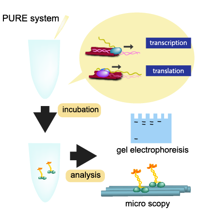
<img src="http://openwetware.org/images/e/e7/Method2.png" style="float:left">
PURE system, which stands for "Protein synthesis Using Recombinant
Elements", is a reconstituted cell-free protein synthesis system
developed in Ueda-lab (Shimizu et al., 2001). This reaction system
consists of proteins, ribosome, amino acids and NTPs, which are
necessary for transcription, translation and energy regeneration.
Shimizu Y. et al., Cell-free translation reconstituted with purified
components. Nat Biotechnol, 19, 751-755 (2001).
2-1. Expression of kinesin using PURE system
<img src="http://openwetware.org/images/0/04/KaseiFigure_2-1.png" >
Figure 2-1. PURE system, which stands for "Protein synthesis Using Recombinant Elements", is a reconstituted cell-free protein synthesis system developed in Ueda-lab (Shimizu et al., 2001). This reaction system consists of proteins, ribosome, amino acids and NTPs, which are necessary for transcription, translation and energy regeneration. Using PUREfrex system, which is the improved version of the original PURE sytem, we suceeded in expressing the kinesin. And HaloTag or SNAPTag protein were fused to the C-terminal end of kinesin. After 3h incubation at 37 'C, specific fluorescent ligand (TMR for HaloTag and ATTO532 for SNAPTag) were added to the reaction mixer. Samples were analyzed by 12% SDS-PAGE. Arrows indicate the position of expressed kinesin.
2-2. Observation of PURE system expressed kinesin movement.
Single-molecule images were visualized by a total internal reflection
fluorescence microscope (TIRF; Miyazono et al. 2010).
Miyazono Y. et al., Strain through the neck linker ensures processive
runs: a DNA-kinesin hybrid nanomachine study. EMBO J 29, 93-106 (2010)
<img src="http://openwetware.org/images/5/5c/KaseiFigure_2-3.png" width=700 height=320 style="float:center;">
Figure 2-2. PURE system expressed kinesin can walk. To examine the motile activity of PURE system expressed kinesin, we observed the kinesin movement at single molecule level. (A) A kymograph showed that PURE system expressed kinesin walked processively. (B) Speed (0.83 +/- 0.27 um/s) is slightly faster than that of E. coli expressed kinesin (0.66 +/- 0.18 um/s). (C) Run length (0.66 um) is slightly longer than that of E. coli expressed kinesin (0.42 um). From these results, we conclude that tha motile activity of PURE expressed kinesin is similar with that of E. coli expressed kinesin.
<a href="#tttop">Back to top</a>
<a name="r3">3. Cargo was attached to the DNA structure.</a>
<img src="http://openwetware.org/images/f/f6/Result_scheme3.png" style="float:left">
<img src="http://openwetware.org/images/6/64/White.PNG" width=40 height=400 style="float:left">
This section describes the cargo is attached to the tube which is in an open form.
3-1. Gel electrophoresis of the biotin conjugated DNA tile and Alexa-532 labeled Streptavidin.
3-2. DNA-tile can bind to NeutrAvidin beads.
method
<img src="http://openwetware.org/images/d/d2/KaseiMethod3.png" >
3-1. Gel electrophoresis of the biotin conjugated DNA tile and Alexa-532 labeled Streptavidin.
<img src="http://openwetware.org/images/1/1b/Figure_3-1.png" >
Figure 3-1. Gel electrophoresis of the biotin conjugated DNA tile and Alexa-532 labeled Streptavidin.
The SA only lane (lane2) showed a broad band of SA in SA image (left) and no signal in the DNA tile image (right). In contrast, after the addition of DNA tile into the SA solution (lane4), sharp a band corresponding to the DNA tile band appeared. These results indicate that biotin labeled staples were incorporated into the DNA tile.
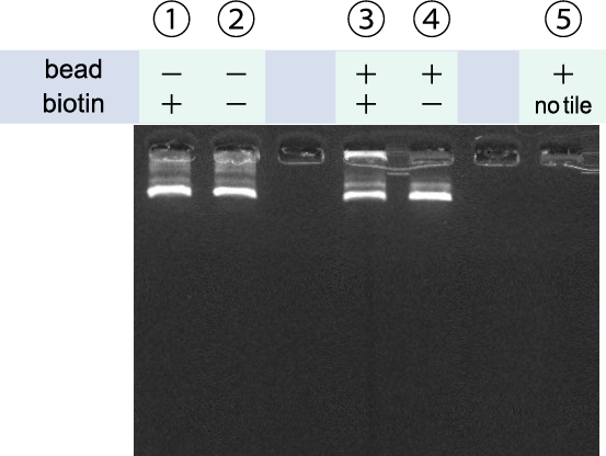
3-2. DNA-tile can bind to NeutrAvidin beads.
<img src="http://openwetware.org/images/4/4b/Figure_3-2_new.png" >
Figure 3-2. DNA-tile can bind to NeutrAvidin beads.
To change the storage buffer to reaction buffer, beads were washed with "1 x Tile buffer" several times. Then washed beads were mixed with DNA-origami tile, having Halo-Tag ligands for protein anchoring and Cy5 fluorophore for detection. We used two types of DNA-tile. One has 4 biotin site for NA binding, and the other did not. Prior to mixture with beads, both types of DNA-tile showed single band(arrow head), indicating that the electrophoretic mobility of the two types are similar. In contrast, after mixing with beads, only the DNA-tile having biotins showed upward shifted band(arrow), coincide with the beads position. These results indicated that the biotinylated DNA-tile can specifically bind to the beads.
<a href="#tttop">Back to top</a>
<a name="r4">4. Kinesin bound to the DNA structure.</a>
<img src="http://openwetware.org/images/c/c3/Result_scheme4.png" style="float:left">
<img src="http://openwetware.org/images/6/64/White.PNG" width=40 height=400 style="float:left">
This section describes the DNA tube with a cargo is transported by the kinesin binded to the tube.
4-1. Kinesin-SNAPf fusion protein can bind to the SNAPfTag-Ligand incorporated DNA tile.
4-2. Kinesin with Halo Tag bound to the DNA tile with Halo Tag Ligand.
method
<img src="http://openwetware.org/images/3/38/KaseiMethod4.png" >
4-1. Kinesin-SNAPf fusion protein can bind to the SNAPfTag-Ligand incorporated DNA tile.
<img src="http://openwetware.org/images/5/5a/KaseiReulstFigure_4-1.png" >
Figure 4-1. Gel electrophoresis of the DNA tile with SNAPfTag Ligand and SNAPfTag-kinesin.
Alexa647-labeled SNAPfTag-Kinesins were mixed with Cy3-labeled DNA origami tiles (rectangle type), and incubated for 2 hours at room temperature. Concentration of the SNAPfTag-kinesin was 20 times higher than that of SNAPfTag handle incorporated into DNA tile. While there are no binding of SNAPfTag-kinesin to the HaloTag ligand incorporated DNA tile (lane 4), there are clear band of SNAPfTag-kinesin at the position of DNA tile (lane 2; label ratio roughly 70 %), suggesting the specific
binding of SNAPfTag-kinesin to SNAPfTag incorporated DNA tile.
4-2. Kinesin-Halo fusion protein bound to the HaloTag-Ligand incorporated DNA tile.
<img src="http://openwetware.org/images/f/f5/KaseiFigure_4-2.png" >
Figure 4-2. Gel electrophoresis of the DNA tile with HaloTag Ligand and HaloTag-kinesin.
Cy5-labeled HaloTag-Kinesins were mixed with Cy3-labeled DNA origami tiles (rectangle type), and incubated for 4 hours at room temperature. Concentration of the HaloTag-kinesin was 10 times higher than that of HaloTag handle incorporated into DNA tile. While there are no binding of HaloTag-kinesin to the plane DNA tile (HaloTag ligand-less tile; lane 4), there are clear band of HaloTag-kinesin at the position of DNA tile (lane 3; label ratio of 32 %), suggesting the specific binding of HaloTag-kinesin to HaloTag incorporated DNA tile.
4-3. kinesin with DNA tile ran longer than kinesin only
To examine the effect of keep kinesins in line, we observed the movement of kinesin - Rectangle DNA origami tile (Rothemund 2006) complex along the microtubule filament. Each bright fluorescent spots indicate the Cy5 dye labeled individual kinesin-tile complex (see movie) <img src="http://openwetware.org/images/6/64/White.PNG" width=40 height=190 style="float:left"> <img src="http://openwetware.org/images/4/47/Wiki_gif.gif" width=81 height=192 style="float:left;" >
<img src="http://openwetware.org/images/a/ac/121015_midterm_copy.034.jpg" width=200 height=192 style="float:left;" >
<img src="http://openwetware.org/images/1/1e/121015_midterm_copy.035.jpg" width=200 height=192 style="float:left;" > <img src="http://openwetware.org/images/3/31/121015_midterm_copy.036.jpg" width=200 height=192 style="float:left;" > >
Figure 4-3-1. Figure 4-3-2. Figure 4-3-3. The results show that the kinesin-tile complex can ran longer than
kinesin only (Figure 4-3-1, 4-3-2, 4-3-3). Similar results were reported
very recently (Derr et al. 2012).
Rothemund PW., Folding DNA to create nanoscale shapes and patterns.
Nature 440: 297.302 (2006)
Derr ND. et al., Tug-of-War in Motor Protein Ensembles Revealed with a
Programmable DNA Origami Scaffold Science Epub ahead of print 11 October
2012)
<a href="#tttop">Back to top</a>
<a name="r5">5. Kinesins synthesized by Pure System transported the DNA structure.</a>
<img src="http://openwetware.org/images/0/09/Result_scheme5.png" style="float:left">
<img src="http://openwetware.org/images/6/64/White.PNG" width=40 height=400 style="float:left">
This section is under construction. We tried hard but haven’t succeeded yet.
<a href="#tttop">Back to top</a>
</body> </html>
