BME103:T930 Group 2 l2
| Home People Lab Write-Up 1 Lab Write-Up 2 Lab Write-Up 3 Course Logistics For Instructors Photos Wiki Editing Help | ||||||||||||||||||||||||||||||||||||||
OUR TEAM
LAB 2 WRITE-UPThermal Cycler Engineering
ProtocolsMaterials
1. Gather materials and assemble as shown in apparatus/manual
DNA Measurement Protocol 1. Create 8 DNA template samples that will be the focus of the investigation and place them in the PCR Machine allowing them to complete the process and replicate. 2. Transfer each sample independently into its designated Eppendorf Tube with the 400mL buffer completely. Ensure that a single pipette is used per sample. 3. After setting up the fluorimeter, place 2 drops of SYBR Green onto the teflon slide followed by 2 drops of the sample you wish to use. (Again use the same pipette used to transfer the sample) 4. Align up the drop so the light is passing through it. 5. Take pictures of the drop using a camera or smartphone, these will later be uploaded to ImageJ for analysis. 6. Each slide is capable of handling 5 individual samples so simply place the drops in an empty space and repeat the process. 7. Also run drops from the scintillation vial as blanks using the same process. 8. Upload the pictures taken into Image J. 9. To analyze the images subtract the INTDEN measurement form the background from the INTDEN measurement form the drop and repeat for all trials. Research and DevelopmentBackground on Disease Markers
rs35685286 [Homo sapiens] GGATGAAGTTGGTGGT--GAGGCCCTGG[A/G]CAGGTTGGTA--TCAAGGTTACAAGAC Chromosome 11- single nucleotide variation http://www.ncbi.nlm.nih.gov/projects/SNP/snp_ref.cgi?rs=35685286
rs34430836 [Homo sapiens] AGGTGCTAGGTGCCTT--TAGTGATGGC[C/G]TGGCTCACCT--GGACAACCTCAAGGG Chromosome 11- single nucleotide variation http://www.ncbi.nlm.nih.gov/projects/SNP/snp_ref.cgi?rs=34430836
Sickle cell anemia is an inherited blood disorder characterized primarily by chronic anemia and periodic episodes of pain. The underlying problem involves hemoglobin, a component of red blood cells. Hemoglobin molecules in each red blood cell carry oxygen from the lungs to body organs and tissues and bring carbon dioxide back to the lungs. In sickle cell anemia, the hemoglobin is defective. After hemoglobin molecules give up their oxygen, some may cluster together and form long, rod-like structures. These structures cause red blood cells to become stiff and assume a sickle shape. Unlike normal red cells, which are usually smooth and donut-shaped, sickled red cells cannot squeeze through small blood vessels. Instead, they stack up and cause blockages that deprive organs and tissues of oxygen-carrying blood. This process produces periodic episodes of pain and ultimately can damage tissues and vital organs and lead to other serious medical problems. Normal red blood cells live about 120 days in the bloodstream, but sickled red cells die after about 10 to 20 days. Because they cannot be replaced fast enough, the blood is chronically short of red blood cells, a condition called anemia.
Sickle cell anemia is an autosomal recessive genetic disorder caused by a defect in the HBB gene, which codes for hemoglobin. The presence of two defective genes (SS) is needed for sickle cell anemia. If each parent carries one sickle hemoglobin gene (S) and one normal gene (A), each child has a 25% chance of inheriting two defective genes and having sickle cell anemia; a 25% chance of inheriting two normal genes and not having the disease; and a 50% chance of being an unaffected carrier like the parents. Source: http://www.ornl.gov/sci/techresources/Human_Genome/posters/chromosome/sca.shtml
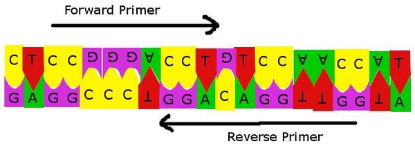
rs35685286 [Homo sapiens] Primer--CTCCGGGACCTGTCCAACCAT Reverse Primer-- GAGGCCCTGGACAGGTTGGTA
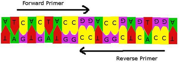
rs34430836 [Homo sapiens] Primer--ATCACTACCGGACCGAGTGGA Reverse Primer--TAGTGATGGCCTGGCTCACCT
This image illustrates the primer attaching to the reverse primer. The primer that is created attaches to the section of DNA that is mutated and is then able to be replicated with PCR, because it will allow the Taq polymerase to begin attaching nucleotides.
This image illustrates the primer attaching to the reverse primer. The primer that is created attaches to the section of DNA that is mutated and is then able to be replicated with PCR, because it will allow the Taq polymerase to begin attaching nucleotides. Bayesian Information
Affected Gene: HBB hemoglobin, beta Population Diversity: About 250 million people, and about 300,000 infants are born with a major hemoglobinopathies every year Probability of having mutation: 4.5%- of the world population [Source: Angastiniotis M, Modell B, Englezos P, Boulyzhenkov V. Prevention and control of hemoglobinopathies. Bull World Health Organ. 1995; 73: 375-386. - See more at: http://www.ispub.com/journal/the-internet-journal-of-biological-anthropology/volume-1-number-2/epidemiology-population-health-genetics-and-phenotypic-diversity-of-sickle-cell-disease-in-india.html#e-44]
Affected Gene: HBD hemoglobin, delta Population Diversity: About 250 million people, and about 300,000 infants are born with a major hemoglobinopathies every year Probability of having mutation: 4.5%- of the world population [Source: Angastiniotis M, Modell B, Englezos P, Boulyzhenkov V. Prevention and control of hemoglobinopathies. Bull World Health Organ. 1995; 73: 375-386. - See more at: http://www.ispub.com/journal/the-internet-journal-of-biological-anthropology/volume-1-number-2/epidemiology-population-health-genetics-and-phenotypic-diversity-of-sickle-cell-disease-in-india.html#e-44] | ||||||||||||||||||||||||||||||||||||||


