BME103:T130 Group 7
| Home People Lab Write-Up 1 Lab Write-Up 2 Lab Write-Up 3 Course Logistics For Instructors Photos Wiki Editing Help | |||||||||||||||||||||||||||||||||||||||||||
OUR TEAM
LAB 1 WRITE-UPInitial Machine TestingThe Original Design
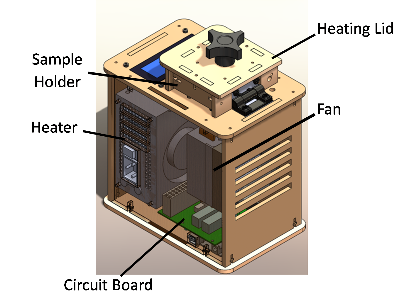
When the PCB board was unplugged from the Open PCR circuit board, the machine's LED display ceased to function. When the white wire that connects the Open PCR circuit board to the 16 tube PCR block was unplugged, the machine's LED display read -40 Celsius. This most likely indicated that the temperature could not be read on the device and that it was instead displaying a null value for the temperature.
October 18,2012; The test program labeled "Simple Test", was performed on machine number 7 to ensure proper function of the Open PCR machine. It ran through two cycles at a full range of temperatures and tested all of the PCR machine's essential systems. No DNA was present for the test run. The test performed without any flaws and the machine proved to be fairly quick at reaching its desired temperature.
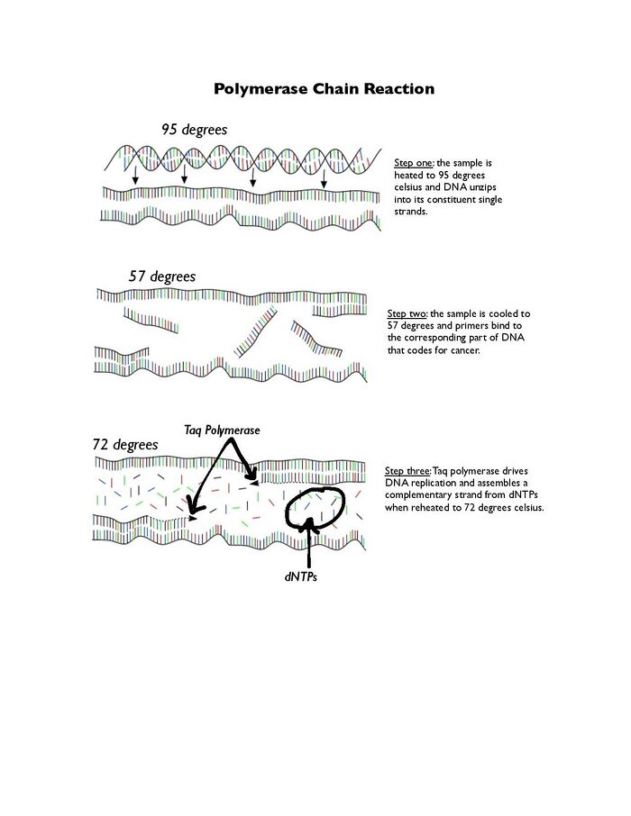
ProtocolsPolymerase Chain Reaction To amplify samples of DNA, the OpenPCR machine was used to perform a Polymerase Chain Reaction (PCR). This technique worked by cycling a mixture of DNA Template, Primers, Taq Polymerase, Magnesium Chloride, and dNTP's through three specific temperatures to create more copies of the desired sequence. After assembling the PCR mixture, the PCR machine was programmed to perform three stages. In the first stage, the samples went through one cycle at 95⁰C for 3 minutes. The purpose of this stage was to initially denature the DNA and allow the primers to act on the DNA. The second stage put the samples through 35 cycles of 95⁰C, 57⁰C, and 72⁰C each for 30 seconds. The purpose of the first part of the second stage is to break apart the hydrogen bonds between the base pairs, denaturing the DNA sequence into two separate strands. The purpose of the low temperature is to allow primers to bind. The purpose of the middle temperature is to create an environment for Taq Polymerase to assemble a new strand that is the desired product of the entire polymerase chain reaction. The last stage, stage three, puts the samples through one cycle of 72⁰C for 3 minutes. There is a final hold of 4⁰C that preserves the DNA. The samples were then taken out of the PCR machine. The target sequence had been amplified a million times and could now be analyzed with less sensitive equipment! To analyze the sequence, see the Fluorimeter section.
To analyze the DNA samples after they've been amplified,
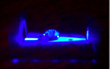
a smartphone with adjustable settings was used to take a photo of the samples. - Exposure to highest setting The Fluorimeter aparatus consists of a hydrophobic Teflon surface on a glass slide. There is an array of 3x10 glass wells on the surface to anchor drops in position for photographing. First, two drops of the cybergreen solution were placed on row 2, column 1&2 (Figure 1). With a sterile pipet two drops of amplified sample DNA were added to the cybergreen solution. It was important to place the DNA on top of the cybergreen in order to achieve the best results and to avoid contamination of the DNA samples. The picture was then photographed in the dark box with the door shut to eliminate excess background light. The next sample was prepared by sucking the liquid off the slide using a 'waste' pipet. The slide was then moved back by two columns for the next sample. This procedure was replicated for each of the patient samples and two slides were used in their entirety. Picture and Analysis: The pictures of each sample were analyzed using the "image j" software to process the images and analyze their content. The pictures, taken with the settings listed above, were upload to the "image j" software. The images were then separated into their composite colors, red, blue, and green. The green portion of the images were then analyzed using the software and its "INTDEN" was recorded.
Interpreting results: The cybergreen soluiton allowed the amount of DNA in a sample to be determined by its "INTDEN". The cybergreen glowed green in the presence of DNA and while this glow was not visable to the naked eye, the smart phone in conjunction with "image j" was able to quantify the luminosity in terms of the "INTDEN". The brighter samples contained more DNA and had a higher "INTDEN" value. This indicated that the sample reacted with the primers better and was cancerous. Research and DevelopmentSpecific Cancer Marker Detection - The Underlying Technology Polymerase Chain Reaction, or PCR, is used to ampllify a specific segment of DNA, in this case the segment known to code for cancer. A primer, which is an artifically synthesized piece of complementary DNA, binds to the desired DNA segment, and the enzyme taq polymerase catalyzes the replication of the DNA strand using dNTPs (free bases). This is repeated over and over to produce many copies of the DNA. Once the copies are made, a fluorescent dye that only bonds to DNA double strands is added to the solution. If a solution that shows fluorescence by using a fluorimeter, then that DNA sample contains the cancer-associated sequence. The single nucleotide polymorphism, or SNP, that is linked to cancer is rs17879961. The DNA base sequence associated with cancer is ACT, while the non-cancer sequence is ATT. PCR works to identify ACT from ATT because the primers, which are complementary to DNA strands containing cancer-positive ACT, will not bind to DNA containing ATT, and instead of DNA double strands, the solution will only contain single strands. The fluorescent dye will only bind to double strands and will identify those.
Reliability through Bayes Law Bayes Law expresses the probability that something is true. In this case, it relates to the probability that a person testing positive for cancer markers actually has cancer.
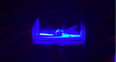
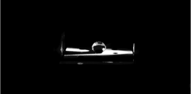
Bayes Law states that the probability of an event, A, given another event, X, is equal to the probability of X given A times the probability of A. This is then divided by the probability of X given A times the probability of A plus the probability of X given not A times the probability of not A. In the case of the cancer markers, events A and X might be the probability that a patient has cancer given that the patient tested positive for the cancer marker. ResultsThese Images were taken using a HTC Evo4G using the settings described under flourimeter measurements. Water and SYBR green Water and SYBR green under split color conditions for green
SYBR green and Calf Thymus
KEY
It is difficult to draw strong conclusions when all of the data is reviewed. The negative control read quite a high value for DNA presence meaning that some sort of error may have occurred. The negative control should have had very low levels of DNA present. To provide better results the experiment should be run again with an emphasis on achieving a lower level of DNA in the negative sample. | |||||||||||||||||||||||||||||||||||||||||||
