BME100 f2018:Group11 T0800 L6
| Home People Lab Write-Up 1 | Lab Write-Up 2 | Lab Write-Up 3 Lab Write-Up 4 | Lab Write-Up 5 | Lab Write-Up 6 Course Logistics For Instructors Photos Wiki Editing Help | ||||||
OUR COMPANY
Our Brand Name LAB 6 WRITE-UPBayesian StatisticsOverview of the Original Diagnosis System This lab involved the testing of 30 samples of DNA from 30 individual patients. The labor was divided between 15 groups of BME 100 students, with approximately five students in each. To acquire higher precision in the results, three replicates were made for each DNA sample. Each replicate sample underwent a polymerase chain reaction to amplify particular segments of the DNA strand; as such, there were 90 total tests run, testing 30 patients for SNP disease. Each sample was treated with DNA polymerase, DNA primers, individual nucleotides and the PCR buffer. These samples were placed in an OpenPCR machine, which allowed the DNA samples to be replicated and amplified. The samples were treated with SYBR Green I stain and placed in a fluorimeter. This allowed an image to be taken of the sample, that would detect the presence of the SNP disease. The images were processed by way of a program called ImageJ. The system was first calibrated by way of a series of calibration solutions. Three photos were taken per sample to increase precision and decrease error. Though many measures were taken to decrease the potential error of the results, substantial error did arise from the fact that this lab was an learning process. The initial process of pipetting the solutions may have been resulted in incomplete transfer of the substances, as this was the first time some of the students used a micropipette. The fluorimeter analysis also provided a number of sources for error. When placing the drop of combined PCR reaction mix and SYBR Green stain, we failed to adjust the positioning of the drop corresponding to the different samples, until halfway through the process. This may have contaminated the samples before and while they were analyzed. We noticed an outlier in our first patient's data that heavily skewed the result, leading us to form a conclusion of 'inconclusive' for that patient. Our class of 15 groups had totals of 11 positive conclusions, 16 negative conclusions, and three inconclusive conclusions.
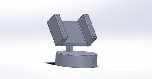
Intro to Computer-Aided Design3D Modeling Having used Soidworks in my BME 182 course, I had some experience in using the sketching and extruding tools, which were two of the tools I utilized in forming our design Additionally, I used a tool known as a chamfer, which cuts into a solid structure at an angle. Once I had the three main parts of the product created I assembled them. Since the regions of the parts I would be attaching were not ends or edges, I had to add sketches of circles to the desired spots. These circles acted as markers in the assembly process, indicating where I wanted the parts to attach to one another. When using Solidworks in my BME 182 class, I had always had an image to base my product design on. When forming our own product for that class, I used a teammates sketch of the device; however, for this product, no one had made a design. We did have an idea that we wanted our product to look like a cell phone dashboard mount, so I researched an image of one such device and used it as a guide for how to create our product. Our Design
Feature 1: ConsumablesThe consumables used in this investigation will remain unchanged as we found no issues when using them. The supplies provided were sufficient, effective, and easy to use. These materials included PCR mix, primer solution, SYBR Green solution, buffer, micropipette tips, PCR tubes, eppendorf tubes, and fluorimeter slides. Feature 2: Hardware - PCR Machine & FluorimeterThe OpenPCR machine and fluorimeter will be used according to the original protocol for this investigation. No major changes were made to the use of the machinery themselves. The top fault which we identified in regards to the system with the Fluorimeter was the phone stand. When working with the previous phone stand, there would be problems regarding the height the phone needed to be put at in order to achieve the correct angle for the picture to be taken. There were also problems with the stability of the phone stand while taking the pictures. For instance, if we pressed the button to take the pictures with too much pressure, the phone would fall and we would have to reset the entire system. We addressed this problem by creating our own stand that is adjustable. It has a stable wider base in order to ensure that there is no chance the phone will fall over and will remain still while photographs are being taken, and minimize the chances of the phone falling. The clamps holding the phone will be adjustable, so as to accommodate larger phones. The grip will be able to be raised and lowered to get the phone at the right angle, and the phone will be able to be tilted while held in the clamps. During the duration of the experiment, our group did not discover any faults in regards to the PCR machine. During the time in which our group was using the PCR machine, it was effective in heating the samples in order for the segments of the DNA to be amplified, which is the main purpose the PCR machine. |
||||||


