BME100 f2015:Group6 1030amL6

From OpenWetWare
Jump to navigationJump to search
BME 100 Fall 2015 Home
People
Lab Write-Up 1 | Lab Write-Up 2 | Lab Write-Up 3
Lab Write-Up 4 | Lab Write-Up 5 | Lab Write-Up 6
Course Logistics For Instructors
Photos
Wiki Editing Help


BIOEDGE INCORPORATED

Caroline Dolt(s)
Kamrie Ickes(s)
Rex Moore
Connor Beck
Name: Chricitan B. Adane
Andres X. Gandara


LAB 6 WRITE-UP

Bayesian Statistics

Overview of the Original Diagnosis System

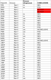
In order to properly analyze the disease-associated SNP 17 teams of 6 students were each given 2 of the 34 patients to analyze. Each group performed OpenPCR on the samples to determine if the disease-associated SNP was present. In order to minimize error each group was given three samples for each patient to analyze. Also, positive and negative controls were provided to ensure that all groups were basing their data on the same values for a positive and negative conclusion. For the fluorimeter tests three images were taken for each droplet which used the same amount of SYBR Green and sample of patient solution. When analyzing the ImageJ the same circle was used to analyze all the droplets which allowed for less variation to ensure more accurate data. The data for 30 patients was succesfull in finding a conclusion. However only 16 of the diagnoses were accurate in analyzing the presence, or lack thereof, of the disease associated SNP. The class produced only two inconclusive results and two patients did not receive data. The class data table was included to make it easier to see the data being discussed. Some problems that were encountered were that it was difficult to take accurate photos of the droplets because the camera would take a burst of pictures which had a flash which could have affected the SYBR Green. Also, the camera was at different lengths from the droplet which slightly influenced the ImageJ which would have changed the data, causing some of the conclusions be inaccurate.

What Bayes Statistics Imply about This Diagnostic Approach


The PCR replicates were generally reliable for calculations 1 and 2. The second set of calculations was more reliable because the probability of a negative final test conclusion given a negative diagnostic signal was close to 90%. However, for the probability of a positive final test conclusion given a positive PCR reaction was only around 80%. The reliability for both calculations is an acceptable number but it could be desired that the number for each would be closer to 100%. The probabilities show that the likelihood of a false test conclusion is fairly small since each test conclusion was greater than 75%. The calculations show that the PCR replicates are reliable in determining if the disease SNP is present or not. However, there is room for improvement which would make the already reliable test even more reliable.


PCR is very reliable when it comes to predicting if the disease will not develop. When given a negative final test conclusion the probability of not developing the disease is upwards of 90% which means that the PCR machine is very reliable when it comes to predicting that the disease will not develop. On the other hand the PCR machine was less reliable when it came to predicting the probability of a disease when a positive test was given. The PCR machine only showed less than a 25% chance of the disease developing even though a positive test was shown. The PCR machine was not very reliable when predicting the development of the disease but was very accurate when it came to predicting the disease not developing.

One possible source of error could be mistakes that were made within Image J which would have influenced the results. Another possible source of error could be that the phone taking the pictures would do a burst of photos which still used flash even though flash was turned off which could have affected the results of the experiment. A third possible source of error could be that while taking the photos the camera was too far from the droplet which could have changed the values produced in Image J which would affect the data in a negative aspect.

Intro to Computer-Aided Design

TinkerCAD
TinkerCAD is an online 3D modeling tool used to rebuild a part of the PCR machine. The parts were uploaded from Dr. Haynes website and put into the create space. For our piece, we chose to assemble the lid. The tool allows you to move the pieces up and down, around, change color, or add basic geometric shapes from the site. We also used the site to make the logo for our company, shown below.


Our Design


Description of image


For our design, we decided to make the PCR system smaller and battery powered. This makes it portable, less expensive, and open to a larger market. This design was chosen to make the device available for more professionals. With a battery powered device, the PCR machine can be used in more scenarios were electricity is not abundant such as Doctors Without Borders as well as other philanthropic organizations. Our OpenPCR design is different because it will be smaller, less expensive and battery powered which allows for it to be used in more markets.


Feature 1: Consumables

  • micropipetter tips
  • biohazard disposal bag
  • PCR Disease Primers
  • PCR tubes
  • Glass Slides
  • SYBR Green solution
  • Buffer controls

The materials are shipped in a cold container to prevent degradation or reaction within the solutions. The SYBR Green will be in darkened tubes to limit the amount of light that enters the tube which could degrade SYBR Green. The micropipetter tips will be sealed in a container to prevent any possible contamination. The PCR tubes and the glass slides will each be kept in separate sealed containers to avoid possible contaminants. Upon arrival the disease primers, SYBR Green and buffer controls should be placed in a lab refrigrator taking extra precautions to keep SYBR Green covered to prevent degradation. Our design improves on the previous one because it allows for all the consumables needed to be available at the same time as well as prevent possible contamination by keeping the tips, slides, and tubes sealed.

Feature 2: Hardware - PCR Machine & Fluorimeter

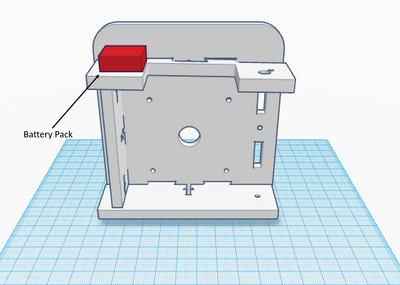
The changes we made were both to the PCR machine. A battery pack was added (shown in red) such that the PCR machine is now self-sufficient on power. In addition, the PCR machine was made smaller yet still holds 16 PCR tubes. These changes collectively make the PCR machine smaller, more portable, and cheaper. Making it easier for researchers who need this technology on the field and making it a viable option not only for those who can not afford an advanced one, but a viable option for those who do, but need a portable version for there research.