20.109(F11): Mod 2 Day 6 Readouts of DNA, Protein
Readout DNA
Introduction
Western Analysis
The mutant phenotypes you're following might be explained by changes in phosphatasing activity of Cph8. They could also be explained by changes in protein expression. For example, if the protein's concentration is 1/10th that of wild type, then it might decrease the pool of kinased OmpR, resulting in lower LacZ activity when the cells are grown in the light even though this would have ~ nothing to do with the phosphatasing reaction rates. Thus it's important to show that the protein still exists in the cell (at a minimum) and that its expression is about the same as the wild type protein (ideally). With a Western analysis, you will compare the expression levels of the wild type and mutant proteins, detecting the various Cph8 versions using an antibody that was raised to a His6-tagged version of EnvZ.
There are some important differences in protein and DNA gel electrophoresis. One thing you’ll notice right away is that the gel itself is different. DNA molecules are typically separated thorough an agarose matrix whereas acrylamide is used for proteins. Both are porous sieves that retard molecules based on their length, with smaller molecules moving through the matrix faster than longer molecules. Agarose gels are run horizontally and acrylamide gels are set in the tank vertically but gravity has nothing to do with either separation. Electrical poles draw the charged molecules through the matrix. Unlike DNA, proteins do not have a uniform charge so before electrophoresis they are coated with a charged molecule (called SDS) to add negative charge proportional to their length. You could expect proteins of identical length but folded into different shapes to separate differently (not the desired outcome) so proteins are also unfolded before they are loaded on a protein gel. This is done by boiling them in the presence of a reducing agent, breaking disulfide bridges and denaturing the protein. The last notable difference in DNA and protein electrophoresis is the visualization techniques used to find the molecules once they’ve passed through the gel. Recall that DNA was visualized with Ethidium Bromide, an intercalating dye that changes its fluorescence when bound to DNA. Proteins can be detected with Coomassie stain, which detects abundant proteins in the gel, turning them blue. Other more sensitive techniques are available for staining protein gels (for example silver stain). Finally, a Western blot can be used, as you will today, to transfer the proteins from the gel to a membrane that is later probed with an antibody specific to the protein of interest. In our case we will use a polyclonal antibody to detect the EnvZ portion of Cph8.
Today is a busy day! In addition to running your protein gel and blotting the proteins to a nitrocellulose membrane, you will also look at the sequencing data if it is available as well as prepare bacterial photographs with your new strains.
Protocols
Part 1: Protein Gel (SDS-PAGE)
Each group will run a lane of molecular weight markers, a lane with a positive control for the Western (e.g. an aliquot of purified His6-EnvZ), a lane with the wild type light sensor, and two lanes of mutants from your library screen. Two teams will share one gel.
- Retrieve the bacterial cultures carrying the wild type or mutant light sensors that have been grown.
- To compare intensities between lanes on the protein gel it's necessary that equal numbers of cells be loaded into each well. You'll assess the number of cells in each sample by making a 1:10 dilution of the three strains in Z-Buffer and use the spectrophotometer to measure the density of the samples at a wavelength of 600 nm. This measurement tells you something about the number of cells in a millileter of liquid. For example a reading of 0.7 says the sample has 0.7 OD units of cells / ml.
- Calculate the volume of your cells needed to give 2 OD. Thinking again about a sample that reads 0.7 OD: if you wanted to collect the number of cells equivalent to 2 OD unit, then you would have to collect 2/0.7 = 2.85 ml of that sample to get 2 OD's worth of cells. Heads up: don't forget that your spectrophotometric reading is for a 1:10 dilution of the original (undiluted) samples, so if you go back to the overnight cultures you'll have to take that dilution factor into account.
- Move the calculated volume of cells to well-labeled eppendorf tubes, and spin the tubes in a microfuge for 1 minute to pellet the bacteria. Be sure that each eppendorf is balanced in the microfuge with an opposing eppendorf containing the same volume. You can collect greater than 1.5 ml of cells by dividing the total volume into smaller aliquots that do fit in the eppendorfs, harvesting the cells, removing the supernatants from the pellets and then adding more culture volume to the same eppendorf tube.
- Resuspend each pellet in 100 ul of "EasyLyse" protein extraction solution (a commercial product from a company called Epicentre). Incubate the solutions at room temperature for 5 minutes, then pellet the debris by spinning the tubes in the microfuge (full speed) for 2 minutes. The supernatant is the lysate that you will run on your protein gel.
- Mix 30 ul of the bacterial lysates with 30 ul of 2X sample buffer. Sample Buffer contains glycerol to help your samples sink into the wells of the gel, SDS to coat amino acids with negative charge, BME to reduce disulfide bonds, and bromophenol blue to track the migration of the smallest proteins through the gel. Wear gloves when using sample buffer or your hands will get blue and smelly.
- Prepare your positive control H6-EnvZ protein tube by moving 50 ul of the sample to an eppendorf tube.
- Put lid locks on the eppendorf tubes and boil for 5 minutes.
- Put on gloves. Load the indicated volumes of each sample onto your acrylamide gel in the order below. Once you have loaded a sample from one tube, move it to a different row in your eppendorf tube rack. This will help you keep track of which samples you have loaded.
| Lane | Sample | Volume to load |
|---|---|---|
| 1 | "Kaleidoscope" protein molecular weight standards | 10 ul |
| 2 | H6-EnvZ positive control protein | 40 ul |
| 3 | wild type light sensor | 40 ul |
| 4 | mutant candidate 1 | 40 ul |
| 5 | mutant candidate 2 | 40 ul |
| 6 | "Kaleidoscope" protein molecular weight standards | 10 ul |
| 7 | H6-EnvZ positive control protein | 40 ul |
| 8 | wild type light sensor | 40 ul |
| 9 | mutant candidate 1 | 40 ul |
| 10 | mutant candidate 2 | 40 ul |
10. Once all the samples are loaded, turn on the power and run the gel at 200 V. The molecular weight standards are pre-stained and will separate as the gel runs. The gel should take approximately one hour to run. During that hour, you should work on part two of today's protocol.
11. Wearing gloves, disassemble the electrophoresis chamber.
12. Blot the gel to nitrocellulose as follows:
- Place the gray side of the transfer cassette in a tupperware container which is half full of transfer buffer. The transfer cassette is color-coded so the gray side should end up facing the cathode (black electrode) and the clear side facing the anode (red).
- Place a ScotchBrite pad on the gray side of the cassette.
- Place 1 piece of filter paper on top of the ScotchBrite pad.
- Place your gel on top of the filter paper.
- Place a piece of nitrocellulose filter on top of the gel. The nitrocellulose filter is white and can be found between the blue protective paper sheets. Wear gloves when handling the nitrocellulose to avoid transferring proteins from your fingers to the filter.
- Gently but thoroughly press out any air bubbles caught between the gel and the nitrocellulose.
- Place another piece of filter paper on top of the nitrocellulose.
- Place a second ScotchBrite pad on top of the filter paper.
- Close the cassette then push the clasp down and slide it along the top to hold it shut.
- Place the transfer cassette into the blotting tank so that the clear side faces the red pole and the gray side faces the black pole.
13. Two blots can be run in each tank. When both are in place, insert the ice compartment into the tank. Fill the tank with buffer. Be sure the stir bar is able to circulate the buffer. Connect the power supply and transfer at 100 V for one hour. You can use this time to complete the other parts of today's protocol.
14. After an hour, turn off the current, disconnect the tank from the power supply and remove the holders. Retrieve the nitrocellulose filter and confirm that the pre-stained markers have transferred from the gel to the blot. Move the blot to blocking buffer (TBS-T +5% milk) and store it in the refrigerator until next time.
Part 2: DNA sequence analysis
If the data from GENEWIZ is available for you to examine, continue with this analysis.

We have provided two ways for you to look at the sequencing data. There are several other programs that are also useful and if you have a favorite, please feel free to use it.
- OPTION 1: Align DNA with a plasmid editor, Ape
- Log in to GENEWIZ using the user name: nkuldell AT mit DOT edu and the pswd: be20109
- View or download the sequence files that are associated with your mutants. These should open up as either a text file, which you can copy into the ApE program that's on your laptop or as an ApE file directly. If there were ambiguous areas of your sequencing results, these will be listed as "N" rather than "A" "T" "G" or "C." It's fine to include Ns in the steps listed below.
- If you've started a new ApE file for your mutants, be sure to name it something more meaningful than the default "New DNA"
- Compare the mutant sequence to the wild type. The wild type sequence is available in several forms on the front page of this module.
- The ApE program has an "align sequences" option for you to compare two files. Since the wild type and the mutant candidates were sequenced with the same primer, this should be a reasonably straightforward comparison.
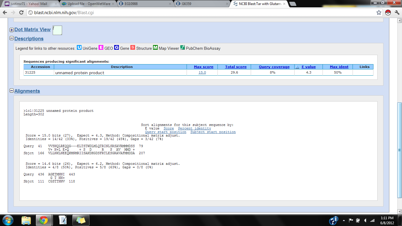
- Print and save a screenshot of the relevant alignment (using shift/command/4 or the Grab program under utilities), and draw conclusions about the alignment in your notebook. You might want to email the alignment screen shot to yourself or post it to your wiki userpage.
- OPTION 2: Align DNA with "bl2seq" from NCBI

BLAST is an acronym for Basic Local Alignment Search Tool, and can be accessed for free through the National Center for Biotechnology Information (NCBI) home page
- Log in to GENEWIZ using the user name: nkuldell AT mit DOT edu and the pswd: be20109
- View or download the sequence files that are associated with your mutants. These should open up as either a text file, which you can copy into the ApE program that's on your laptop or as an ApE file directly. If there were ambiguous areas of your sequencing results, these will be listed as "N" rather than "A" "T" "G" or "C." It's fine to include Ns in the steps listed below.
- If you've started a new ApE file for your mutants, be sure to name it something more meaningful than the default "New DNA"
- Paste the mutant's sequence into the "Sequence 1" box at the BLAST2 sequences site. The alignment program can be accessed through the NCBI BLAST page or from this link.
- Paste the pCph8 sequence from here or here into the "Sequence 2" box.
- Align the sequences. Matches will be shown by lines between the aligned sequences.
- Print and save a screenshot of the relevant alignment (using shift/command/4 or the Grab program under utilities), and draw conclusions about the alignment in your notebook. You might want to email the alignment screen shot to yourself or post it to your wiki userpage.
Identify Amino Acid changes with Sequence Manipulation Suite
Once you have identified the variations in the sequences, if there are any, then you'll want to know what amino acids they correspond to. You can use the Sequence Manipulation Suite to help you translate the region of interest in all 3 reading frames. The correct translation frame should not have stop codons in the wild type sequence and should have the expected A G V S H sequence (= K-P+ hotspot) in the translation of the wild type Cph8. Alternatively you can use the sequence information from the front page of this module to identify the reading frame and use the genetic code to find your mutations. Either way, you should print and save a screenshot of the relevant translation, and draw conclusions about the amino acid changes in your notebook. You might want to email the translation screen shot to yourself or post it to your wiki userpage.
Part 3: Bacterial Photograph
Review the protocol presented earlier in this module.
Part 4: OPTIONAL: Plan additional experiments
You can take the experimental reins over these next few days. As you've collected data for these mutants, there may be additional experiments that you've thought of that you'd like to try. Time to plan them now and get the reagents you'll need. Let us know what you're thinking about doing and we'll try to make it happen.
DONE!
For Next Time
- To be turned in: You must calculate the expected length of the Cph8 protein using the DNA sequence that's here. If you don't already have a favorite program for doing this calculation, try to use the suite of DNA analysis programs that are here. Express the size of the protein both in # of amino acids and also in kilodaltons. The latter will be useful as you examine your Western blot next time.
- NOT to be turned in: You should update the Materials and Methods section that you are writing for your research article to include the experiments you have performed today.
Reagents
- Epicentre EasyLyse Solution
- BioRad Ready Gel 4-15% Tris-HCl Gel, cat #161-1158
- SDS-PAGE Loading Dye
- 60 mM Tris, pH 6.8
- 2% SDS
- 1% glycerol
- 0.01% bromphenol blue
- 5% beta-mercaptoethanol
- Running Buffer
- 25 mM Tris
- 192 mM Glycine
- 0.1% SDS
- Transfer Buffer
- 25 mM Tris
- 192 mM glycine
- 20% v/v methanol
- TBS-T Tris-Buffered Saline + Tween + milk
- 20 mM Tris, pH 7.5
- 500 mM NaCl
- 0.1% Tween
- 5% milk
- H6EnvZ + control protein

