Fluorescence microscope pictures by myself
- Heather 11:17, 13 November 2008 (EST):
I also wrote about these a little in today's blog post. All these photos are of cephalic human neural crest cells discussed over the last couple of months in the notebook. Also check out this link for cell compartments (ER vs Golgi?).
 alpha SMA (red, Alexa Fluor 555); CNPase not very visible from brief passage on confocal. Look again on Tuesday.
 alpha SMA (red, Alexa Fluor 555), superimposed with green/blue autofluorescence.
 TuJ1 (invisible at Alexa Fluor 633) + beta 2 microglobulin (Alexa Fluor 555), superimposed with green/blue autofluorescence.
 alpha SMA (red, Alexa Fluor 555)
 alpha SMA (red, Alexa Fluor 555), superimposed with blue autofluorescence only.
- Stempan stem cell medium (PAN)
 alpha SMA (red, Alexa Fluor 555), superimposed with green/blue autofluorescence. 20x instead of others at 40x.
 alpha SMA (red, Alexa Fluor 555), 20x.
 TuJ1 (invisible at Alexa Fluor 633) + beta 2 microglobulin (Alexa Fluor 555), superimposed with green/blue autofluorescence.
 PGD2 doesn't seem to stain much (in red, Alexa Fluor 546). Red, green and blue perfectly superimpose. 40x.
 alpha SMA (red, Alexa Fluor 555), superimposed with green/blue autofluorescence. Note vesicles, as if secreted from Golgi eg. lysosomes, peroxisomes or melanosomes.
- 401 Panserin medium. The one with all the melanocyte-like things with phase-dark vesicles. Anything marked "nucleus" refers to a non-stained oval structure.
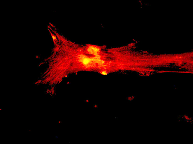
 alpha SMA (red, Alexa Fluor 555), 40x.
 alpha SMA (red, Alexa Fluor 555), 40x.
 alpha SMA (red, Alexa Fluor 555), 40x.
- Heather 11:17, 13 November 2008 (EST):
|  Genomics of human neural crest cells


