Biomod/2013/Todai/Result
<html> <head> <meta name="viewport" content="width=1200">
<style>
</style> </head>
<body>
<figure>
<img src="http://openwetware.org/images/9/9c/Logo-OCKver.png" width=730px height=128px>
</figure>
 <a href="#TOP">
   <figure>
     <img src="http://openwetware.org/images/b/b1/Return-top-0828new.png" width:60px height:60px>
   </figure>
 </a>
</body> </html>
<html> <head> <title>Result-Todai nanORFEVRE-</title> <style>
figure.figure-left {
 position:relative;
 left:-35px;
 }
.res-conclusion {
 font-size:120%;
 font-weight:bolder;
 color:blue;
 position:relative;
 left:30px;
 margin-top:10px;
 }
</style>
</head>
<body>
<a name="Result"> Result</a>
<figurestyle="position:relative;left:-10px;">
<img src="
http://openwetware.org/images/1/15/Todai_Projectsteps_map.png" width=720px height=480px >
</figure>
<a name="Contents"> Contents</a>
<a href="#STEP1">STEP 1: DNA strands assemble to form designed structures</a>
- <a href="#Optimize_the_condition_to_assemble_OCK">1)Optimize the condition to assemble OCK</a>
- <a href="#Conformation_of_the_3D_structure_of_OCK_by_TEM">2)TEM imaging of the 3D structure of OCK</a>
<a href="#STEP2">STEP 2: Penetration into the membrane</a>
- <a href="#Flotation_assay">1)Flotation asssay</a>
- <a href="#Preparation_of_GUVs">2)Preparation of GUVs</a>
<a href="#STEP3">STEP 3: Recognition of cancer-specific proteins</a>
- <a href="#Optimization_of_aptamer_lock_system">1) Optimization of aptamer-lock system</a>
- <a href="#Embedding_of_recognition_system_to_OCK">2) Embedding of recognition system to OCK</a>
<a href="#STEP4">STEP 4: Oligomerization in solution</a>
- <a href="#Oligomerization_by_streptavidin-biotin_complex">1)Oligomerization by streptavidin-biotin complex</a>
- <a href="#Tem_imaging_of_OCK_dimers_by_streptavidin-biotin_complex">2)TEM imaging of OCK dimers connected by streptavidin-biotin interaction</a>
- <a href="#Oligomerization_by_Click_reaction">3)Oligomerization by Click reaction</a>
<a name="Oligomeric_Cell_Killer_(OCK)"> Oligomeric Cell Killer (OCK)</a>
<article>
<a name="STEP1"> STEP 1: DNA strands assemble to form designed structures</a>
<a name ="Optimize_the_condition_to_assemble_OCK"></a>1)Optimize the condition to assemble OCK
    <figure>
        <iframe style="float:left; margin:0;margin-right:-10px;margin-bottom:10px; position:relative;left:-20px;" width="420" height="315" src="//www.youtube.com/embed/1ci93_QI6QA" frameborder="0" allowfullscreen></iframe>
</figure>
The DNA nanostructure, <a target="_bramk" href="http://openwetware.org/wiki/Biomod/2013/Todai/Design#Oligomeric_Cell_Killer " style="color:#e00000"> "Oligomeric Cell Killer" </a> , was designed to achieve our goal(--><a href="http://openwetware.org/wiki/Biomod/2013/Todai/Project">Project</a>).
The result of simulation by "CanDo[1]" showed the shape and flexibility of OCK. To know the optimum condition of the structure assembly, we did experiments in three conditions as follows.
- At different concentration of MgCl2
- At different incubate temperature
- At different length of incubate time
       (<a target="_blank" href="http://openwetware.org/wiki/Biomod/2013/Todai/Experiment#Protocols" style="color:#e00000;">
       protocols
       </a>
       )
Optimum concentration of MgCl2
<figure>
        <img src="http://openwetware.org/images/a/af/OOCK_Optimize_MgConc.png" width=640px height=360px style="position:relative; left:10px">
           <figcaption> Agarose-gel electrophoresis to research the optimum concentration of MgCl2
</figcaption>
</figure>
Fast-migrating species upon agarose-gel electrophoresis was yielded at 10~20mM MgCl2 condition. At higher MgCl2 concentration, a sub-band, which might be a dimer, appeared.
--->> Optimum concentration of MgCl2: 10mM
Optimum incubate temperature
<figure>
          <img src="http://openwetware.org/images/9/95/OOCK_Optimize_Temp-Todai.png" width=640px height=360px >
          <figcaption>
          Agarose-gel electrophoresis to research the optimum temperature
          </figcaption>
</figure>
Fast-migrating species upon agarose-gel electrophoresis was yielded at 52.0 °C.
--->> Optimum temperature : 52.0 °C
To decide optimum length of incubate time
<figure>
          <img src="http://openwetware.org/images/9/94/OOCK_Optimize_Time-Todai.png" width=640px height=360px >
          <figcaption>
          Agarose-gel electrophoresis to research the optimum time
          </figcaption>
</figure>
The band for 3h is fast migrated and sharp.
--->> Optimum incubate time : 3h
<a name="Conformation_of_the_3D_structure_of_OCK_by_TEM"></a>2) TEM imaging of the 3D structure of OCK
Gel electrophoresis cannot visualize the 3D structure of OCK, so it was confirmed by Transmission electron microscopy (TEM). (<a target="_blank" href="http://openwetware.org/wiki/Biomod/2013/Todai/Experiment#Protocols" style="color:#e00000;"> protocols </a> )
</article>
TEM imaging of OCK
<figure>
        <img src="http://openwetware.org/images/e/ea/Monomer-Todai.png" width=480px height=360px>
           <figcaption> TEM image of OCK
           Three monomers of OCK were observed in this figure.

           </figcaption>
</figure>
TEM images confirm that our OCK has two domains. Comparing the observed structure to our design, one domain match the shape and size to plane-like domain. And the other domain matches to stick-like domain. Furthermore, in close watching the images, DNA well, which exists one side of plane-like domain, could be detected.
</article>
<a name="STEP2"> STEP 2: Penetration into the membrane</a>
<article>
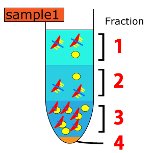
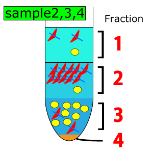
<a name="Flotation_assay"></a>1) Flotation assay
OCK was designed to penetrate lipid bilayer. However, it is difficult to conclude the penetration of OCK. Therefore, we first did flotation assay to detect the interaction of OCK with lipid. (<a target="_blank" href="http://openwetware.org/wiki/Biomod/2013/Todai/Experiment#Protocols" style="color:#e00000;"> protocols </a> )
<figure>
<img src="http://openwetware.org/images/6/67/Todai_Fraction2.png" width=300px height=300px>
</figure>
<figure>
<img src="http://openwetware.org/images/e/e1/Todai_Fraction1.png" width=300px height=300px>
</figure>
<figure>
        <img src="http://openwetware.org/images/c/ca/Todai_result_step2_fluolescence.JPG" width=450px height=350px>
           <figcaption> The fluorescence intensity of NIL (in liposome) in each fraction

           The result of fluorescence spectrophotometer (JASCO, FP-6500) showed that liposome distributed mostly in fraction 3(lower layer).
           </figcaption>
      </figure>
          <figure>
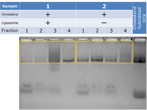
        <img src="http://openwetware.org/images/f/fe/FlotationOCK12gel-Todai.png" width=480px height=360px>
           <figcaption> 1% Agarose gel electrophoresis of each fraction in sample 1, 2
           </figcaption>
      </figure>
          <figure>
        <img src="http://openwetware.org/images/0/0d/FlotationOCK34gel-Todai.png" width=480px height=360px>
           <figcaption> 1% Agarose gel electrophoresis of each fraction in sample 3, 4
           </figcaption>
      </figure>
          <figure>
<img src="http://openwetware.org/images/f/ff/450px_FloatingOCKprofile-Todai.jpg" width=450px height=300px>
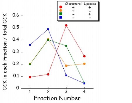
<figcaption>The ratios of OCK in each fraction were analyzed by the density of band.</figcaption>
</figure>
With the condition of cholesterol +/ liposome+, the peak fraction was No.3, which coincide with peak fraction of liposome. In contrast, lacking of cholesterol or liposome, OCK exist mainly in fraction No.2. As the peak fraction of OCK shifted from fraction No.2 to No.3, with the attachment of cholesterol and existence of liposome, we concluded that OCK stack in liposome.
--> OCK stack in liposome.
</article>
<article>
<a name="Preparation_of_GUVs"></a>2) Preparation of GUVs
GUV, Giant Unilamellar Vesicle, was prepared to visualize the sticking of OCK in membrane. The comparasion between the fluorescence of OCK (Cy5) and GUV(NIL, Nile Red) was expected to suggest the sticking. (<a target="_blank" href="http://openwetware.org/wiki/Biomod/2013/Todai/Experiment#Protocols" style="color:#e00000;"> protocols </a> )
<figure>
<img src="http://openwetware.org/images/b/be/GUVconfocal_scale-Todai.png" width=450px height=350px>
</figure>
GUVs were observed with confocal laser scanning microscope (Carl Zeiss, LSM 5 Exciter). As the tracer of GUVs, 0.1 mol% Nile Red (Ex 553 nm, Em 637 nm) was used. About 10 um of GUVs were observed.
</article>
<article>
<a name="STEP3"> STEP3: Subunits recognize cancer-specific proteins. </a>
<a name="Optimization_of_aptamer_lock_system"></a>1) Optimization of aptamer-lock system
<figure>
<img src="http://openwetware.org/images/5/50/Todai_Recognition_ideal_mod.png" width=480px height=270px>
</figure>
We did pilot study of oligomerization process triggered by membrane protein recognition. We used cholesterol modified PDGF as model membrane protein, as DNA origami embedded aptamer system recognizing PDGF was reported (Douglass et al. (2012)).
Our simplified model lock system is consisted with two steps: 1) Blocking of streptavidin binding to biotin by steric hindrance. Our lock system consists of two strands: biotin attached strands (biotin strands) and aptamer attached strands (aptamer strands). These two strands hybridize each other in inactive form and hide biotin moiety from the streptavidin by steric hindrance effect. 2) Upon binding of ligands (PDGF in this study) to aptamer strands, the complementary strand(biotin strands) is released from the DNA aptamer, because ligands take over the DNA strands of DNA aptamer from the complementary strands, and biotin can now bind to streptavidin. Therefore, the cancer cell recognition and OCK oligomerization are achieved simultaneously in the future study.
</article>
Integration of aptamer strands into DNA origami tile
<figure>
<img src="http://openwetware.org/images/d/d3/Todai_intofapt_1.png" width=600px height=450px>
       </figure>
                                 <figure>
<img src="http://openwetware.org/images/6/6c/Todai_intofapt_2.png" width=600px height=450px>
       </figure>
                    <figure>
        <img src="http://openwetware.org/images/7/7f/Tile-ins.png" width=480px height=360px>
           <figcaption>
</figcaption>
</figure>
We confirmed the integration of aptamer attached strands (aptamer strands) into rectangle DNA origami tile (rect-tile).
Responsibility of aptamer
<figure>
<img src="http://openwetware.org/images/4/42/Todai_Recognition_tile_B_PDGF_mod.png" width=600px height=450px>
      </figure>
      <figure>
        <img src="http://openwetware.org/images/f/f5/Figure9_10-Todai.png" width=480px height=270px>
           <figcaption>
</figcaption>
</figure>
We confirmed the responsibility of aptamer sequence embedded in rect-tile (shown above). The position of Cy5-PDGF band coincided with that of DNA tile, showing that the aptamers work also on rect-tile. Furthermore, the linker length between aptamer sequence and staple sequence, the latter staple sequence is embedded into rect-tile, does not affect the binding ability of aptamer to PDGF.
Blocking capability of lock system by aptamer
<figure>
<img src="http://openwetware.org/images/9/96/Todai_Recognition_tile_B_SA_mod.png" width=600px height=450px>
</figure><tbody> </tbody>
       <figure>
         <img src="http://openwetware.org/images/3/30/Figure11_12-Todai.png" width="360px" height="240px">
</figure> |
       <figure  style="position:relative;left:-50px;">
         <img src="http://openwetware.org/images/2/24/Koyama_131027_1-Todai.JPG" width="240px" height="240px">
</figure> |
We confirmed the blocking capability of our lock system for streptavidin
binding (left figure, the image of gel electrophoresis). Our lock system consists of two strands:
biotin attached strands (biotin strands) and aptamer attached strands
(aptamer strands). These two strands hybridize each other in inactive
form and hide biotin moiety from the streptavidin by steric hindrance
effect. We confirmed this blocking capability by mixing Cy3 labeled
streptavidin with lock system embedded rect-tile. Data indicates that
the slight blocking capability upon shorten the polyT linker between
aptamer sequence and staple sequence. Recently, we tried other sequence
and have better results, which may be presented in the Jamboree in Boston.
Optimum embedding condition of our lock system into rect-tile
<figure>
<img src="http://openwetware.org/images/e/ec/Todai_intofapt_3.png" width=600px height=450px>
       </figure>
      <figure>
        <img src="http://openwetware.org/images/c/c8/Tile_double_insertion-Todai.png" width=480px height=360px>
           <figcaption>
</figcaption>
</figure>
Next, we optimize the embedding condition of our lock system into rect-tile. This time full length of biotin strands were used instead of truncated ones used in above figure. Data indicate that the integrate efficiency of both biotin strands and aptamer strands into rect-tile is independent of the incubate temperature.
We improve our lock system everyday. Don't miss our presentation in Jaboree in Boston !
<a name="Embedding_of_recognition_system_to_OCK"></a>2) Embedding of recognition system to OCK
To embed recognition system to OCK, we equiped PDGF aptamer used in rect-tile to OCK and confirmed the association of aptamer and PDGF . (<a target="_blank" href="http://openwetware.org/wiki/Biomod/2013/Todai/Experiment#Protocols" style="color:#e00000;"> protocols </a> )
Recognition of PDGF by DNA aptamer on OCK
<figure>
<img src="http://openwetware.org/images/c/c3/OCK_PDGFWeb-Todai.png" width=320px height=180px>
</figure>
-->PDGF was recognized by the aptamer of OCK
<article>
<a name="STEP4"> STEP 4: Oligomerization in solution</a>
<a name="Oligomerization_by_streptavidin-biotin_complex"></a>1) Oligomerization by streptavidin-biotin complex
Biotins are equipped to OCK for oligomerization. The experiment which confirmed that streptavidins induced oligomeriation. (<a target="_blank" href="http://openwetware.org/wiki/Biomod/2013/Todai/Experiment#Protocols" style="color:#e00000;"> protocols </a> )
</article>
Streptavidins induced oligomerization
<figure>
<img src="http://openwetware.org/images/5/5b/Streptavidin_dimer-Todai.png" width=600px height=450px>
<figcaption>The mixing ratio of streptavidin to OCK was equal to 5:3, which means the mixing ratio of streptavidin to biotin was equal to 5:6 in the condition (L+R).</figcaption>
      </figure>
    </article>

    <article>
<a name="Tem_imaging_of_OCK_dimers_by_streptavidin-biotin_complex"></a>2) TEM imaging of OCK dimers connected by streptavidin-biotin interaction
Dimers of OCKs were also imaged by TEM to confirm the bands observed in the experiment 3.1) originated from the dimers. (<a target="_blank" href="http://openwetware.org/wiki/Biomod/2013/Todai/Experiment#Protocols" style="color:#e00000;"> protocols </a> )
</article>
       <figure>
         <img src="http://openwetware.org/images/c/c1/Dimerv2-Todai.png" width="300px" height="300px" >
       </figure>
|
       <figure>
         <img src="http://openwetware.org/images/5/53/Dimer_2v2-Todai.png" width="300px" height="300px" >
       </figure>
|
Dimers of OCKs were observed in this experiment and two of them were shown above.
-->The dimerization by streptavidin-biotin complex was confirmed.
<article>
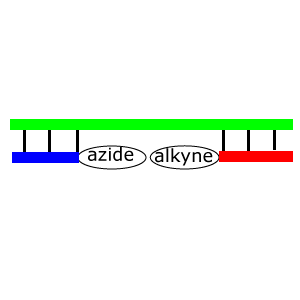
<a name="Oligomerization_by_Click_reaction"></a>3) Oligomerization by Click reaction
Azide and alkyne, which function as a reactive group of click reaction, are also equiped to OCK. It demands Cu+ as catalyst, but too high concentration of Cu+ (cation) might denaturate OCK like Mg2+. Therefore, we optimized the concentration of Cu+ to OCK first, and then the optimum Cu+ concentration to click reaction was investigated. (<a target="_blank" href="http://openwetware.org/wiki/Biomod/2013/Todai/Experiment#Protocols" style="color:#e00000;"> protocols </a> )
a) Optimum concentration of CuSO4 to OCK
<figure>
<img src="http://openwetware.org/images/5/5e/CuAlive-Todai.png" width=600px height=450px>
</figure>
-->Optimum concentration of CuSO4: 625 uM or less
b) Optimum concentration of CuSO4 to click reaction
<figure>
<img src="http://openwetware.org/images/9/94/ClickResult-Todai.png" width=600px height=450px>
</figure>
Product of click reaction appeared over 375 uM CuSO4 concentration. Combining with the stability data, we decided to use 625 uM CuSO4 condition.
--> Optimum concentration of CuSO4: 625 uM
</article>
<article>
<a name="Cupper-free_click_reaction"></a>4) Copper-free click reaction
Click reaction demands copper catalyst, which works as a toxine in human body. Therefore, we studied about copper-free click reaction for the application to human body. (<a target="_blank" href="http://openwetware.org/wiki/Biomod/2013/Todai/Experiment#Protocols" style="color:#e00000;"> protocols </a> )
a) Optimum concentration of CuSO4 to OCK
       <figure>
         <img src="http://openwetware.org/images/2/25/Gelphoto1-Todai.png" width="400px" height="225px">
       </figure>
|
       <figure  style="position:relative;left:-50px;">
         <img src="http://openwetware.org/images/2/2f/Yatagai_131027_1.JPG" width="225px" height="225px">
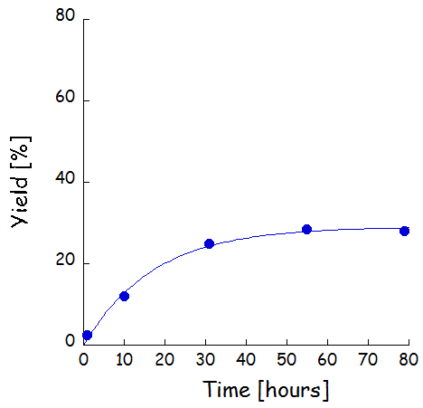
<figcaption> The reaction rate of copper-free click reaction with no accelerator </figcaption> </figure> |
We first measured the Cu-free click reaction in solution (without any catalyst nor accelerator). The association time at 2 uM oligonucleotide condition was 17.1 h, and appearent association time was estimated as 8.1 [1/M/s].
       <figure>
         <img src="http://openwetware.org/images/4/45/Gelphoto2-Todai.png" width="400px" height="225px">
       </figure>
|
       <figure  style="position:relative;left:-35px;">
         <img src="http://openwetware.org/images/2/23/YatagaiclickStA-Todai.png" width="225px" height="225px">
       </figure>
|
       <figure>
         <img src="http://openwetware.org/images/c/c5/Gelphoto3kai-Todai.png" width="360px" height="270px">
       </figure>
|
       <figure style="position:relative;left:-35px;">
         <img src="http://openwetware.org/images/c/c6/Yatagaiclickhybri-Todai.png" width="270px" height="270px">
</figure> |
               <figure  style="position:relative;left:-45px;">
         <img src="http://openwetware.org/images/a/a2/Yatagai_131027_2.JPG" width="270px" height="270px">
</figure>
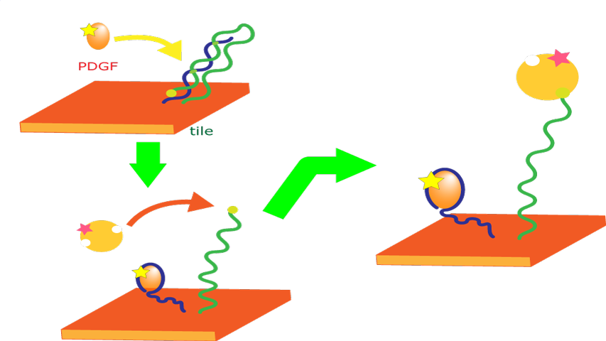
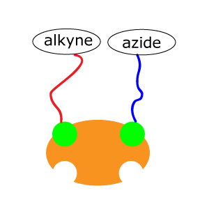
We add accelerator, which can work as a scaffold and make alkyne and azide reactive group close. Acceleration of the click reaction was observed. In other words, azide and alkyne reactive groups do not react each other in solution, but easy to react each other after proximally-positioned. This character is very suitable to prevent non-specific oligomerization, while accelerating the specific oligomerization in OCK.
Note: Streptavidin has 4 identical subunits. So we can not control the binding order of alkyne and azide oligo with streptavidin method. Therefore, vicinity subunit may have identical reactive groups (e.g. alkyne-alkyne or azide-azide, instead of alkyne-azide or azide-alkyne), and may reduce the yield.
--> Cu-free click reaction has suitable character for specific oligomerization.
</article>
<a name="Reference"> Reference</a>
       <a name="proref-1">
       [1] CanDo(<a href="http://cando-dna-origami.org/usersguide">http://cando-dna-origami.org/usersguide</a>)
       </a>
<footer style="position:relative;left:400px"> Copyright © Todai nanORFEVRE, all rights reserved. </footer>
</body> </html>
