Applications of 3D Printing - Microfluidics
Two of the factors driving innovation in 3D printing for microfluidics are channel size and resolution,1.
Exploration into additive materials using other materials such as concrete and biomaterials has also been of interest2.
There are also some new methods of 3D printing that are being developed.
High Resolution 3D Printing Using a Customized Resin and Photolithography Printer Using an adapted stereolithography (SLA) printer and a series of custom-formulated resins, a lab was able to create microchannels at up to 18x20 um resolution3. This method has not been reproduced in literature and definitely has yet to be scaled up, but it holds promise for the future of 3D printing microfluidic circuits.
(Will include some figures of the channel dimensions for scale from the paper)
3D printing is a crucial tool for the developers of microfluidic devices. Not all 3D printing techniques are used by microfluidic developers. "the term “3D printing”—considered to be synonymous with “solid freeform fabrication”—refers to a family of additive-based manufacturing techniques. Importantly, not all 3D printing techniques are suitable for microfluidics. The most widely used 3D printing techniques with relevance to microfluidics are selective laser sintering" (Au, Anthony K, Wilson Huynh, Lisa F. Horowitz, and Albert Folch). The different methods of 3D printing in microfluidics include Fabrication by Stereolithography, Fabrication by Selective Laser Sintering, Fabrication by photo polymer inkjet printing, fabrication by fused deposition modeling, and fabrication by laminated object manufacturing. These different methods of 3D printing have different levels of resolution and automation.
How does 3D printing play an important role in the fabrication of microfluidics devices?
There are some key functions of microfluidic device that includes separation of liquids and samples detection and fluid manipulation. By using 3D printing the time needed for the fabrication process is greatly reduced and it can be done with just one machine and fully automated which allows the process to be easily replicated. The 3D printing offers the opportunity to fabricate the whole microfluidic device in a single step without the need of the assembly step.
Stereolithography (SLA) was the first commercialized 3D printing machine. Thermoplastics photopolymers are used as the base material for SLA fabrication. The photopolymer is in a liquid state before fabrication. A UV laser was used to scan the material and hardened by using low powered, highly focused laser. The platform is lowered by a distance equal to layer thickness (usually 0.05-0.15mm) and then the laser moves on to the subsequent layer and repeat the process again until the desired product is finished. The excess polymer in each layer stays in liquid state and rinsed off the platform. A major advantage of SLA fabrication is the very high precision of the surface resolution.
A good example of a microfluidic device created by the SLA is the immunomagnetic flow assay on-a-chip designed by Lee and team [1]. This experiment is a great example of how works that were previously restricted to laboratory due to their size can now be studied with the aid of 3D printing. The commercialized 3D Viper SLA system printed a cylindrical 3D micro-channel named as a High Capacity Efficient Magnetic O-shaped Separator(HEMOS). (Figure 1)
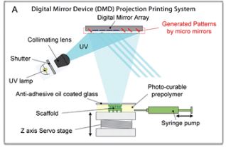
Digital Micromirror Device-based Projection Printing (DMD-PP) is another type of 3D printing technology. It is a projection system that uses a controllable digital mirror to reflect laser light in an entire plane which then allows the curing of an entire layer at a time. (Figure 2) Compared to SLA system the DMD-PP the building structure is bottom-up different from the SLA which requires the stage to move as well as the laser. An example of microfluidic device created using the DMD-PP is the 3D in vitro microchip which is made of hydrogel with a honeycomb structure which allows the microchip to mimic the 3D vascular morphology of an in vivo micro environment.
References
1Au, A. K., Huynh, W., Horowitz, L. F. & Folch, A. 3D-Printed Microfluidics. Angew. Chemie - Int. Ed. 55, 3862–3881 (2016). DOI: 10.1002/anie.201504382 2Ngo, T. D., Kashani, A., Imbalzano, G., Nguyen, K. T. Q. & Hui, D. Additive manufacturing (3D printing): A review of materials, methods, applications and challenges. Compos. Part B Eng. 143, 172–196 (2018). DOI: 10.1016/j.compositesb.2018.02.012 3Gong, H., Bickham, B. P., Woolley, A. T. & Nordin, G. P. Custom 3D printer and resin for 18 μm × 20 μm microfluidic flow channels. Lab Chip 17, 2899–2909 (2017). DOI: 10.1039/C7LC00644F 4Bose, S., Ke, D., Sahasrabudhe, H. & Bandyopadhyay, A. Additive manufacturing of biomaterials. Prog. Mater. Sci. 93, 45–111 (2018). DOI: 10.1016/j.pmatsci.2017.08.003 5Kelly, B. E. et al. Volumetric additive maufacturing via tomographic reconstruction. Science (80-. ). 126, 21 (2019). DOI: 10.1126/science.aau7114
W. Lee, D. Kwon, B. Chung, G. Y. Jung, A. Au, A. Folch and S. Jeon, Anal. Chem., 2014, 86, 6683–6688.
Soft Matter, 2012, 8, 4946 DOI: 10.1039/C2SM07354D
Lab Chip, 2015, 15, 3627 DOI: 10.1039/C5LC00685F
R. Liska, M. Schuster, R. Inführ, C. Turecek, C. Fritscher, B.Seidl, V. Schmidt, L. Kuna, A. Haase and F. Varga, J. Coat.Technol. Res., 2007, 4, 505–510.
C. W. Hull, Journal, 1986.


