Etchevers:Notebook/Genomics of hNCC/2008/10/22
 Genomics of human neural crest cells Genomics of human neural crest cells
|
<html><img src="/images/9/94/Report.png" border="0" /></html> Main project page <html><img src="/images/c/c3/Resultset_previous.png" border="0" /></html>Previous entry<html> </html>Next entry<html><img src="/images/5/5c/Resultset_next.png" border="0" /></html> |
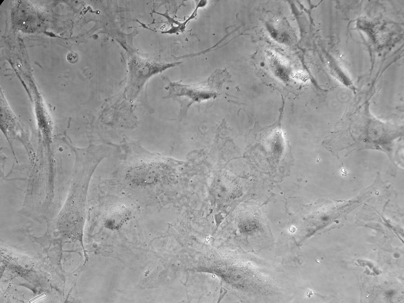
Petit bout de papierLooked at rabbit cornea for the male 980; it may have been the control rabbit; the R and L corneas looked identical. Some auto-fluorescence in collagen layers (PAF fixed, no anti-aldehyde treatment), scar not obvious to find in either case either. May need to cut more eyes, may need to deparaffinate - but I don't think so. Took photos of different cultures after fixation on light microscope.
| |


