File:CFL in vitro.png
From OpenWetWare
Jump to navigationJump to search
Size of this preview: 800 × 471 pixels. Other resolution: 2,639 × 1,554 pixels.
Original file (2,639 × 1,554 pixels, file size: 808 KB, MIME type: image/png)
Summary
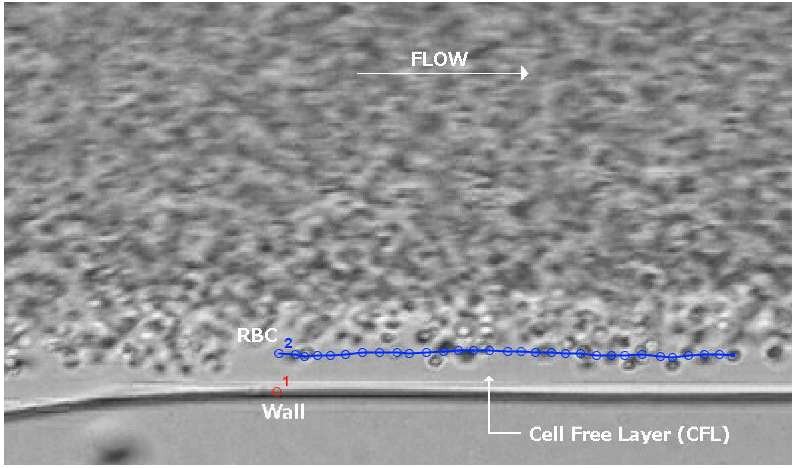
RBCs flow through a PDMS microfluidic channel and form an RBC-FL. The blue line tracks a single RBC across frames. The image was created by overlaying frames of a captured video. Pinto, E.; Faustino, V.; Rodrigues, R. O.; Pinho, D.; Garcia, V.; Miranda, J. M.; Lima, R. A Rapid and Low-Cost Nonlithographic Method to Fabricate Biomedical Microdevices for Blood Flow Analysis. Micromachines 2015, 6 (1), 121–135. https://doi.org/10.3390/mi6010121. is licensed under CC BY 4.0
File history
Click on a date/time to view the file as it appeared at that time.
| Date/Time | Thumbnail | Dimensions | User | Comment | |
|---|---|---|---|---|---|
| current | 12:54, 30 April 2024 | | 2,639 × 1,554 (808 KB) | Nkokkula (talk | contribs) | RBCs flow through a PDMS microfluidic channel and form an RBC-FL. The blue line tracks a single RBC across frames. The image was created by overlaying frames of a captured video. Pinto, E.; Faustino, V.; Rodrigues, R. O.; Pinho, D.; Garcia, V.; Miranda, J. M.; Lima, R. A Rapid and Low-Cost Nonlithographic Method to Fabricate Biomedical Microdevices for Blood Flow Analysis. Micromachines 2015, 6 (1), 121–135. https://doi.org/10.3390/mi6010121. is licensed under CC BY 4.0 |
You cannot overwrite this file.
File usage
The following page uses this file:
