User:Christian Ortiz/Notebook/Biology 210 at AU
March 22, 2015 - CO Zebrafish Experiment
Introduction: The purpose of this experiment was to see the effects that alcohol has on embryo's development. We are able to use zebrafish because based off Dawin's Theory of Evolution, these mammals have a common ancestry. This explains why zebrafish embryos are somewhat similar to human embryos in terms of development. The hypothesis is that we should see the zebra fish have physical abnormalities such as smaller eyes and shorter bodies as well as reduced motor skills such as not being sensitive when the petri dish is touched.
Material/Methods: Two petri dishes were collected and labeled. One was labeled control and the other was labeled test group. A 10 mL volumetric pipette was used to fill the control petri dish with 20 mLs of Deer park water and the test group petri dish with 20 mLs of ethanol (1.5% Alcohol). Using a modified transfer pipette, just had the tip clipped off, 20 translucent embryos were transferred to each dish from the pool of embryos. Observations were made and data was collected. 5 days later 10 mLs of water was removed from the control petri dish and 25 mLs of fresh water was added to the petri dish. As for the test group, 10 mLs of alcohol was removed and 25 mL of new alcohol solution was added. During this time, empty egg cases were also removed and any interesting dead samples were saved in paraformaldhyde. On day 7, 5 mLs of water was removed from the control group and 5 mLs of fresh water was added. On day 7, the embryos were feed one drop of paramecium. As for the test group, 5 mLs of alcohol was removed and 5 mLs of new alcohol solution was added. Between day 7 and 14, 5 mLs of each petri dish solution was removed and 10 mLs of their respected solution was added. Also after day 7, the zebrafish were fed two drops of paramecium after we changed the water and alcohol each time we saw them. When day 14 came around final observations and measurements were made.
Conclusion:
The results supported part of the hypothesis stated. The physical differences were there as the alcohol group had smaller eyes but yet they were still smaller. At one point it seemed like the control group stopped growing. In the development point of view, the alcohol really affected the test group as they took longer to develop. In terms of movement, the test group was more responsive to taps on the petri dish but these were different movements. The test group would move really fast and take sharp turns. Compared to the control group that world react enough to acknowledge the taps but not let it completely freak them out. Lastly, the test group had a slower heart rate compared to the control group. The overall result was that the test group slowly kept dying as the days went by. This shows that alcohol played a huge role in mortality and development. If a mother drinks during pregnancy they should expect to see serious abnormalities.
February 18, 2015 - CO
Observing invertebrates in the Manicured Grasslands.
Introduction: The purpose of this lab was to understand the complexity of invertebrates then use the same skills to analyze invertebrates in the Manicured Grasslands. Invertebrates are extremely diverse, for example the Cnidaria and the Ctenophora are which is an animal with radial symmetry meaning a sphere-like structure. An example of Cnidaria is the jellyfish. Other structures vary, for example instead of radial symmetry some animals have bilateral symmetry which consist of a head and a tail. In relation to the transect, the samples collected are soil samples which means most of the invertebrates studied were arthropod, micro arthropod, and different types of worms. The reason for these specific types of organisms being found in the soil is because these are soil animals known as predators. They're predators in the way that they suck fluids from the roots, or feast on decaying plant material. This benefits the ecosystem because it's a way to reuse decomposing plants and animal material.
Material/Methods:
To analyze the transect, the Berlese Funnel was taken apart and a pipette was used to transfer the liquid from the sample tube to the petri dishes. One sample was taken from the top 10 mls of the tube and placed in a labeled petri dish. The second sample came from the bottom 10 mls of the tube and transferred to the second petri dish. Each petri dish had their own three samples put on separate slides for analysis. When making the slides the group tried to pick up visible organisms. A dissecting microscope was used to help give a better view of the organisms. When an organism was spotted, the group used the "Hope College Leaf Litter Arthropod Key" to identify the invertebrate.
Data/Observations:
The first table is gonna show the overall characteristics of the invertebrates observed.
The size range of the organisms is 5.5 mm. The last organism, who's Phylum was Anthropoda, was the largest organism measuring in at 6mm. The smallest organism was the 4th organism, who's Phylum was Nematoda, measuring in at 0.5 mm. The most common organism in the leaf litter were the unsegmented worms which belong to the Nematoda Phylum.
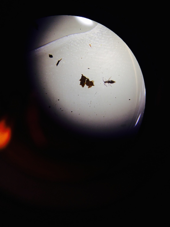
This was an image of organism number 5.

This was an image of the unsegmented worm. It's a little difficult to see because it is hiding next to the debris. The worm is located directly in the center.

This was an image of organism number 1.

Based on the the conditions of the transect and what it has to offer it safe to assume that squirrels, rats, bees, humming birds, and sparrows all use the transect.
The next image is a food web of all the groups of organisms in the transect.
Conclusion:
The Manicured Grasslands showed a very interesting diversity of organisms. Community is seen in different species occupying this transect varying from rats to unsegmented worms. The carrying capacity for this transect is quite small mainly because of what it has to offer. It has no water source which is essential for all living organisms. All this transect has to offer is plants and the little organisms it has with the plants such as insects. In terms of trophic levels, the humming bird, rat, sparrow and the squirrel are on top. This is mainly because of their complexity and what they feed of off. Followed by worms, bees, and insects. These are in the middle because they feed off of bacteria or plants but are still prey to the previous organisms mentioned.
February 10, 2015 - CO The Characteristics of Plants from the Manicured Grasslands and the Importance of Fungi.
Introduction: It is known that land plants evolved from bryophytes which are moss type plants. As time passed by, tracheophytes were formed from bryophytes and the main difference here being the tracheophytes's vascular system. As more time passed plants evolved to flowering plants which are known for spreading pollen. The purpose of the first half of the experiment was to observe plants and characterize them for presence of vascularization, their specialized structures, and mechanism of reproduction. The second part of the lab required observation of fungi. Fungi do not ingest their food rather they absorb nonliving organic matter or feed on living organic mater. Fungi have three divisions Zygomycota, Basidiomycota, and Ascomycota. The overall purpose for this lab was to understand the functions of Plants and Fungi
Material/Methods:
For the Plant portion of the lab, the group was given a Ziploc bag to collect five plant samples from the transect. The five samples had to be diverse and also old leaves and branches were encouraged. The samples were taken back to the lab for observation. Observations taken for the samples were their shape, size, and the cluster arrangement of the leaves. The seeds of the samples were then identified as either monocot or dicot and whether there was any evidence of flowers or spores. The second part of the lab asked to observe and classify samples of fungi.
Data/Observations: Table one shows an overall description of the samples collected from the transects.
Sample #1 was a branch off the chopped bushes near the cement step. It was a monocot and a fibrous root system. This sample belonged to the Seedless Plants group when fully grown. It is believed to be a Pteridophyta (fern).


Sample #2 was a green and thing sample. Its veins were parallel and the leaf should venation. This monocot belongs to the Seedless Plants group.


Sample #3 was a stem on the bush. It seemed to be a stem for a flowering plant. The vascular bundles were usually arranged in a ring. This dicot belongs to the Angiosperms Seed Plants.


Sample #4 was the grass that covers most of the transect. The veins were usually parallel and it showed leaf venation. This monocot belongs to the Seedless Plants group.

Sample #5 was the small bulb found in the soil between the bushes. This dicot should a fibrous root system. This is believed to belong to the Angiosperms Seed Plants when fully grown.


Aside from the samples, the leaf litter seemed to only be around the soil area were the bushes were. This is because of the climate change that made the bushes loose their leaves.

The second part of the lab was observing the fungi samples provided. One thing to keep in mind was Fungi sporangia, this is a hyphae grown upward which forms a dome like structure. These are most commonly known as spores, these Fungi sporangia are important because it is the site of meiosis.
The following images are the images of the samples.
 Sample 1
Sample 1 looks like mold based on the fact that it has spores. This makes this fungi belong to the Ascomycota group.
Sample 2 is a fungus based on the highly organized structure. This belongs to the Basidomycota group.
Sample 3 is a fungus based on the zygospore which is a thick walled structure. This belongs to the Zygomycota group.
The next image is a drawing of what was seen under the microscope for sample number 2.

In conclusion, the transect showed to have a diversity of plants in such a small area. Results showed that the reproduction mechanism was based off what type of group the plant belonged to. To better this experiment, it is recomended to collect samples when they are in their form. This means collecting sample around the right time of year, this allows for a more accurate perspective of the type of plants the transect has to offer. For the fungus, there was a pattern were most of the fungus had highly organized structure.
February 4th, 2015 - CO
Observing a niche at AU
Introduction: The purpose of this experiment was to identify bacteria with DNA sequences as well as observe antibiotic resistance. This experiment had a focus on studying prokaryotes that are grouped in the Domain Bacteria. These Archaea typically grow in extreme environments. For this reason there was no expectation to see any Achaea species grow on the agar plate since it's not an extreme environment. To identify the prokaryotes staining techniques were used as well as the basic bacterial cell shapes. The stain for bacteria is known as the Gram stain. Bacteria that are gram-positive have peptidoglycan and that's why they are able to retain the dye. Bacteria that do not retain the dye are known as gram-negative and typically have less peptidoglycan. Gram-positive bacteria will stain blue while gram-negative will stain pink. The hypothesis for this experiment was that the tetracycline plates would have less bacteria.
Material/Methods: There was a colony count for each of the plates before all the procedures. Four samples were taken, two from the nutrient agar plate and two from the tetracycline plate. One sample of each plate would be used for a wet mount so that it could be put under a microscope. The other pair of samples would go through staining. For the wet mount, a loop was sterilized over a flame then used to scrap up some of the bacteria from a plate and placed on a slide that had water. After the cover slip was placed over the drop it was put under the microscope for observation. For the gram stain, a loop was sterilized over a flame then used to scrape up bacteria from a plate and mixed into a drop of water on a slide. A circle would be drawn underneath he sample with a red wax pencil. The slide would then be passed through a flame three times with the bacteria side facing against the flame. When the sample was placed on a tray, the bacteria sample was covered with crystal violet for one minuter. It was later rinsed using a wash bottle that contained water. Once washed it was covered with Gram's iodine mordant for one minuter then rinsed again with the wash bottle. The sample was decolorized by putting 95% alcohol on it for 15 seconds. The smear was then covered with safranin stain for 25 seconds and rinsed off with the wash bottle. The sample was ready for observation so a kimwipe helped clean up the excess water.
Data/Observations: Before any procedure was done, one last observation of the Hay Infusion Culture was taken after three makes of being made. The Hay Infusion still looked the same but there was a new smell that seemed like mold. Also, there seemed to be more white residue.

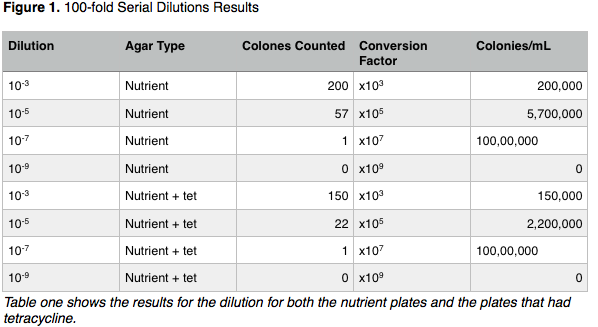
The colonies showed the bacteria as expected where the plate that had tetracycline would have less colonies.
The main difference between the plates with vs without antibiotics, in terms of colonies, was that the plates with antibiotics had less bacteria. Also, the plates with antibiotics seemed to have yellow bacteria ll around were as the other set of plates would have bacteria with white on it. This goes to show that tetracycline prevents a certain number and apparently type of bacteria to grow when in its presence. Species that grow in the presence of tetracycline are insensitive to the antibiotic. The plates with green lettering are the ones with the antibiotic and the ones with red lettering do not have antibiotics.
The characteristics of the bacteria varied as table 2 is going to demonstrate.
Figure 3 shows a drawing of the organisms seen.
The next three images are photographs of what was actually seen under the microscope.
The most interesting part of the entire experiment is the result of the bacteria in plate T 10↑-7. It grew a spider web type of organism.
Besides that everything came out as expected.
Conclusion:
The stains proved to help identify bacteria a lot faster. It was only difficult to identify the bacteria without the stain because everything look the same. The data does support the hypothesis somewhat. It's clear through table one that the tetracycline plate had less bacteria. It also contained consistent bacteria, where most of the colonies in the tetra plates shared the same characteristics. The spiderweb looking bacteria raised questions about something going wrong or possibly having a bacteria that was insensitive to the antibiotic. To improve this experiment one should stain the bacteria first in order to find it or task someone to only look for the bacteria without the stain. The wet mount took a lot of time to identify a bacteria.
Work Cited:
Chopra, I., & Roberts, M. (2001). Tetracycline Antibiotics: Mode of Action, Applications, Molecular Biology, and Epidemiology of Bacterial Resistance. Microbiology and Molecular Biology Reviews, 65(2), 232–260. doi:10.1128/MMBR.65.2.232-260.2001
January 28, 2015 - CO
Algae and Protists in a Transect
Introduction: The purpose of this experiment was to examine any algae and protists in the transects. Algae and protist are part of two large groups of unicellular eukaryotes. The main difference between the two is that algae perform photosynthesis and protists consume nutrients.This project was also meant to teach students about identifying unknowns by using a dichotomous key. The key helps identify organisms based of observations about their size, shape, movement, and color. The hypothesis for this experiment is that all the algae would be near the plant or at the bottom samples.
Material/Methods: The 500 mLs of the Hay Infusion Culture was used for sampling. Using a transfer pipette two samples were taken from the container. One sample was from the top of the container and the second sample was from the bottom of the container. These samples were placed in labeled wet mounts and set under a microscope at both 10x and 40x. The last procedure called for a preparation of a serial dilution. Four 10 mLs tubes with broth were labeled 10^-2, 10^-4, 10^-6, and 10^-8. Then four nutrient agar and four nutrient agar plus tetracycline plates were both labeled 10^ -3, 10^ -5, 10^ -7, and 10^ -9. The four four plates that contained tetracycline were labeled "tet" as well to help differentiate. The original 500 mL of the Hay Infusion had its lid closed and was shaken until everything was mixed. A 100 microliter pipette was used to transfer 100 μL from the culture to the 10 mLs of broth in the tube labeled 10^ -2. 10mLs was then taken from the first test tube to the second test tube labeled 10^ -4 and so on until the last tube, 10^ -8. For the plate serial dilution, 100 μL was taken from the 10^ -2 tube and placed on the nutrient agar plate labeled 10^ -3. The sample was spread using a heated glass "L' shaped rode. Same method would be used for the rest of the plates where 10^ -4 would go into plate 10^ -5, 10^ -6 would go into plate 10^ -7, and 10^ 8 would go into plate 10^ -9. The same method would be used for the "tet" plates. The plates where then placed agar side up in a rack.
Data/Observations: When the lid was taken off the container there was a dirt like smell. The appearance was a rather brown looking color but it was not dirty enough because there was still plenty of visibility. From an aerial view of the liquid, there was mold floating on the water while the dirt and grass sank to the bottom. This image shows a side view of the Hay Infusion Culture.

The samples were taken from the top and bottom of the container. This was important because it would help show the diversity of organisms. Some organisms might prefer hanging around plants and dirt as opposed to the ones that tend to avoid them but still live in the same ecosystem. One reason for this might be that some protist need certain nutrients to consume that only a plant may carry. Were an algae would not need those nutrients because it creates its own.
This next figure shows the the organisms found and their dimensions.
Most of the organisms were motile except for one. All the organism measure between the ranges of 35μL and 250μL. Five of the six organisms were protist, the one that wasn't was a protist had a relationship tie with algae so it was classified as other. That same organism is known for photosynthesis but besides that one all the others were not photosynthesizing. There seemed to be an interesting pattern where little to no algae were found near the bottom of the container.
The next two images are rough sketches of the organisms observed. The last image is a look at the Euplotes found.


Conclusion:
Based on the data, the organisms identified as algae were only at the top of the container. Mostly all the protists organisms where near the plant and at the bottom of the container. This data refutes the hypothesis. Had the Hay Infusion Culture grew for another two months I would predict that the diversity of the organisms would decrease. The frequency of certain organisms should increase meaning that organism that don't have the necessary traits to survive in the ecosystem would die. In terms of improving this experiment, it would be extremely useful to slow down the organisms a lot more.There was numerous times when the organisms would swim away making it hard to measure or identify. Also, taking picture of the organism helped confirm what an organism looked like without having to strain the eye.
January 25, 2015 - CO
Observing a niche at AU
Introduction: The theory of evolution by natural selection is one of the most important theories in biology. To help grasp this theory it was necessary to observe evolution by examining different niches at AU. By observing the characteristics of a niche we are able to see the diversity in an environment. This ecology would help us understand the interactions between multiple organisms in a single environment. The hypothesis for this experiment is that the characteristics in this niche would be rather simple in terms that the diversity of organisms would not get more complex until the sample is under the microscope.
Material/Methods: Each group was assigned a 20 by 20 meter dimension piece of land. A ground sample was taken from the transect and placed in a sterile 50 mL conical tube. This sample would be used to make a Hay Infusion Culture. To make the Hay Infusion Culture the ground sample was placed inside of a plastic jar that contained 500 mLs of deer water. Before closing the jar and mixing gently, 0.1 grams of dried milk was added to the container. Lastly, the lid was taken off and the open jar was placed in the lab room.
Data/Observations:
The transect the group received was a piece of land that that interacted with a sitting area. Its location was in the center of the Quad. From East to West, the transect had grass that lead up an area of bushes. This area of bushes was supported by soil under it but the soil also had its own area. The soil would also meet up with the cement bench. The biotic components of this transect were the bushes, soil, and grass. The abiotic components of the transect was the cement bench. This image is a sketched aerial-view of the transect with both biotic and abiotic factors. 
Conclusion: Based on the fact that there was few abiotic and biotic factors it is safe to say that the hypothesis was right. From an aerial view, there was very little diversity interactions between the components. The Hay Infusion will probably make everything a lot of more complicated. To improve this experiment I would have preferred taking a picture as opposed to sketching out the landscape. This would allow for more accurate representation of how the transect looked.


