Talk:20.109(F09): Mod 2 Day 6 Readouts of DNA, Protein
Information about genetic screen
T/R lab
INFORMATION TO BE FILLED IN BY EACH GROUP
| team color | library screened (K+ or P+) | Approx # of colonies examined (i.e., the # of colonies you looked at to find your 2 candidates) |
Seq data | b-gal data | Notes |
| Red | P+ | Mutant 2 carries two mutations: A553I and H557A |
|
* Borrowed Mutant 2 from Green candidate 2 | |
| Orange | K+ | 2 | Mutant 1 did not generate sequence data. Mutant 2 carries two mutations: G554V, V555S, S556A |
WT without HA Light
Average= 567
Dark
Average= 961
WT-HA Tagged Light
3 replicates: 221.9531881 -- 212.4645892 -- 233.3603896
Average= 222.592722
STDEV= 10.46257002
Dark
3 replicates: 1588.465298 -- 174.3015663 -- 168.576792
Average= 643.7812188
STDEV= 818.1254186
Mutant 2 Light
3 replicates: 521.467 -- 526.889 -- 507.7317
STDEV= 9.87505632370193
Average= 518.696264320771
Dark
3 replicates: 684.342 -- 668.203 -- 684.615
STDEV= 9.39776470423833
Average= 679.053216786013
|
|
| Yellow | P+ | 2 | |||
| Green | |||||
| Blue | |||||
| Pink | K+ | 2 | |||
| Purple | |||||
| Silver |
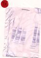
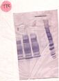
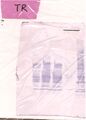
All mutant candidates were digested with a cocktail of Nde and Mlu in 1X NEB3 for 45 minutes at 37° then loaded on a 1% agarose gel (1XTAE)
_TR_Mod2_agarose1.png)
| Lane | Team | Sample |
|---|---|---|
| 1 | 1Kb DNA Ladder | |
| 2 | Red | candidate 1 Nde/Mlu digest |
| 3 | candidate 2 Nde/Mlu digest | |
| 4 | blank | |
| 5 | 1Kb DNA Ladder | |
| 6 | Orange | candidate 1 Nde/Mlu digest |
| 7 | candidate 2 Nde/Mlu digest | |
| 8 | blank | |
| 9 | 1Kb DNA Ladder | |
| 10 | Yellow | candidate 1 Nde/Mlu digest |
| 11 | candidate 2 Nde/Mlu digest | |
| 12 | 1Kb DNA Ladder | |
| 13 | Green | candidate 1 Nde/Mlu digest |
| 14 | candidate 2 Nde/Mlu digest |
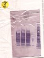
_TR_Mod2_agarose2.png)
| Lane | Team | Sample |
|---|---|---|
| 1 | 1Kb DNA Ladder | |
| 2 | Blue | candidate 1 Nde/Mlu digest |
| 3 | candidate 2 Nde/Mlu digest | |
| 4 | 1Kb DNA Ladder | |
| 5 | Pink | candidate 1 Nde/Mlu digest |
| 6 | candidate 2 Nde/Mlu digest | |
| 7 | 1Kb DNA Ladder | |
| 8 | Purple | candidate 1 Nde/Mlu digest |
| 9 | candidate 2 Nde/Mlu digest | |
| 10 | blank | |
| 11 | 1Kb DNA Ladder | |
| 12 | Control with stuffer fragment | Nde/Mlu digest |
| 13 | Control without stuffer fragment | Nde/Mlu digest |
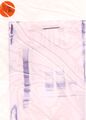
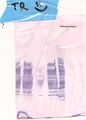
Here is your Western data. If you'd like to see the blots themselves, they'll be in the lab at the front bench.
The "?" after your team color are associated with teams who labeled their containers on the lids rather than the tubs. These blots were possibly mis-designated during the washing and scanning of these blots since the lids were separated from the tubs.
If you'd like to note which of the 2 preparations of primary antibody you used, you can do so in the label under the blot's image
-
 Red Western, probed with mouse raised monoclonal anti-HA, detection with Goat antimouse + AP
 -
 Orange Western, probed with mouse raised monoclonal anti-HA, detection with Goat antimouse + AP
 -
 Yellow? Western, probed with mouse raised monoclonal anti-HA, detection with Goat antimouse + AP
 -
 Green? Western, probed with mouse raised monoclonal anti-HA, detection with Goat antimouse + AP
 -
 Blue? Western, probed with mouse raised monoclonal anti-HA, detection with Goat antimouse + AP
 -
 Pink Western, probed with mouse raised monoclonal anti-HA, detection with Goat antimouse + AP
 -
 Purple? Western, probed with mouse raised monoclonal anti-HA, detection with Goat antimouse + AP
_TR_Western_red.jpg)
_TR_Western_Orange.jpg)
_TR_Western_Yellow%3F.jpg)
_TR_Western_Green%3F.jpg)
_TR_Western_blue%3F.jpg)
_TR_Western_pink.jpg)
_TR_Western_purple%3F.jpg)
W/F lab
Digest Sample info
Please fill in the info below for your team for others to refer to:
| team color | library screened (K+ or P+) | Approx # of colonies examined | Seq data | b-gal data | Notes |
| Red | |||||
| Orange | |||||
| Yellow | |||||
| Green | |||||
| Blue | P+ | 2 red group's Colonies |
| ||
| Pink | K+ | 2 | :( | Mutant 1 Light (Avg): 144 Mutant 1 Dark (Avg): 143 Mutant 2 Light (Avg): 327 Mutant 2 Dark (Avg): 301 | |
| Purple | Using Red Group's Data (sequencing failed) | N/A | |||
| Silver |
Digest Images
Candidates were digested for 55' at 37 °C and run on a 1% agarose gel. Each gel was photographed at two different exposure times - use whichever one makes your DNA easier to see. Also see the second T/R gel for how control DNA (with and without stuffer fragment) runs.
| Gel 1 Lane | Gel 1 Sample | Gel 1 Lane | Gel 1Sample |
| 1 | 1 Kb marker | 6 | Green Cand 1 |
| 2 | Red Cand 1 | 7 | Green Cand 2 |
| 3 | Red Cand 2 | 8 | |
| 4 | 9 | ||
| 5 | 1 Kb marker | 10 |
| Gel 2 Lane | Gel 2 Sample | Gel 2 Lane | Gel 2Sample |
| 1 | 1 Kb marker | 6 | Pink Cand 1 |
| 2 | Blue Cand 1 | 7 | 1 Kb marker |
| 3 | Blue Cand 2 | 8 | Purple Cand 1 |
| 4 | 1 Kb marker | 9 | Purple Cand 2 |
| 5 | Pink Cand 1 | 10 |


Western images
Below are your Westerns. (Please crop out your image for use in the research article.) Some information may be lost in the digital images, so feel free to look at the original blots after journal club to get a good sense of where your bands are.

