User:Philip S. Sumberaz/Notebook/Biology 210 at AU: Difference between revisions
No edit summary |
Sarah Knight (talk | contribs) No edit summary |
||
| (23 intermediate revisions by 2 users not shown) | |||
| Line 1: | Line 1: | ||
*'''Philip S. Sumberaz''':'''March 3, 2015''' | |||
''Purpose:'' The purpose of this lab experiment was to use the sequenced 16S RNA bacterium gene to find a genomic match to a real life species so as to compare this result of a known phenotype to the observations that were made earlier in the transect analysis. | |||
''Materials and Methods:'' Given our RNA sequences through saved computer data, the sequences were put through a Nucleotide BLAST program online in an attempt to find a matching sequence to the ones found in the bacteria colonies. The BLAST program reports similar genotypes via percent accordance, and the species scientific name. Following the supposed identification, the name was search on the internet to find the phenotype of the species; from there, a comparison can be made. In addition to the initial forward analysis, the sequence was also backwards analysed for a possible different result. | |||
''Data and Observations:'' In the initial forward sequence comparison, the RNA sequence came back with an 85% similarity rate with the RNA sequence of Chryseobacterium. This is actually a genus of bacterium; however, this is not wholly disappointing. Among the genus, most, if not all species have a phenotype description of small golden rods. This phenotype matches, roughly, the observed traits of the colony this sequence was for originally. The species observed for this sequence was #2 on the table a few entries below, for the week this lab was originally performed. While it is true that a coccus shape was observed, the microscopes could have been too blurry or low of magnification to make the shape out at the incredibly small size these bacterium were. It is worth mentioning that the reverse analysis of the RNA sequence returned a Psuedomoans result, but at only a 59% match rate. Furthermore, the Psuedomonas genus does not share the brilliant golden color that was most characteristic of this colony. The second colony, being the #4 row on the table a few entries ago, had a poor sequencing result that could not reliably return a matching genus or species within any reasonable percentage of matching. | |||
Source Reference: | |||
Vandamme, P., Bernardet, J., Segers, P., Kersters, K., & Holmes, B. (1994). NOTES: New Perspectives in the Classification of the Flavobacteria: Description of Chryseobacterium gen. nov., Bergeyella gen. nov., and Empedobacter nom. rev. International Journal of Systematic Bacteriology, 827-831. | |||
'''4.7.15''' | |||
You need to include your sequences in your notebook entry and the image of the gel. | |||
SK | |||
*'''Philip S. Sumberaz''':'''February 26, 2015''' | |||
''Purpose:'' The purpose of this experiment is to compare zebrafish embryonic development in water to development within estrogen treated water and observe how the treated water effects the fish. | |||
''Materials and Methods:'' Using petri dishes, electric pipettes, transfer pipettes, tap water, zebrafish embryos, and estrogen treated water, we began setting up the experiment. Using transfer pipettes with enlarged ends (made via cutting off the original opening), zebrafish embryos were drawn out of the containers of water and inserted into their respective petri dish. In each petri dish, one for a control and the other for the estrogen treated water, 20 embryos were put into them. After inspecting the embryos to see if all of them were alive initially, we observed them under the microscope further the identify what stage in their development they were in. Upon checking in with the fish in a 24-hour period, any dead eggs were to be removed from the petri dishes and the stages of development were to be recorded. Another inspection of the fish occurred 72-hours after incubation. Further deceased embroys were to be removed and the stages recorded again. Furthermore, the solution both petri dishes held was to be replaced from 10 mL to 25 mL, using new solution. Paramecium was also added to the petri dishes to act as a food source for the developing fish. | |||
''Data and Observations:'' The Observations are listed below in a table. One occurrence to note is that by the fourth day of the experiment, the entire control group had died in normal water. Needing a control group for when the experiment concludes, another 20 zebrafish embryos were chosen at similar stages in development and are going to be used as control for the remainder of the experiment. | |||
{| {{table}} | |||
| align="center" style="background:#f0f0f0;"|'''Day''' | |||
| align="center" style="background:#f0f0f0;"|'''Dish Variable''' | |||
| align="center" style="background:#f0f0f0;"|'''# Living Eggs''' | |||
| align="center" style="background:#f0f0f0;"|'''# Dead Eggs''' | |||
| align="center" style="background:#f0f0f0;"|'''# Living Larvae''' | |||
| align="center" style="background:#f0f0f0;"|'''# Dead Larvae''' | |||
| align="center" style="background:#f0f0f0;"|'''Length''' | |||
| align="center" style="background:#f0f0f0;"|'''Stage''' | |||
|- | |||
| 0||Control||20||0||0||0||1mm||All in zygote stage | |||
|- | |||
| ||Estrogen Dose||20||0||0||0||1mm||Mostly zygote stage, some Fertilized, one two-cell | |||
|- | |||
| 1||Control||6||14||0||0||1.5mm||Bud Stage and 2-Somite | |||
|- | |||
| ||Estrogen Dose||10||10||0||0||2mm||4-Somite and 8-Somite | |||
|- | |||
| 4||Control||0||5||0||1||2.5mm||20-Somite and 25-Somite | |||
|- | |||
| ||Estrogen Dose||0||4||6||0||4 cm||Early-Larvae Stages | |||
|- | |||
| | |||
|} | |||
'''4.7.15''' | |||
You are missing conclusions for this project. The layout should be the same as all previous lab book entries. | |||
SK | |||
'''2.20.15''' | |||
Excellent invertebrate entry. | |||
No vertebrate observations or food web. | |||
'''SK''' | |||
*'''Philip S. Sumberaz''':''February 18, 2015'' | |||
''Purpose:'' The purpose of this week's lab time was to observe and note the invertebrates living in the samples taken from the transect that were cultivated through a Berlese Funnel. In addition to observing the invertebrates found within these samples, known invertebrates such as earthworms, crabs, spiders, and other species were observed. In contrast, vertebrates were also observed in short order after observations were made. Furthermore, the direction in which we would take our Zebrafish experiments was determined. | |||
''Materials and Methods:'' If the stills and known species were to be observed, one could use the provided microscopes and sample organisms to make observations about general size, shape, and other features of invertebrates. Outside of that, the main focus of this lab was to observe invertebrates found within the alcohol trap at the bottom of the Berlese Funnel. Detaching the tube from the bottom of the funnel structure, and pouring half of the sample into one petri dish and half into another; and using a dissection microscope to observe the organisms. Organisms found were recorded into the lab notebook. If three to five different organisms could not be found, a sample of alcohol trap from another lab section from the same transect was to be observed. Vertebrates that may have influences on the transect were observed or figured after the observations on invertebrates were concluded. | |||
''Data and Observations:'' The first thing that was done was the observation of some worms, or different classes of coelomates (including acoelo and psuedocoelo). The worms did not appear terribly different; meaning they all seemed to be around the same size and look like "worms". They, of course, have their differences; however, the most interesting being how they moved differently. While all three slink around the ground slowly, they move in accordance to their internal structures and how they have membranes to aid with movement. The Acoelomates and Psuedocoelomates do not have full membrane linings of their bodies, so they shrink their back ends forward, and then extend their front end; whereas the Coelomates almost "slither" as they move forward. This is partially due to their interiors being more protected against movement. Following the observations of different Coelomates, we observed the Berlese Funnel organisms. The three entries found are listed in hard-copy lab notebook, and a picture of the page will be provided below. Only three could be found, even using separate section samples of the fifth transect. The size range within what we found was only about 1-1.5 mm up to 2 mm, but ranges could certainly vary amongst the different invertebrates that can be found. Upon figuring and observing vertebrates on and around the transect, it has been concluded that Blue Jays, Dark-Eyed Juncos, Humans, Squirrels, and the occasional domestic dog. All these species benefit from the water, open space, and air from the transect. Other abiotic factors that are present are pollutants, like forks and paper products discarded on the quad, and stone mural have affects on organisms as well. Biotic factors present include the invertebrates living there, the grass, the trees, the rosebushes, and the weeds. Whether these organisms have a profound affect, or are just a passing point of attention, they are living influences on the transect. | |||
[[Image: Springtails.JPG]]http://openwetware.org/images/a/ae/Data_Table_Invertebrates.JPG | |||
'''2.20.15''' | |||
Very good notebook entry. Good data table. Drawings of plants are pretty good. | |||
'''SK''' | |||
*'''Philip S. Sumberaz''':''February 12, 2015'' | |||
''Purpose:'' Continuing our analysis of our transects, this week's lab period was spent collecting and observing plant life within our transects. On top of observing them, we studied their characteristics and attempted to classify them based on their evolutionary complexity. Furthermore, samples from the transect were collected and prepared to create a Berlese Funnel for next week's lab. | |||
''Materials and Methods:'' The first thing that we went to do as a transect group was, once again, visit our transects. This time however, we had a Ziploc bag and were instructed to find five different plant life samples. With a bag of plant samples and some soil in hand, it was back into the lab to begin analyzing the plant life. The first criteria they were observed for was vascularization, or how they gather and transport required resources to continue living. Some simple plants, like algae or mosses, simply collect water and needed resources through contact and absorption. A slightly more complex form of this comes in the structure rhizoids, which are hair-like protrusions from the plant that increase its surface area and allow it to better collect resources. More complex plants like flowering plants have vascular systems called xylem and phloem, which act as metaphorical arteries and veins in humans. They use pressure to move water and sugars throughout the plant. Following this, microscopes and dissecting scopes were used to view specialized structures, or the lack thereof. Structures such as guard cells, cuticles, and others are structures that serve a specified purpose for the plant. These structures are an indication of evolutionary complexity, as are xylem and phloem. Lastly, the plant's reproductive systems were studied and categorized between either monocot or dicot. Characteristics such as seed cotyledon, vascular organization, and petal geometry are indicators of the categories, and all samples were studied for these traits. (Spring 2015 General Biology Lab Manual). | |||
''Data and Observations:'' Many of the lab's observations were listed in a data table, and a picture of the table will be provided below. Regardless, a few points will be reiterated. First, the "flower" sample came from a dead rose bush, thus the flower had only dry, brittle leaves and a wilted bud to look at. The weeds collected from the transect came from the edge of the mulch bedding, as they try to access the nutrient rich soil. They were all tangled between the grass lawn and the mulch bed. The oak leaf originated from an oak tree that is technically outside the transect, but one of its many branches reaches out over the transect, explaining the leaf's presence. Unfortunately, no seeds or other reproductive plant characteristics were able to be recovered from the transect. Many of the samples from our transect are angiosperms, which makes sense, as many plants that people like to look at, like flowering plants and trees, are angiosperms. Furthermore, no fungus samples were able to be collected from manicured lawn, again, a result that is unsurprising. (Apologies for the size and poor quality of pictures... I am, regrettably, no artist.) | |||
http://openwetware.org/images/c/cb/Plant_Life_Chart.JPG | |||
'''2.10.15''' Very good notebook entry. Could have included data from serial dilution results table. To make a table in OWW, try using http://excel2wiki.net to convert excel tables to a format for OWW. '''SK''' | |||
*'''Philip S. Sumberaz''':'''February 4, 2015''' | |||
''Purpose:''As a sort of continuation off of last weeks lab period, this weeks time was spent observing the bacteria that grew on the controlled augar plates over the past seven days. The bacteria were observed and different characteristics were noted before they were used to begin a PCR cycle for next weeks lab. | |||
''Materials and Methods:'' The most apparent material in this lab were living colonies of bacteria that the groups grew in the last week; originating from the Hay Infusion culture that was disposed of this week. In addition to these two items, many others were used. Transfer pipettes, wire hooks, glass observation slides, cover slips, magnification oil, compound microscopes, and the Biology 210 Lab manual were all used to observe and identify individual bacteria within extracts from single colonies that were selected to be used during the entire lab. The wire hook was used to take some of the colony off the augar, then placed onto a wet-mounted slide. A cover slip was applied and the slide was placed on the microscope to be observed. When the 100x magnification was to be used, the oil had to be applied to keep the field of view clear; as well as to keep the lens from being damaged. Observation of the bacteria colonies were included in the lab manual Table 2. After the observations were made, the different bacteria samples were Gram Stained. During the process; using Crystal Violet, Gram's Iodine Mordant, 95% Alcohol solution, Safranin, and water; the peptidoglycan within a bacteria's cell wall is stained a violet purple. If the cell wall contains a lot of peptidoglycan, then the bacteria will appear purple. Alternatively, if there is a relatively low amount of peptidoglycan is present, then the bacteria cells will appear pink. Finally, the colonies were taken from once more to create a PCR container. First, a sample of the colony was taken and put in 100 micro-liters of water. They were then incubated a 100 degrees C for 10 minutes, before being centrifuged for 5 additional minutes. Afterwards, 5 micro-liters of supernatant from the centrifuged sample is added to a mixture of 20 micro-liters of primer and water that has dissolved the PCR bead in a PCR tube. (Spring 2015 Biology 210 Lab Manual) | |||
''Data and Observations:''Prior to being disposed of, the Hay Infusion had a more pungent swamp smell, as well as a thick green coloring and coating on the surface of the water. This change is most likely due to new, or previously under-represented microorganisms that were able to grow more in the changed environment of the culture. Moving on to the bacterium cultures; the differences between the augar plates within the same set are relatively mild in terms of expectation. Meaning that there are simply less bacteria colonies as the concentration is reduced, as one would very much expect. However, the differences between the tetracycline augar plates and pure augar plates are quite pronounced. Where on the normal 10^-3 (the highest concentration) plate the number of colonies is well over a countable number and estimated to be in the thousands, as well as having quite a bit of diversity between all the colonies; the TET plate had only two or three different colonies and about 250 total colonies. These results indicate that there are very few types of bacteria that can live in the presence of this antibiotic within the group's transect. Fungus was only able to grow on an augar plate that did not have antibiotics present; within the results that were collected. Tetracycline inhibits the bacteria's ability to reproduce, therefore allowing the bacterium's short life cycle to do its work for it; eliminating the function by which bacteria colonies stay alive and develop. However, bacterium that are resistant to tetracycline thrive in the antibiotic, and become resistant to it through evolution. The table within my hard-copy lab notebook will be included below. | |||
http://openwetware.org/images/0/07/10%5E-3_Regular_Plate.JPG | |||
http://openwetware.org/images/b/b0/10%5E-3_TET_Plate.JPG | |||
http://openwetware.org/images/6/62/Hard_Copy_Observations.JPG | |||
'''2.4.15''' Very good entry. Well organized and lots of relevant detail. '''SK''' | |||
*'''Philip S. Sumberaz''':'''January 29, 2015''' | *'''Philip S. Sumberaz''':'''January 29, 2015''' | ||
''Purpose:'' The purpose of this lab period the actions taken in it were to observe the life that developed inside of the group's Hay Infusion Culture that originated from the transect that was assigned the week prior, which also happens to be the transect the first lab notebook entry concerns. Furthermore, multiple Petri dishes were prepared for the upcoming lab to study bacterial life that exists within the Hay Infusion and how they will develop and grow within two different environments. | ''Purpose:'' The purpose of this lab period the actions taken in it were to observe the life that developed inside of the group's Hay Infusion Culture that originated from the transect that was assigned the week prior, which also happens to be the transect the first lab notebook entry concerns. Furthermore, multiple Petri dishes were prepared for the upcoming lab to study bacterial life that exists within the Hay Infusion and how they will develop and grow within two different environments. | ||
''Materials and Methods'' One of the main materials in this lab was the Hay Infusion Culture from the previous week. Materials used in the process of observing life inside of it were: transfer pipettes, observation slides, cover slips, proto-slow, microscopes, and Dichomotous Keys. The samples to be observed were taken from the surface of the Hay Infusion and the bottom of the Hay Infusion using the transfer pipettes. The samples taken from the culture were then placed on glass slides, and following the added drop of proto-slow, the cover slip was applied. The microscope was used to find organisms living inside the cultures, and the Dichomotous Keys were used to identify the organisms. After identifying a satisfactory number of organisms, the Hay Infusion was covered and shaken-up. Four test tubes containing sterile broth were gathered and labeled 10^-2, 10^-4, 10^-6, and 10^-8. Four Petri dishes with sterile augar were acquired and labelled 10^-3, 10^-5, 10^-7, and 10^-9; the same being done to four separate dishes with Tetracycline infused augar plates. (The tetracycline dishes were also labeled "TET"). Then, using a P-1000 micropippetor, 100 micro-liters of the mixed Hay Infusion Culture were added to the four tests tubes. Then 100 micro-liters was taken from the Infusion and added to the 10^-2 test tube. Another 100 micro-liters was taken from this test tube and added to the 10^-4 tube, and so on until 10^-8. Following that, another 100 micro-liter sample was taken from the 106-2 tube and added to the 10^-3 Petri dishes (both 100 micro liter samples, separately); intuitively, this is done for the remaining three sets of dishes. The broth is then spread using sterilized metal spreaders and left for a week to develop. (Biology 210 Spring 2015 Lab Manual) | ''Materials and Methods'' One of the main materials in this lab was the Hay Infusion Culture from the previous week. Materials used in the process of observing life inside of it were: transfer pipettes, observation slides, cover slips, proto-slow, microscopes, and Dichomotous Keys. The samples to be observed were taken from the surface of the Hay Infusion and the bottom of the Hay Infusion using the transfer pipettes. The samples taken from the culture were then placed on glass slides, and following the added drop of proto-slow, the cover slip was applied. The microscope was used to find organisms living inside the cultures, and the Dichomotous Keys were used to identify the organisms. After identifying a satisfactory number of organisms, the Hay Infusion was covered and shaken-up. Four test tubes containing sterile broth were gathered and labeled 10^-2, 10^-4, 10^-6, and 10^-8. Four Petri dishes with sterile augar were acquired and labelled 10^-3, 10^-5, 10^-7, and 10^-9; the same being done to four separate dishes with Tetracycline infused augar plates. (The tetracycline dishes were also labeled "TET"). Then, using a P-1000 micropippetor, 100 micro-liters of the mixed Hay Infusion Culture were added to the four tests tubes. Then 100 micro-liters was taken from the Infusion and added to the 10^-2 test tube. Another 100 micro-liters was taken from this test tube and added to the 10^-4 tube, and so on until 10^-8. Following that, another 100 micro-liter sample was taken from the 106-2 tube and added to the 10^-3 Petri dishes (both 100 micro liter samples, separately); intuitively, this is done for the remaining three sets of dishes. The broth is then spread using sterilized metal spreaders and left for a week to develop. (Biology 210 Spring 2015 Lab Manual) | ||
''Data and Observations'' The Hay Infusion had a faint marshy smell, and has some green shoots between the surface and the bottom layer. Furthermore, there was a white mold-like substance on the top of the culture. Samples were taken from the bottom and top of the culture, both around the edges of the beaker. The usefulness of taking samples from both "open" water and near plants is that the organisms vary. Those in more "open" space need to have a higher ability to retain resources, or create their own; while organisms found near plants most likely consume materials made by other organisms, such as the plant life. The organisms Panderina and Ameoba Proteus were found in the surface sample while the organism Colpidium was found the culture floor sample. The Panderina were about 30-35 micrometers in length, while the Ameoba Proteus was closer to 420 micrometers. The Colpidium were around 80 micrometers in length. Had this Hay Infusion been allowed to sit undisturbed for a longer period of time, the life inside of it would be come more developed so long as the amount of resources within the jar could support the growing population of life. This concept is known as the carrying capacity for an ecosystem. Overall, the samples would have more life in them, but the point is that life would not continue to grow forever, there would be a point fairly soon when the amount of resources within the ecosystem meets a balancing point with the amount of life it is trying to support. | ''Data and Observations'' The Hay Infusion had a faint marshy smell, and has some green shoots between the surface and the bottom layer. Furthermore, there was a white mold-like substance on the top of the culture. Samples were taken from the bottom and top of the culture, both around the edges of the beaker. The usefulness of taking samples from both "open" water and near plants is that the organisms vary. Those in more "open" space need to have a higher ability to retain resources, or create their own; while organisms found near plants most likely consume materials made by other organisms, such as the plant life. The organisms Panderina and Ameoba Proteus were found in the surface sample while the organism Colpidium was found the culture floor sample. The Panderina were about 30-35 micrometers in length, while the Ameoba Proteus was closer to 420 micrometers. The Colpidium were around 80 micrometers in length. Had this Hay Infusion been allowed to sit undisturbed for a longer period of time, the life inside of it would be come more developed so long as the amount of resources within the jar could support the growing population of life. This concept is known as the carrying capacity for an ecosystem. Overall, the samples would have more life in them, but the point is that life would not continue to grow forever, there would be a point fairly soon when the amount of resources within the ecosystem meets a balancing point with the amount of life it is trying to support. | ||
http://openwetware.org/images/7/78/Ameoba_Proteus.jpg | http://openwetware.org/images/7/78/Ameoba_Proteus.jpg | ||
Latest revision as of 10:45, 7 April 2015
- Philip S. Sumberaz:March 3, 2015
Purpose: The purpose of this lab experiment was to use the sequenced 16S RNA bacterium gene to find a genomic match to a real life species so as to compare this result of a known phenotype to the observations that were made earlier in the transect analysis.
Materials and Methods: Given our RNA sequences through saved computer data, the sequences were put through a Nucleotide BLAST program online in an attempt to find a matching sequence to the ones found in the bacteria colonies. The BLAST program reports similar genotypes via percent accordance, and the species scientific name. Following the supposed identification, the name was search on the internet to find the phenotype of the species; from there, a comparison can be made. In addition to the initial forward analysis, the sequence was also backwards analysed for a possible different result.
Data and Observations: In the initial forward sequence comparison, the RNA sequence came back with an 85% similarity rate with the RNA sequence of Chryseobacterium. This is actually a genus of bacterium; however, this is not wholly disappointing. Among the genus, most, if not all species have a phenotype description of small golden rods. This phenotype matches, roughly, the observed traits of the colony this sequence was for originally. The species observed for this sequence was #2 on the table a few entries below, for the week this lab was originally performed. While it is true that a coccus shape was observed, the microscopes could have been too blurry or low of magnification to make the shape out at the incredibly small size these bacterium were. It is worth mentioning that the reverse analysis of the RNA sequence returned a Psuedomoans result, but at only a 59% match rate. Furthermore, the Psuedomonas genus does not share the brilliant golden color that was most characteristic of this colony. The second colony, being the #4 row on the table a few entries ago, had a poor sequencing result that could not reliably return a matching genus or species within any reasonable percentage of matching.
Source Reference: Vandamme, P., Bernardet, J., Segers, P., Kersters, K., & Holmes, B. (1994). NOTES: New Perspectives in the Classification of the Flavobacteria: Description of Chryseobacterium gen. nov., Bergeyella gen. nov., and Empedobacter nom. rev. International Journal of Systematic Bacteriology, 827-831.
4.7.15 You need to include your sequences in your notebook entry and the image of the gel. SK
- Philip S. Sumberaz:February 26, 2015
Purpose: The purpose of this experiment is to compare zebrafish embryonic development in water to development within estrogen treated water and observe how the treated water effects the fish.
Materials and Methods: Using petri dishes, electric pipettes, transfer pipettes, tap water, zebrafish embryos, and estrogen treated water, we began setting up the experiment. Using transfer pipettes with enlarged ends (made via cutting off the original opening), zebrafish embryos were drawn out of the containers of water and inserted into their respective petri dish. In each petri dish, one for a control and the other for the estrogen treated water, 20 embryos were put into them. After inspecting the embryos to see if all of them were alive initially, we observed them under the microscope further the identify what stage in their development they were in. Upon checking in with the fish in a 24-hour period, any dead eggs were to be removed from the petri dishes and the stages of development were to be recorded. Another inspection of the fish occurred 72-hours after incubation. Further deceased embroys were to be removed and the stages recorded again. Furthermore, the solution both petri dishes held was to be replaced from 10 mL to 25 mL, using new solution. Paramecium was also added to the petri dishes to act as a food source for the developing fish.
Data and Observations: The Observations are listed below in a table. One occurrence to note is that by the fourth day of the experiment, the entire control group had died in normal water. Needing a control group for when the experiment concludes, another 20 zebrafish embryos were chosen at similar stages in development and are going to be used as control for the remainder of the experiment.
| Day | Dish Variable | # Living Eggs | # Dead Eggs | # Living Larvae | # Dead Larvae | Length | Stage |
| 0 | Control | 20 | 0 | 0 | 0 | 1mm | All in zygote stage |
| Estrogen Dose | 20 | 0 | 0 | 0 | 1mm | Mostly zygote stage, some Fertilized, one two-cell | |
| 1 | Control | 6 | 14 | 0 | 0 | 1.5mm | Bud Stage and 2-Somite |
| Estrogen Dose | 10 | 10 | 0 | 0 | 2mm | 4-Somite and 8-Somite | |
| 4 | Control | 0 | 5 | 0 | 1 | 2.5mm | 20-Somite and 25-Somite |
| Estrogen Dose | 0 | 4 | 6 | 0 | 4 cm | Early-Larvae Stages | |
4.7.15 You are missing conclusions for this project. The layout should be the same as all previous lab book entries. SK
2.20.15 Excellent invertebrate entry. No vertebrate observations or food web. SK
- Philip S. Sumberaz:February 18, 2015
Purpose: The purpose of this week's lab time was to observe and note the invertebrates living in the samples taken from the transect that were cultivated through a Berlese Funnel. In addition to observing the invertebrates found within these samples, known invertebrates such as earthworms, crabs, spiders, and other species were observed. In contrast, vertebrates were also observed in short order after observations were made. Furthermore, the direction in which we would take our Zebrafish experiments was determined.
Materials and Methods: If the stills and known species were to be observed, one could use the provided microscopes and sample organisms to make observations about general size, shape, and other features of invertebrates. Outside of that, the main focus of this lab was to observe invertebrates found within the alcohol trap at the bottom of the Berlese Funnel. Detaching the tube from the bottom of the funnel structure, and pouring half of the sample into one petri dish and half into another; and using a dissection microscope to observe the organisms. Organisms found were recorded into the lab notebook. If three to five different organisms could not be found, a sample of alcohol trap from another lab section from the same transect was to be observed. Vertebrates that may have influences on the transect were observed or figured after the observations on invertebrates were concluded.
Data and Observations: The first thing that was done was the observation of some worms, or different classes of coelomates (including acoelo and psuedocoelo). The worms did not appear terribly different; meaning they all seemed to be around the same size and look like "worms". They, of course, have their differences; however, the most interesting being how they moved differently. While all three slink around the ground slowly, they move in accordance to their internal structures and how they have membranes to aid with movement. The Acoelomates and Psuedocoelomates do not have full membrane linings of their bodies, so they shrink their back ends forward, and then extend their front end; whereas the Coelomates almost "slither" as they move forward. This is partially due to their interiors being more protected against movement. Following the observations of different Coelomates, we observed the Berlese Funnel organisms. The three entries found are listed in hard-copy lab notebook, and a picture of the page will be provided below. Only three could be found, even using separate section samples of the fifth transect. The size range within what we found was only about 1-1.5 mm up to 2 mm, but ranges could certainly vary amongst the different invertebrates that can be found. Upon figuring and observing vertebrates on and around the transect, it has been concluded that Blue Jays, Dark-Eyed Juncos, Humans, Squirrels, and the occasional domestic dog. All these species benefit from the water, open space, and air from the transect. Other abiotic factors that are present are pollutants, like forks and paper products discarded on the quad, and stone mural have affects on organisms as well. Biotic factors present include the invertebrates living there, the grass, the trees, the rosebushes, and the weeds. Whether these organisms have a profound affect, or are just a passing point of attention, they are living influences on the transect.
 http://openwetware.org/images/a/ae/Data_Table_Invertebrates.JPG
2.20.15 Very good notebook entry. Good data table. Drawings of plants are pretty good. SK
- Philip S. Sumberaz:February 12, 2015
Purpose: Continuing our analysis of our transects, this week's lab period was spent collecting and observing plant life within our transects. On top of observing them, we studied their characteristics and attempted to classify them based on their evolutionary complexity. Furthermore, samples from the transect were collected and prepared to create a Berlese Funnel for next week's lab.
Materials and Methods: The first thing that we went to do as a transect group was, once again, visit our transects. This time however, we had a Ziploc bag and were instructed to find five different plant life samples. With a bag of plant samples and some soil in hand, it was back into the lab to begin analyzing the plant life. The first criteria they were observed for was vascularization, or how they gather and transport required resources to continue living. Some simple plants, like algae or mosses, simply collect water and needed resources through contact and absorption. A slightly more complex form of this comes in the structure rhizoids, which are hair-like protrusions from the plant that increase its surface area and allow it to better collect resources. More complex plants like flowering plants have vascular systems called xylem and phloem, which act as metaphorical arteries and veins in humans. They use pressure to move water and sugars throughout the plant. Following this, microscopes and dissecting scopes were used to view specialized structures, or the lack thereof. Structures such as guard cells, cuticles, and others are structures that serve a specified purpose for the plant. These structures are an indication of evolutionary complexity, as are xylem and phloem. Lastly, the plant's reproductive systems were studied and categorized between either monocot or dicot. Characteristics such as seed cotyledon, vascular organization, and petal geometry are indicators of the categories, and all samples were studied for these traits. (Spring 2015 General Biology Lab Manual).
Data and Observations: Many of the lab's observations were listed in a data table, and a picture of the table will be provided below. Regardless, a few points will be reiterated. First, the "flower" sample came from a dead rose bush, thus the flower had only dry, brittle leaves and a wilted bud to look at. The weeds collected from the transect came from the edge of the mulch bedding, as they try to access the nutrient rich soil. They were all tangled between the grass lawn and the mulch bed. The oak leaf originated from an oak tree that is technically outside the transect, but one of its many branches reaches out over the transect, explaining the leaf's presence. Unfortunately, no seeds or other reproductive plant characteristics were able to be recovered from the transect. Many of the samples from our transect are angiosperms, which makes sense, as many plants that people like to look at, like flowering plants and trees, are angiosperms. Furthermore, no fungus samples were able to be collected from manicured lawn, again, a result that is unsurprising. (Apologies for the size and poor quality of pictures... I am, regrettably, no artist.)
http://openwetware.org/images/c/cb/Plant_Life_Chart.JPG
2.10.15 Very good notebook entry. Could have included data from serial dilution results table. To make a table in OWW, try using http://excel2wiki.net to convert excel tables to a format for OWW. SK
- Philip S. Sumberaz:February 4, 2015
Purpose:As a sort of continuation off of last weeks lab period, this weeks time was spent observing the bacteria that grew on the controlled augar plates over the past seven days. The bacteria were observed and different characteristics were noted before they were used to begin a PCR cycle for next weeks lab.
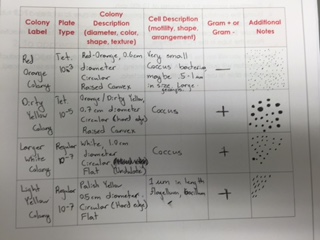
Materials and Methods: The most apparent material in this lab were living colonies of bacteria that the groups grew in the last week; originating from the Hay Infusion culture that was disposed of this week. In addition to these two items, many others were used. Transfer pipettes, wire hooks, glass observation slides, cover slips, magnification oil, compound microscopes, and the Biology 210 Lab manual were all used to observe and identify individual bacteria within extracts from single colonies that were selected to be used during the entire lab. The wire hook was used to take some of the colony off the augar, then placed onto a wet-mounted slide. A cover slip was applied and the slide was placed on the microscope to be observed. When the 100x magnification was to be used, the oil had to be applied to keep the field of view clear; as well as to keep the lens from being damaged. Observation of the bacteria colonies were included in the lab manual Table 2. After the observations were made, the different bacteria samples were Gram Stained. During the process; using Crystal Violet, Gram's Iodine Mordant, 95% Alcohol solution, Safranin, and water; the peptidoglycan within a bacteria's cell wall is stained a violet purple. If the cell wall contains a lot of peptidoglycan, then the bacteria will appear purple. Alternatively, if there is a relatively low amount of peptidoglycan is present, then the bacteria cells will appear pink. Finally, the colonies were taken from once more to create a PCR container. First, a sample of the colony was taken and put in 100 micro-liters of water. They were then incubated a 100 degrees C for 10 minutes, before being centrifuged for 5 additional minutes. Afterwards, 5 micro-liters of supernatant from the centrifuged sample is added to a mixture of 20 micro-liters of primer and water that has dissolved the PCR bead in a PCR tube. (Spring 2015 Biology 210 Lab Manual)
Data and Observations:Prior to being disposed of, the Hay Infusion had a more pungent swamp smell, as well as a thick green coloring and coating on the surface of the water. This change is most likely due to new, or previously under-represented microorganisms that were able to grow more in the changed environment of the culture. Moving on to the bacterium cultures; the differences between the augar plates within the same set are relatively mild in terms of expectation. Meaning that there are simply less bacteria colonies as the concentration is reduced, as one would very much expect. However, the differences between the tetracycline augar plates and pure augar plates are quite pronounced. Where on the normal 10^-3 (the highest concentration) plate the number of colonies is well over a countable number and estimated to be in the thousands, as well as having quite a bit of diversity between all the colonies; the TET plate had only two or three different colonies and about 250 total colonies. These results indicate that there are very few types of bacteria that can live in the presence of this antibiotic within the group's transect. Fungus was only able to grow on an augar plate that did not have antibiotics present; within the results that were collected. Tetracycline inhibits the bacteria's ability to reproduce, therefore allowing the bacterium's short life cycle to do its work for it; eliminating the function by which bacteria colonies stay alive and develop. However, bacterium that are resistant to tetracycline thrive in the antibiotic, and become resistant to it through evolution. The table within my hard-copy lab notebook will be included below.
http://openwetware.org/images/0/07/10%5E-3_Regular_Plate.JPG http://openwetware.org/images/b/b0/10%5E-3_TET_Plate.JPG
http://openwetware.org/images/6/62/Hard_Copy_Observations.JPG
2.4.15 Very good entry. Well organized and lots of relevant detail. SK
- Philip S. Sumberaz:January 29, 2015
Purpose: The purpose of this lab period the actions taken in it were to observe the life that developed inside of the group's Hay Infusion Culture that originated from the transect that was assigned the week prior, which also happens to be the transect the first lab notebook entry concerns. Furthermore, multiple Petri dishes were prepared for the upcoming lab to study bacterial life that exists within the Hay Infusion and how they will develop and grow within two different environments.
Materials and Methods One of the main materials in this lab was the Hay Infusion Culture from the previous week. Materials used in the process of observing life inside of it were: transfer pipettes, observation slides, cover slips, proto-slow, microscopes, and Dichomotous Keys. The samples to be observed were taken from the surface of the Hay Infusion and the bottom of the Hay Infusion using the transfer pipettes. The samples taken from the culture were then placed on glass slides, and following the added drop of proto-slow, the cover slip was applied. The microscope was used to find organisms living inside the cultures, and the Dichomotous Keys were used to identify the organisms. After identifying a satisfactory number of organisms, the Hay Infusion was covered and shaken-up. Four test tubes containing sterile broth were gathered and labeled 10^-2, 10^-4, 10^-6, and 10^-8. Four Petri dishes with sterile augar were acquired and labelled 10^-3, 10^-5, 10^-7, and 10^-9; the same being done to four separate dishes with Tetracycline infused augar plates. (The tetracycline dishes were also labeled "TET"). Then, using a P-1000 micropippetor, 100 micro-liters of the mixed Hay Infusion Culture were added to the four tests tubes. Then 100 micro-liters was taken from the Infusion and added to the 10^-2 test tube. Another 100 micro-liters was taken from this test tube and added to the 10^-4 tube, and so on until 10^-8. Following that, another 100 micro-liter sample was taken from the 106-2 tube and added to the 10^-3 Petri dishes (both 100 micro liter samples, separately); intuitively, this is done for the remaining three sets of dishes. The broth is then spread using sterilized metal spreaders and left for a week to develop. (Biology 210 Spring 2015 Lab Manual)
Data and Observations The Hay Infusion had a faint marshy smell, and has some green shoots between the surface and the bottom layer. Furthermore, there was a white mold-like substance on the top of the culture. Samples were taken from the bottom and top of the culture, both around the edges of the beaker. The usefulness of taking samples from both "open" water and near plants is that the organisms vary. Those in more "open" space need to have a higher ability to retain resources, or create their own; while organisms found near plants most likely consume materials made by other organisms, such as the plant life. The organisms Panderina and Ameoba Proteus were found in the surface sample while the organism Colpidium was found the culture floor sample. The Panderina were about 30-35 micrometers in length, while the Ameoba Proteus was closer to 420 micrometers. The Colpidium were around 80 micrometers in length. Had this Hay Infusion been allowed to sit undisturbed for a longer period of time, the life inside of it would be come more developed so long as the amount of resources within the jar could support the growing population of life. This concept is known as the carrying capacity for an ecosystem. Overall, the samples would have more life in them, but the point is that life would not continue to grow forever, there would be a point fairly soon when the amount of resources within the ecosystem meets a balancing point with the amount of life it is trying to support.
http://openwetware.org/images/7/78/Ameoba_Proteus.jpg
1.27.15
Good first lab book entry. Need to include some more detail eg. Hay Infusion set-up and organize into: Purpose, Materials, Observations and Conclusions.
SK
- Philip S. Sumberaz:January 25, 2015
I was assigned to Group Number 5 in Section 5 Biology 210 lab. Our group 5 goes to the quad outside the Hurst building, around the middle of the lawn. Our transect includes mostly manicured grass lawn, and has some mulched soil for gardened plants around the middle of the quadrangle center. Part of the cement wall from the center mural space comes into the transect from the west side. Some rose bushes, or at least thorny plants, grow in the soil; on top of some frilly bushes. The ground itself is fairly flat, and at the time, quite wet due to the recent precipitation. Abiotic factors in our transect include: Snow, the cement wall, dirt, woodchips, and water; Biotic factors include: Grass, Bushes, Leaves, Sticks, and Bugs.