User:Matthew Kelly/Notebook/Biology 210 at AU: Difference between revisions
No edit summary |
|||
| Line 1: | Line 1: | ||
---- | ---- | ||
== '''Algae and Protists Lab January 20th-27th, 2016''' == | == '''Bacteria, Algae and Protists Lab January 20th-27th, 2016''' == | ||
| Line 61: | Line 61: | ||
[[Image:table2.2.jpg]] | [[Image:table2.2.jpg]] | ||
Table 2.2: Gram | Table 2.2: Gram Stain Table | ||
Each bacteria sample was taken at 40x magnification. | Each bacteria sample was taken at 40x magnification. | ||
[[Image:Agar-5Example.jpg]] | |||
Figure 2.1: Gram stained bacteria sample on wet mount from the agar plate of a dilution of 1 x 10^-5 | |||
[[Image:Agar-9Example.jpg]] | |||
Figure 2.2: Gram satined bacteria sample on wet mount from the agar plate of a dilution of 1 x 10^-7 | |||
[[Image:tet-5Example.jpg]] | |||
Figure 2.3: Gram satined bacteria sample on wet mount from the tet+ plate of a dilution of 1 x 10^-7 | |||
[[Image:Tet-9Example.jpg]] | |||
Figure 2.1: Gram stained bacteria sample on wet mount from the tet+ plate of a dilution of 1 x 10^-9 | |||
== '''Conclusions and Future Directions:''' == | == '''Conclusions and Future Directions:''' == | ||
Revision as of 13:03, 5 February 2016
Bacteria, Algae and Protists Lab January 20th-27th, 2016
Purpose:
The purpose of this lab was to identify the microscopic protists that were living in transect 2.
Materials and Methods:
We created a Hay Infusion and allowed it to sit for one week. We created wetmount slides from samples from 3 different sections of the Hay Infusion jar exactly one week after creating the Hay Infusion. We looked at the samples under 40x magnification. We used a dichotomous key to identify protists in the sample slides.
Data and Observations:
Hay Infusion Description:
The Hay Infusion jar was comprised of plant matter, creek water, soil, and rocks. The jar had a strong odor of decomposing plants and mold. This was different from the prior weak, which was less pungent. The water was tinted brown last week, but as of this week the sediment has settled and the water is more clear. The water itself appear to have a plastic like film on the surface. The plant matter settled towards the bottom of the jar and appeared to compress over the two week period. The water was still and mold appeared to be growing along the sides of the top layer. In order to observe all of the possible protists, we took samples from the top of the water, the middle of the water, and the bottom of the water where the sediment had settled. Finding protists in the samples from the top and middle proved difficult. However, the sample from the bottom provided many protists.
Image2.1: Picture of the Hay Fusion from Transect 2
Protists Samples:
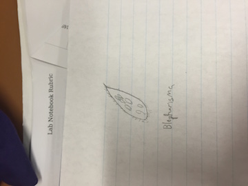
In the top and middle samples, we could not confidently identify any protists. However, we did identify 3 different protists in the bottom sample. The first identified was Blepharisma. It was not very motile organism with a brown tint. The sketch of the Blepharisma is pictured below.
Image2.2: Drawing of the observed Blepharisma
The next organism we identified was a Chilomonas. Unlike the Blepharisma, the Chilomonas was clearly motile. It measured to be 75 micrometers. The sketch of the Chilomonas is pictured below.
Image2.3: Drawing of the observed Chilomonas
The final organism we identified was a Paramecium. It was motile and measured at 2.5 micrometers.
Image2.4: Drawing of the observed Paramecium
Bacteria:
The bacteria and fungi results from the agar and tetracycline plates are displayed below.
Table 2.1: 100-fold serial dilution results
Table 2.2: Gram Stain Table
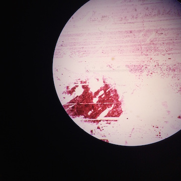
Each bacteria sample was taken at 40x magnification.
Figure 2.1: Gram stained bacteria sample on wet mount from the agar plate of a dilution of 1 x 10^-5
Figure 2.2: Gram satined bacteria sample on wet mount from the agar plate of a dilution of 1 x 10^-7
Figure 2.3: Gram satined bacteria sample on wet mount from the tet+ plate of a dilution of 1 x 10^-7
Figure 2.1: Gram stained bacteria sample on wet mount from the tet+ plate of a dilution of 1 x 10^-9
Conclusions and Future Directions:
January 13th, 2016
Aerial Diagram of Transect 2
Image1.1
Abiotic factors:
-Water
-Rocks
-Soil
-Irrigation System
-Bricks
Biotic Factors:
-Trees
-Ferns
-Squirrels
-Grass
-Worms
Transect Description:
This transect begins with grass, rocks, ferns, and trees. These lead into a flowing creek that holds hundreds of rocks, ranging in size from massive rocks to pebbles. Typically, the larger rocks are towards the outside the creek, partially submersed in the water. the pebbles line the inside of the creek between the large rocks. On the opposing side of the creek, there is more grass, ferns, trees, and some moss on top of rocks. In the creek are rocks and broken branches. The weather appears to be heavily impacting life in this transect. Many plants seem to be dying and fallen leaves cover the majority of the transect. While it seems quiet now, I predict that it will be lively in the spring.
Transect 2 is a habitat with unique abiotic diversity for campus habitats. Within the transect there is a flowing creek, giving it a water source for both plants and animals. The rocks provide coverage for organisms looking to escape sunlight and to hide from potential predators. The rich soil with a consistent water source allows plant life to thrive in this transect. The abundance of plant life also allows this transect to provide a mix of different animal life, both predators and prey. On top of that, the water is also a home for a diverse group of organisms that are not visible to the naked eye.


