User:Kenia Fernandez-Ortiz/Notebook/Biology 210 at AU: Difference between revisions
No edit summary |
No edit summary |
||
| Line 18: | Line 18: | ||
http://openwetware.org/images/8/88/Screen_Shot_2014-03-22_at_4.56.17_PM.png | http://openwetware.org/images/8/88/Screen_Shot_2014-03-22_at_4.56.17_PM.png | ||
http://openwetware.org/images/f/f3/Screen_Shot_2014-03-22_at_4.56.31_PM.png | http://openwetware.org/images/f/f3/Screen_Shot_2014-03-22_at_4.56.31_PM.png | ||
Discussion: We found that the retinoid acid embryos were lighter in color and they died off quicker. What this could mean for humans is that during embryo development, it it is exposed to too much vitamin A, it could lead too paler skin, maybe some disease related to skin pigmentation, and eye development. it could also increase the mortality rate. There were many parts of the lab we couldn't get data for because it was too difficult. | Discussion: We found that the retinoid acid embryos were lighter in color and they died off quicker. What this could mean for humans is that during embryo development, it it is exposed to too much vitamin A, it could lead too paler skin, maybe some disease related to skin pigmentation, and eye development. it could also increase the mortality rate. There were many parts of the lab we couldn't get data for because it was too difficult. | ||
Kenia Michelle Fernandez- Ortiz | Kenia Michelle Fernandez- Ortiz | ||
---- | ---- | ||
Latest revision as of 14:04, 22 March 2014
3/22/2014 Embryology Lab
Intro- It is very important to study this topic because nearly every vertebrate and invertebrates goes through embryonic development. It is a very sensitive stage and when it is influenced by outside elements, deformities can occur. We decided to test the effects of Retinoic Acid (vitamin A) on developing embryos (to see if any deformities occurred). Before starting the embryogenesis experiment, we read a scientific paper that essentially conducted the same experiment in order to get a better sense of what types of data we were most likely to collect. This article kept many controlled variables like temperature, pH, number of zebrafish in each petri, and amount of Retinoic acid given to the experimental group. All of these things we also kept constant and we took notes on similar independent variables. Deformities that were observed varied a bit from the reading, but overall it was close. Zebrafish are a model organism for studying embryogenesis because it has been proven that zebrafish and mammalian embryos have similar reactions to environmental effects.
Materials and Methods- •Day 1: This day we set up two petri dishes. One with 10mL of water (for the control group) and another with 10mL of retinoic acid. We then put 20 embryos in each petri dish giving us a total of 40 embryos.
•Day 3: For both the controlled and experimental group we counted for the number of hatched, unhatched, and dead embryos. We observed swimming ability and measured the heartbeat of hatched and unhatched embryos. In order to measure the heart beat we counted the heartbeat for 10 seconds and then multiplied that number by 6 (because of 60 seconds in a minute). The color, length, and shape of the tail was observed and measured. Eye pigmentation, eye size, and eye movement were observed. Yolk absorption was measured by either a small, medium, or large scale and we also staged them to the best of our ability. All of these observations were done for both the control and tested groups. After we made observations we changed the water by taking out 7 pipet fulls of water or retinoic acid and putting in 10mLs of either one.
•Day 5: For this day we counted how many eggs were hatched, how many were dead and alive, and we measured the heartbeat using the same methods used on day 1. We also noted swimming ability and tail movement by seeing if they twitched a lot when they swam. We noted yolk absorption (small, medium, large) and eye pigmentation and movement. We also observed the development of the body and tail. We looked for deformities or irregularity in color. We checked for deformities in fins and tail and we measured the tail length and eye diameter. After we made observations we changed the water by taking out 7 pipet fulls of water or retinoic acid and putting in 10mLs of either one.
•Day 7: We checked (and measured) for deformities in the tail, fins, eye diameter, and body structures. We also noted heart rate and how many had hatched.
•Day9: We checked for the same protocols as above.
•Day 11: Checked for the same protocols http://openwetware.org/images/8/88/Screen_Shot_2014-03-22_at_4.56.17_PM.png http://openwetware.org/images/f/f3/Screen_Shot_2014-03-22_at_4.56.31_PM.png
Discussion: We found that the retinoid acid embryos were lighter in color and they died off quicker. What this could mean for humans is that during embryo development, it it is exposed to too much vitamin A, it could lead too paler skin, maybe some disease related to skin pigmentation, and eye development. it could also increase the mortality rate. There were many parts of the lab we couldn't get data for because it was too difficult.
Kenia Michelle Fernandez- Ortiz
3/2/2014
Lab 6: Bacteria Found in Hay Infusion
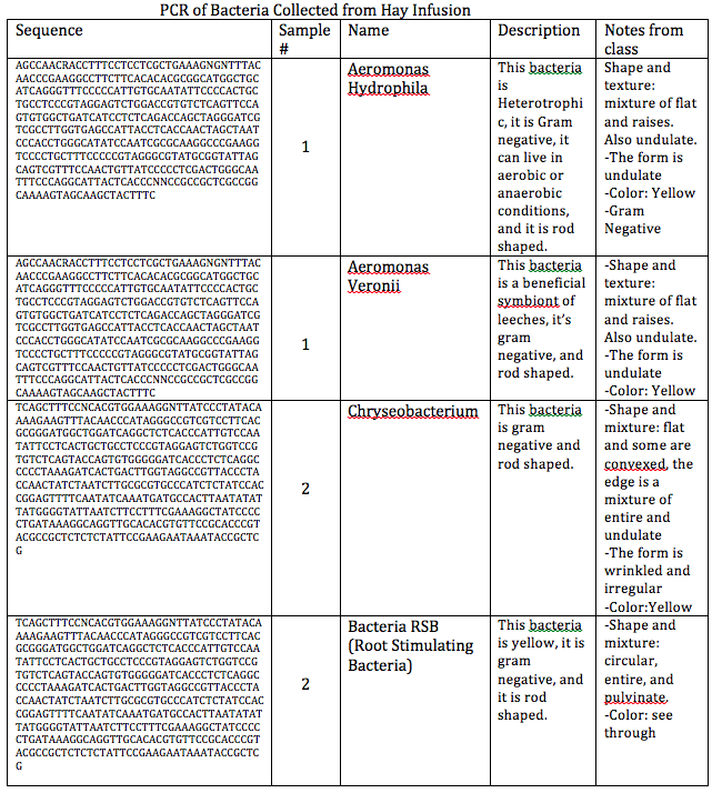
Objective- The objective for this lab was to identify bacteria found in our hay infusion by obtaining the DNA sequence from the PCR reaction.
Methods and Materials- See lab 2 and 3.
Results- http://openwetware.org/images/5/51/Screen_Shot_2014-03-03_at_12.38.35_AM.png
Conclusion- This lab made me see that our observations were really close to the PCR information. This lab also lets me see that there are organism living in areas that I never though organisms would live in.
2/24/2014
Lab 5: Invertebrates
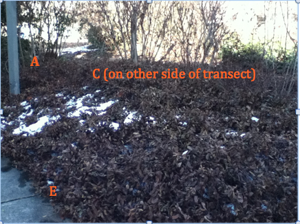
Objective- The objective for this week’s lab is to learn about the invertebrate’s structure and how it evolved into more complicated systems. We are able to learn about invertebrates more closely by examining the one’s we collected from our transect/Berlese Funnel experiment. The purpose of the Berlese Funnel was so that we could see invertebrates that are in hibernated form. My prediction is that we will see many invertebrates because there is an abundance of soil and shrubbery plants in our transect in addition to the fact that not a lot of people pass through there. Our Transect could be home to many different kinds of invertebrates.
Materials and Methods- 1) The first procedure we did was observe Acoelomate, Pseudocoelomates, and Coelomates. We observed Planaria (Acoelomate), that were fed egg yolk, under the dissecting microscope and then we looked at a cross sectional slide under a microscope. 2) The next step was to observe the nematodes (pserdocoelomates) under a dissecting microscope as well as of a cross sectional slide under a microscope. 3) Lastly, we observed Annelida and it’s internal organs as well as it’s muscle layer. 4) The next procedure we did was observe the invertebrates from our Berlese Funnel that we collected from our transect. We took the preserved contents from the flask of the Berlese Funnel and transferred it into a petri dish. 5) What we did was use a dichotomous key to classify the invertebrates.
Raw Data-
Planaria: The movement was slow but steady, it is liking a stretching and contracting sort of movement. It wiggles a little and it's main way of swimming is by doing a folding movement with its body. This movement relates to their body structure because they are flat (hence the name flat worms). For the color: some are brown and some are black.
http://openwetware.org/images/3/3d/IMG_0974.jpg Photo of Planaria ( Left side is dissecting microscope drawing, right side is the cross sectional drawing)
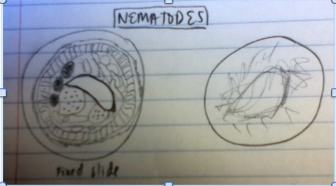
Nematodes: It rarely moves and when it does it has a twitchy sort of movement. They wiggled a lot. This movement relates to their body structure because they have bilateral symmetry. There color is grayish black.
http://openwetware.org/images/0/0e/Screen_Shot_2014-02-26_at_3.24.32_PM.png
Drawing of a Nematode (left: cross-sectional drawing, right: dissecting microscope drawing)
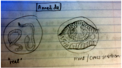
Annelida:They move by stretching and contracting and they also wiggle. This type of movement relates to their body structure because earthworms have a mesoderm, an ectoderm, and an endoderm. They are a light brown color.
http://openwetware.org/images/b/ba/Screen_Shot_2014-02-26_at_3.26.18_PM.png
Drawing of Annelida (left: drawing of the earthworm, right: fixed/cross-sectional drawing)
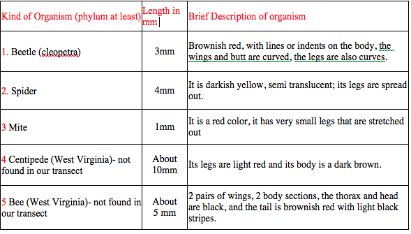
Chart of Invertebrates in Transect
http://openwetware.org/images/6/64/Screen_Shot_2014-02-26_at_3.45.39_PM.png
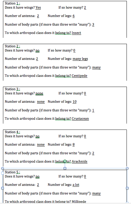
Red Question: The size of the range of the organisms we measure was about 2mm. The outlier ( the longest) was about 10mm. The mites were the smallest kind and the centipede and the bee are the largest. The most common organisms we found in the litter were mites.
Invertebrate worksheets http://openwetware.org/images/5/51/Screen_Shot_2014-02-28_at_7.20.58_PM.png http://openwetware.org/images/8/88/Screen_Shot_2014-02-28_at_10.09.00_PM.png
Classification, Biotic, and Abiotic factors of potential Organisms in Transect http://openwetware.org/images/1/1e/Screen_Shot_2014-02-28_at_11.24.38_PM.png
Food Web of Organisms in Transect http://openwetware.org/images/1/10/Screen_Shot_2014-02-28_at_11.43.06_PM.png
Conclusion- Overall, I there weren't as many invertebrates as I had expected. It seems that with the less diversity of plants the less diversity of invertebrates there is. At least this could be true for our transect. For next time, we could collect dirt from multiple areas in the transect so that we could see is there is more diversity. We could also see if there is a correlation between where the dirt is and what type of invertebrate lives there. This means we'd have to do multiple Berlese Funnels.
Kenia Michelle Fernandez Ortiz
2/24/2014 Lab 4: Plant and Fungi
Objectives- the overall objectives for this lab were to study plants (their characteristics and their diversity). We are also looking at the characteristics of fungi and their importance to the environment. In this lab we will collect 5 samples of plants from our transect and try and classify them. This experiment will help us not only determine what types of plants live in our transect, but we’ll also be able to make connections between the plants environment and its specialized characteristics. For example, we’ll be able to understand why a shrub can live in our transect. My prediction is that we won’t find a variety of plants because we had mostly shrubs and dead leaves.
Materials and Methods- 1) The first thing we did was fill up three large Ziplock bags with samples from our transect. We filled up the first bag with leaf litter. We filled up the first bag with approximately 500g of soil. The soil had to be from a spot that looked like it was soft, with dead leaves (which we had plenty of), and looked dug up a little. This would be to prepare for next weeks lab. With this soil we set up a Berlese Funnel to look for invertebrates 2) In the second bag we put samples of 5 plants (which we cut off carefully) so that we could examine and classify them in lab. We also found “flowers” or seeds that were about flower (this we put in the third bag). The results are in Table 1. 3) After we characterized the plants, we looked at plant specialization and reproduction. For plant specialization we examined the leaves of mosses and angiosperms. Then we looked at a diagram of the reproductive cycle of bryophytes (mosses) and of angiosperms. Then, we characterized whether the leaves in our transect were monocot or dicot. 4) Lastly, we observed fungi under the microscope.
http://openwetware.org/images/7/71/Screen_Shot_2014-02-26_at_9.31.43_AM.png http://openwetware.org/images/1/1c/Screen_Shot_2014-02-26_at_9.42.45_AM.png http://openwetware.org/images/9/97/Screen_Shot_2014-02-26_at_9.42.52_AM.png http://openwetware.org/images/6/61/Screen_Shot_2014-02-28_at_11.46.42_PM.png http://openwetware.org/images/1/18/IMG_0873.jpg this is a picture of the plants in our transect. Starting from the left there is the unknown pant, under that is the shrub, then there is the Pacific Madrone, the Honey Locust, and the American Holly.
Red Question: the plants in our in our transect that are dicot are the American Holly, the Pacific Madrone, the shrub, and the unknown plant. The Honey Locust is the only monocot because it has long narrow leaves. There was a sort of budding flower type thing on one of the branches from the skinny trees. They were small and dry to tell if they had evidence of flowers or spores. But from what I could see, it doesn't have spores on them but there is evidence of flowers. Look at picture below:
http://openwetware.org/images/6/6e/IMG_0880.JPG
Red Question: Fungi Sporangia are what grow on bread when it gets moldy. They're black dots at the end of the fuzzy stuff that grows on bread. They are important because it is how the fungus asexually reproduces. The fungi drawn below is a part of the Ascomycota group. It was basically comprised of dark circles with tiny hair-like extensions coming out of them.
http://openwetware.org/images/1/16/IMG_0970.jpg
Conclusion- In the end we did have some plants in out transect that we could observe, there just wasn't a variety of plants. In the future I think we should either extend the transect to include more plants or try to do this more towards the spring to that we can have more variety of plants to observe. Overall, we did see a few plants that helped us make the connection between the environment and the plants structures.
2/15/2014 Lab 3: Microbiology and Identifying Bacteria with DNA
Objective- The objective for this week’s lab was to identify and discover the different bacteria. Three shapes classify bacteria: bacillus (rod shaped), coccus (circularly shaped), and spirillum (spiral shaped). There is also a stain that helps characterize bacteria. This is called the Gram stain. A bacterium is said to be gram-positive if the cell retains the violet dye and gram-negative if it stains pink. If the cell is gram-positive it means the bacteria has a thick peptidoglycan layer in the cell walls. If it is gram-negative it means it has less peptidoglycan in the cell walls. Another objective was to observe antibiotic resistance of tetracycline and to understand how DNA sequences are used to identify organism (the PCR reaction was used for this). I believe that the peptidoglycan wall has a lot to do with antibiotic resistance. I have heard that applying too much hand sanitizer is bad for you because it kills the good bacteria, but it will be very interesting to learn exactly why that is.
Materials and Method- 1) The first thing we did when we entered lab was check on our hay infusions for any changes in the niches. 2) Then, we looked at the dilutions that we made last week and we counted how many colonies where on each agar plate (with and without tetracycline). We used a conversion factor to get the colonies per mL number. 3) Then, observed prokaryotes under the microscope. We observed prepared slides of bacteria and wet mound slides of gram stained bacteria from the nutrient agar and from the tetracycline. 3) In order to get a better understanding of what these cells look like we observed the prepared slides that showed spirillus, bacillus, and coccus shaped bacteria. 4) Then we took two very small colony samples from the nutrient agar plate and one small sample from the tetracycline plate. In total it was three colony samples. These are used for the PCR chain reaction to see how DNA sequences are used to classify bacteria. 5) Next, we took another sample of the colonies from the agar to observe under the microscope 6) Then, we gram stained the colony. It was a long process. First, we labeled the slides, and then we passed the slide three times under a flame. Then, we put a crystal violet dye on it and let it sit for one minute. After that we rinsed it and put the iodine mordant and let it sit for a minute. Then, we decolorized the slide by rinsing it with 95% alcohol for 20 seconds. Lastly, we observed the final product using a microscope. 7) The last thing we did in lab was set up the PCR reaction by putting one colony in 100 microliters of water in a test tube and incubating it to 100 degrees Celsius for 10 minutes. After this we put it in the centrifuge to separate the DNA.
Questions in red from the lab manual for procedure I- • I think that Archaea could grow in the agar plates because they are a form of bacteria. • The appearance or smell of the hay infusion might change week after week because the bacteria are breaking down the vegetation and more microbes are growing,
Observations and Data- This data helped us better visualize the colonies and see about how many there were and how they looked like.
Procedure I:
http://openwetware.org/images/a/a5/Screen_Shot_2014-02-22_at_8.27.50_AM.png
http://openwetware.org/images/4/4b/Screen_Shot_2014-02-22_at_8.37.12_AM.png
Red Questions- There is no bacterial growth on the tetracycline plates (the white fuzzy stuff is bacteria). This indicates that the bacteria aren’t immune to the tetracycline and the tetracycline is effective. The effect of tetracycline on the total number of bacteria is that it reduces the number of bacteria (our plates don’t have any fungi). Only one species of bacteria is unaffected by tetracycline. Tetracycline works by inhibiting protein synthesis. It changes the cytoplasm and causes nucleotides to come out of the cell. This doesn’t the kill the cell (http://www.chm.bris.ac.uk/motm/tetracycline/antimicr.htm
http://openwetware.org/images/f/f2/Screen_Shot_2014-02-22_at_8.41.57_AM.png
Conclusion- This lab was very helpful at teaching us cell morphology and the difference between nutrient agar and tetracycline growth. I understood that by using too much antibacterial soap you’re killing the bad bacteria, but then some bacteria are left behind that aren’t effected, their DNA changes so they become anti-biotic resistant.
Kenia Michelle Fernandez Ortiz
2/9/2014 Lab 2: Identifying Algae and Protists
Objective- Our objectives for this lab were to analyze, study, and understand the characteristics of Protists and Algae and to understand how to use that dichotomus key. We also observed our hay infusion cultures and prepared serial dilutions. I believe that in the hay infusions we will find a lot of organisms that couldn’t be seen with the naked eye. This experiment will help us see if there are any organisms because the contents we collected in the jar act as a mini “ecosystem”. The serial dilutions will help us further examine the types of organisms in the jar.
Materials and Methods- 1) First, we observed known organisms under the microscope and tried characterizing them using the dichotomus key. We did this twice for practice. 2) Next, we carefully examined our hay infusions for any changes in the ecosystem. We took samples form the surface of the water (where plants were floating) and from the bottom of the jar because those are two different niches where organisms can live on. 3) Then, we drew and measured how the organisms looked like and tried characterizing them with the dichotomus key given. 4) Then, we prepared serial dilutions for next week’s lab by taking 4 test tubes and filling them with 10 mls of water, mixing the contents in the jar, piping out 100 microliters, and putting it into the first test tube which we labeled 2 for dilution 10^-2. Then we took 100 microliters from tube 2 and put it into tube 3 and labeled it 10^-3. This step was repeated two more times. 5) Lastly, we took 100 microliters from tube 2 and put it on a nutrient agar plate which we labeled 10^-3. This step was repeated three more times. One with tetracycline, one with test tube 4 and 6. Observations and data- The top of our culture looks moldy and murky and it smells horrible. Most of the contents are at the bottom of the jar. There is also like a clear film thing on the surface that looks like the top of oatmeal after it cools off. Organisms at the bottom of the jar might not need more oxygen then the ones at the bottom. In the picture you see that the first three organisms were taken from the surface of the jar. The last three were taken from the bottom of the jar. The first organism under “Surface” was taken from the clear filmy substance.
http://openwetware.org/wiki/Image:Lab_2d.jpg
All of the organisms were mobile. Some more than others, for example the paramecium buraria was very mobile, but the actinosphaerium only rotated very slowly in the same place. The arcela moved more like a pseudopod and the bottom two were very mobile. The blepharisma moved slower. The paramecium bursaria meets all the need of life because the reproduce (asexually), and they have DNA, and they eat food to survive. If the hay infusion culture had been observed for another two months I think that the organisms would have died out because there wasn’t a lot of movement in the jar. Most of the dirt and plants were at the bottom. The selective pressures that may have affected our samples was the fact that the jar was clothes, this didn’t let enough oxygen get in.
This is a picture of the serial dilutions we did Basically we took 100 microliters from one test tube and put it into the other ( we repeated this three more times). These dilutions helped us see organisms better; since it is hard to see what’s going on the water is too convoluted.
http://openwetware.org/wiki/Image:Lab_2_dd.jpg
Conclusion- Ultimately, we saw that a lot of organisms were living in our culture. I think that we will get a better idea of what kind of organisms are in the jar when we observe the serial dilutions. I would like to have a better understanding of what selective pressures could kill or affect the life of the organisms.
Kenia Michelle Fernandez-Ortiz
2/6/14, lab 1 notes
Great job! Start working on building a map of your transect to detail your land and where your samples are taken from. We will talk about this more Wednesday
AP
1/15/2014 Lab 1: Niches at AU
Objective- The objective of this lab was to see how biotic and abiotic factors of a niche effect natural selection. The purpose of this lab is to see how organisms can evolve over time. Before looking at our transects we observed three different types of algae that illustrate the Volvocine Line, which will help us later on when we study the organisms in our 20 by 20 foot transects. My hypothesis is that
Materials and Methods- 1. Before going to our transects, we observed three types of algae (Volvox, Chlamydomonas, and Gonium) under the microscope. While observing we noted the number of cells in our field of view, the size of the colony, and any reproductive or functional specialization of cells.
http://openwetware.org/images/thumb/c/c0/Lab_1.jpg/800px-Lab_1.jpg
2. After this we went to go observe our transect. It is located right before the amphitheater and near the Bender Arena. It is surrounded by vegetation and there are benches for people to sit. We observed biotic and abiotic factors on our transect.
3. Next, before going back to the lab we took some vegetation from our transect in a sterile 50ml conical tube so that we could do a hay infusion.
4. In order to do the hay infusion, we added 500ml of water, 0.1 gram of powdered milk, and the contents of our conical (which included wet soil, mulch, dead leaves and tan colored twig looking leaves) tube to a beaker. After this we transported the contents into a jar.
5. Next, we let it sit until the next lab.
Observations and Data-
Biotic factors we observed were one bird, plants, and shrubs. Most of the tall plants where planted more towards the middle of the transect and surrounding it where purplish colored shrubs. Abiotic factors we observed were wet mulch, soil, sunlight, cigarette buts, rocks, a sprinkler, a coca cola paper cup, and two lampposts. It was also very chilly outside and it is not very busy with people walking. Most of the dead leaves were found on the wet mulch under the shrubs, very little were on top of the shrubs.
Conclusion- Ultimately, today’s lab will help us study microorganisms living in our transect. I believe that we will find some bacterial organisms living in our transect next lab. In the future it may be interesting to take a sample of soil deeper in the ground. There may be more organisms living deeper in the soil to observe.
Kenia Michelle Fernandez-Ortiz